Atypical central neurocytoma with leptomeningeal dissemination: a case report
←
→
Page content transcription
If your browser does not render page correctly, please read the page content below
Goyal et al. Journal of the Egyptian National Cancer Institute
https://doi.org/10.1186/s43046-020-00030-8
                                                                           (2020) 32:23
                                                                                                                         Journal of the Egyptian
                                                                                                                         National Cancer Institute

 CASE REPORTS                                                                                                                                      Open Access

Atypical central neurocytoma with
leptomeningeal dissemination: a case
report
Shikha Goyal1*, Tejinder Kataria1, Deepak Gupta1, Aruj Dhyani1, Ishani Mohapatra2 and Karanjit Singh Narang3

  Abstract
  Background: Central neurocytomas represent 0.25–0.5% of all intracranial tumors in adults. Leptomeningeal spread
  is uncommon, and the exact incidence of meningeal spread is unknown due to sparse literature. We present the
  clinical course and management outcome of a case of atypical central neurocytoma with leptomeningeal spread.
  Case presentation: A young gentleman, who initially presented with memory loss, was found to have a right
  intra-axial periventricular mass on imaging. He underwent subtotal resection, and operative histopathology
  suggested a periventricular atypical neurocytoma. In view of subtotal resection, adjuvant focal radiation therapy was
  recommended, but he developed headache and blurring of vision 10 days postoperatively. Contrast enhanced
  craniospinal magnetic resonance imaging (MRI) showed residual primary tumor as well as diffuse leptomeningeal
  spread. Cerebrospinal fluid cytology also showed malignant cells. After tumor board discussion, craniospinal axis
  irradiation was advised and delivered. He remained disease-free for 10 months after radiation therapy, but then
  developed local and spinal recurrence, and offered salvage chemotherapy. His general condition deteriorated
  following chemotherapy with disease progression, and he was subsequently advised best supportive care.
  Conclusion: Leptomeningeal dissemination in atypical neurocytomas portends an aggressive course and adverse
  prognosis; management decisions may need tailoring as per individual presentation.
  Keywords: Atypical neurocytoma, Craniospinal irradiation, Neuronal tumors, MIB-1 labeling index, Case report

Background                                                                            classification included both typical and atypical types. The
Central neurocytomas (CN) represent 0.25–0.5% of all                                  most promising marker for predicting tumor aggressive-
intracranial tumors in adults, mostly affecting patients in                           ness is Ki-67/MIB-1 labeling index (LI) and various cutoffs
third decade and are even more uncommon in children                                   ranging from 2–10% have been proposed [3–5]. The exact
[1]. In their first description of CN in 1982, Hassoun et al.                         incidence of meningeal spread is not known, with only
characterized two tumors that were neuronal on electron                               about 20 cases reported in literature, but a recent analysis
microscopy but resembled oligodendroglioma rather than                                of atypical neurocytoma with malignant behavior revealed
medulloblastoma on light microscopy [2]. The authors                                  their increased propensity for craniospinal axis dissemin-
emphasized on the relatively mature appearing neuronal                                ation [6]. Juratli et al. have described a case that initially
population of tumor cells and the benign clinical course.                             presented with spinal disease at lumbar spine level and
  However, subsequent reports of CN described a subset                                misdiagnosed as “atypical” ependymoma. Over a period of
with a more aggressive clinical course. Neurocytoma                                   20 years, there were multi-level spinal recurrences man-
                                                                                      aged with surgeries, and only later was a cranial (third
* Correspondence: drshikhagoyal@gmail.com                                             ventricular) non-progressive lesion identified [7].
1
 Division of Radiation Oncology, Medanta The Medicity, Gurugram, Haryana
122001, India
Full list of author information is available at the end of the article

                                      © The Author(s). 2020 Open Access This article is licensed under a Creative Commons Attribution 4.0 International License,
                                      which permits use, sharing, adaptation, distribution and reproduction in any medium or format, as long as you give
                                      appropriate credit to the original author(s) and the source, provide a link to the Creative Commons licence, and indicate if
                                      changes were made. The images or other third party material in this article are included in the article's Creative Commons
                                      licence, unless indicated otherwise in a credit line to the material. If material is not included in the article's Creative Commons
                                      licence and your intended use is not permitted by statutory regulation or exceeds the permitted use, you will need to obtain
                                      permission directly from the copyright holder. To view a copy of this licence, visit http://creativecommons.org/licenses/by/4.0/.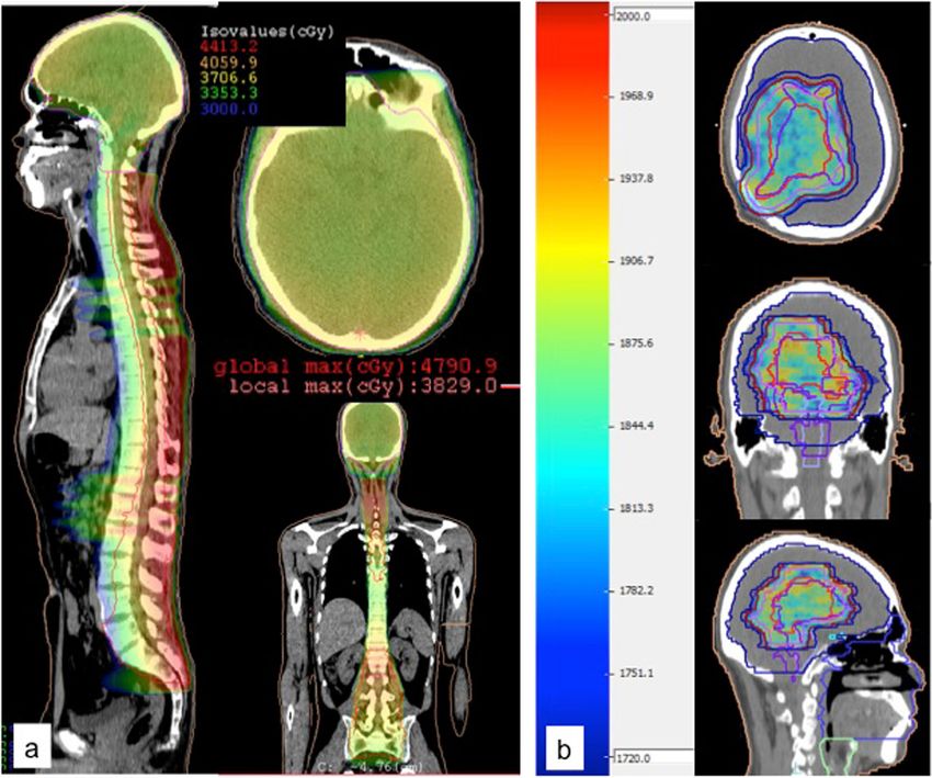
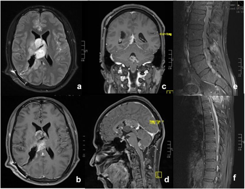
Goyal et al. Journal of the Egyptian National Cancer Institute   (2020) 32:23                                                        Page 2 of 8

Case presentation                                                         like cells in a delicate vascular network, intervening neutro-
A 33-year-old gentleman presented to the neurologist                      phils, calcification, and Homer-Wright Rosettes. Necrosis
with complaints of loss of memory and forgetfulness                       and microvascular proliferation were observed. Several mi-
since 2.5 months, along with recent onset headache and                    toses were discernible with areas of hemorrhage (Fig. 2a–c)
vomiting. His clinical examination showed a Glasgow                       Tumor cells were negative for isocitrate dehydrogenase 1
coma score of 15 with inability to remember recent and                    (IDH-1) and chromogranin, positive for synaptophysin, fo-
some past events and also to perform simple mathemat-                     cally for glial fibrillary acid protein (GFAP), and negative
ical calculations. There were no appreciable cranial                      for epithelial membrane antigen (EMA) and p53. MIB-1 LI
nerve deficits, motor, or sensory loss.                                   (Ki-67) was 8–10% (Fig. 2d–i). A diagnosis of atypical neu-
  A contrast enhanced magnetic resonance imaging                          rocytoma (World Health Organization, WHO grade II),
(MRI) of brain showed a large, fairly defined lobulated                   right ventricle was given (Fig. 2).
heterogeneously enhancing space occupying lesion                             Owing to subtotal excision and high Ki-67, he was ad-
(SOL) with its epicenter in subependymal region of right                  vised adjuvant focal radiotherapy (RT) to residual tumor.
lateral ventricular body, showing gross extensions into                   However, on the tenth postoperative day, he developed
the intraventricular space, thalamus, body of corpus cal-                 sudden onset severe headache, vomiting, and blurring of
losum, and leftward displacement of septum pellucidum.                    vision. MRI of brain was repeated along with a screening
There was mild ventricular dilatation. There was a small                  study of spine with contrast. Residual right ventricular
eccentric non-enhancing cystic necrotic focus. A small                    tumor mass was seen infiltrating into right thalamus,
focus, hypointense on T1 and hyperintense on T2 and                       posterior limb of right internal capsule, and reaching
FLAIR sequences with restricted diffusion on diffusion-                   across the midline into the medial part of the left thal-
weighted imaging (DWI) and heterogeneous post-contrast                    amus, causing mass effect over the third ventricle and
enhancement was seen involving left thalamus. No lepto-                   mild upstream dilatation of the lateral ventricles. There
meningeal enhancement was seen on the brain imaging,                      was abnormal enhancement along the interpenduncular
and thus spine was not imaged preoperatively nor was a                    cistern, prepontine, perimesencephalic basal cistern, Syl-
cerebrospinal fluid (CSF) study performed (Fig. 1). On                    vian fissures, cortical sulci, floor of fourth ventricle, and
magnetic resonance spectroscopy (MRS), the lesion                         tentorium cerebelli, suggesting leptomeningeal spread.
showed a high choline to N-acetylaspartate (NAA) ratio (>                 Diffuse leptomeningeal enhancement was also seen
2). Findings suggested a mitotic lesion, possibly lymphoma                along pial surface of spinal cord and along the nerve
or glioblastoma.                                                          roots of cauda equina. There was no focal intramedul-
  A neurosurgeon was consulted, and only a subtotal                       lary lesion (Fig. 3). CSF cytology returned positive for
tumor excision was possible. Intraoperatively, it was a                   malignant cells. A diagnosis of atypical neurocytoma
greyish, soft, moderately vascular, suckable tumor ex-                    with leptomeningeal spread was made.
tending inside the frontal horn, atrium, and trigone of                      The clinical developments were discussed in the multi-
right lateral ventricle and to the opposite ventricle.                    disciplinary tumor board, and thereafter, the treatment
  Histopathological examination showed sections of                        plan was revised. He received craniospinal irradiation
tumor tissue composed of sheets of oligodendrogioma-                      (CSI) of 36 gray (Gy) using three-dimentional conformal

 Fig. 1 MRI brain (contrast) showed a large, fairly defined lobulated heterogeneously enhancing space occupying lesion with its epicenter in
 subependymal region of right lateral ventricular body, showing gross extensions into the intraventricular space, thalamus, body of corpus
 callosum, and leftward displacement of septum pellucidum, with mild ventricular dilatation. There was a small eccentric non-enhancing cystic
 necrotic focus. A small focus, hypointense on T1 and hyperintense on T2 and FLAIR sequences with restricted diffusion on diffusion-weighted
 imaging (DWI) and heterogeneous post-contrast enhancement was seen involving left thalamus. No leptomeningeal enhancement was seen. a
 Axial T1 contrast sequence. b–d Axial, coronal, and sagittal T2 FLAIR sequences of the brain showing the tumor extensions
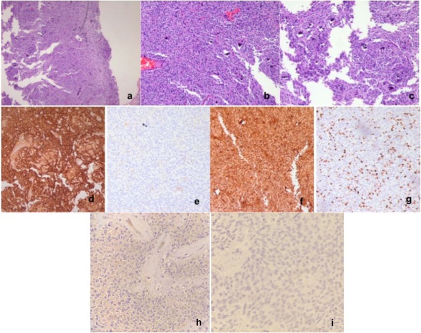
Goyal et al. Journal of the Egyptian National Cancer Institute    (2020) 32:23                                                          Page 3 of 8

 Fig. 2 a Photomicrographs showing sections of tumor tissue composed of sheets of oligodendrogioma-like cells with round to oval nuclei within
 a clear cytoplasm in a delicate vascular network, intervening neutrophils, calcification, and Homer-Wright Rosettes (H&E, 4×). b, c High power
 views (H&E, 10×) showing necrosis, microvascular proliferation, several mitoses, and areas of hemorrhage. d Immunostaining showing diffuse
 positivity for synaptophysin. e Tumor cells are immunonegative for epithelial membrane antigen (EMA). f Focal positivity seen for glial fibrillary
 acid protein (GFAP). g Ki-67 labeling index was 8–10%. h Isocitrate dehydrogenase 1 (IDH-1) and i chromogranin were negative on
 immunostaining. The morphological and immunohistochemical features are those of an atypical neurocytoma

radiotherapy (3D-CRT) with a tumor bed boost of 20 Gy                       posterior subarachnoid space in upper cervical cord at
using volumetric modulated arc therapy (VMAT) (Fig. 4).                     the level of C2 vertebra, along with abnormal leptomen-
He completed the planned treatment course with one                          ingeal enhancement along ventricles and spinal canal.
instance each of grade 2 neutropenia requiring growth factor                (Fig. 6). He received 6 cycles of salvage chemotherapy
support, grade 2 mucositis, and grade 1 nausea/vomiting.                    (cisplatin and etoposide every 3 weeks) but his condition
  He was on regular follow-up with imaging every 3                          deteriorated rapidly thereafter, and after tumor board
months. He was clinically stable but had persistent                         discussion, he was offered best supportive care.
memory deficits. Partial response in the thalamic lesion
and complete resolution of leptomeningeal enhancement
were noted on MRI done at 3 and 6 months (Fig. 5).                          Discussion
After 10 months of completing CSI, he complained of                         Most CNs present with features of increased intracranial
gait imbalance and tendency to fall on right side along                     tension (headache, vomiting), though visual field defects,
with hand tremors while writing. Contrast MRI of brain                      gait abnormalities, and memory changes may also develop.
and spine showed interval development of nodular mass                       Clinical suspicion of CN is rare, and the final diagnosis is
lesions in fourth ventricular outlet and right-sided                        confirmed only with postoperative histopathology.
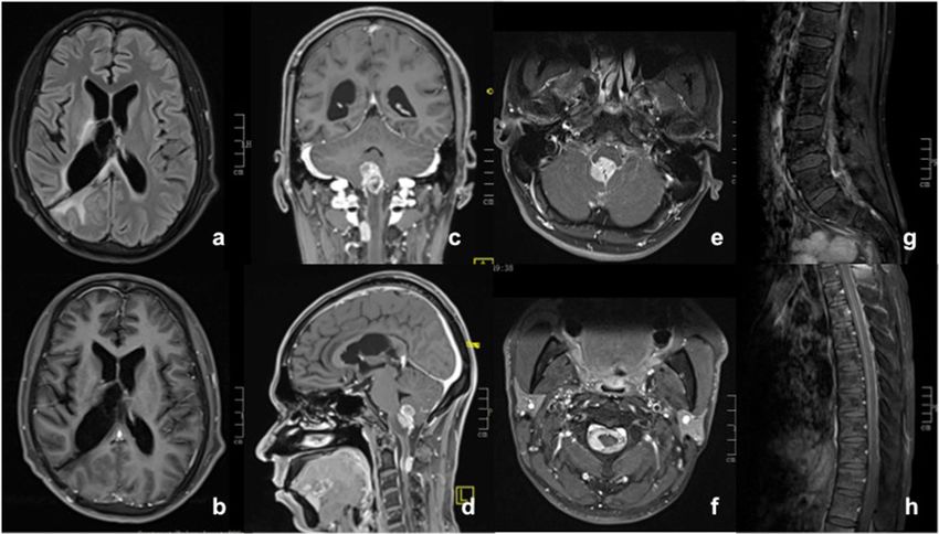
Goyal et al. Journal of the Egyptian National Cancer Institute      (2020) 32:23                                                           Page 4 of 8

 Fig. 3 MRI of brain showed the residual right ventricular tumor mass infiltrating into right thalamus, posterior limb of right internal capsule, and
 reaching across the midline into the medial part of the left thalamus, causing mass effect over the third ventricle and mild upstream dilatation of
 the lateral ventricles. Abnormal enhancement was seen along the interpenduncular cistern, prepontine, perimesencephalic basal cistern, Sylvian
 fissures, cortical sulci, floor of fourth ventricle, and tentorium cerebelli, suggesting leptomeningeal spread. a T2 FLAIR axial section. b–d T1
 contrast axial, coronal, and sagittal images. e, f Diffuse leptomeningeal enhancement along pial surface of spinal cord and along the nerve roots
 of cauda equina without any focal intramedullary lesion

   Tumors arise from subependymal plate of lateral ventri-                   Most cases have a favorable prognosis with benign bio-
cles. Computed tomography (CT) typically shows a ven-                        logical behavior. However, atypical aspects have been re-
tricular space occupying lesion, usually involving lateral                   ported in 20–25% cases [9]. This rare subgroup, termed
ventricles; the mass is well circumscribed with attachment                   “atypical central neurocytomas”, constitues tumors with
to septum pellucidum and enhances with iodinated con-                        non-central location (extraventricular or spinal), older
trast. Approximately 50% tumors have calcification. MRI                      age at presentation, MIB-1 LI over 2–3%, and/or histo-
shows a tumor that is iso- or hyperintense to cerebral cor-                  logic atypia with infiltrative margins, increased mitoses,
tex and enhances with contrast. Usually no draining vein                     necrosis, endothelial or vascular proliferation or cellular
is seen, and the attachments and infiltration are better vi-                 pleomorphism, and biological aggression manifesting as
sualized on MRI than on CT. Role of positron emission                        early recurrence and progression [3, 5, 9]. A higher cut-
tomography (PET) or spectroscopy is uncertain [8].                           off for MIB-1 LI (10%) has been suggested by Qiu-lin
   On light microscopy, typical CNs appear as small                          et al., with a proposal to label them as “anaplastic neuro-
round cells with round nuclei and scant cytoplasm re-                        cytoma, WHO grade III” [4].
sembling oligodendroglioma or ependymoma and often                             Surgical excision is the mainstay of treatment. Gross
stain positive for synaptophysin. Electron microscopy                        total resection (GTR) yields a local control (LC) of 57%
and immunohistochemistry suggest a neuronal origin.                          and 5-year survival (5y-S) of 93% without need for

Goyal et al. Journal of the Egyptian National Cancer Institute    (2020) 32:23                                                         Page 5 of 8

 Fig. 4 (a) Axial, Coronal and Sagittal images of the Craniospinal RT plan showing dose colorwash. A dose of 36 Gy in 20 fractions was planned to
 the entire craniospinal axis using 3DCRT. (b) This was followed by a boost to tumor bed (20 Gy in 10 fractions) using VMAT

adjuvant treatment. For subtotal resection (STR) alone,                    yields comparable control rates but slightly lower incidence
LC and 5y-S drop to 7% and 43%, improving to 70% and                       of complications and higher risk of distant failures, though
78%, respectively, with adjuvant radiation therapy [10].                   the difference is not significant [12].
  Rades and Schild, after an extensive review of 438 patients,               A literature review of 19 atypical cases with malignant
including 365 adults and 87 atypical CNs, with a minimum                   behavior showed MIB-1 LI above 2% in 12 of 14 cases
follow up of 1 year, gave treatment recommendations for                    with available data and above 10% in those progressing
various CN subgroups. No adjuvant therapy is required after                within a year [6]. Seventeen patients had craniospinal dis-
GTR. After STR, adjuvant radiation improves survival in                    semination though CSI was given to 3 patients only after
atypical lesions and in adults but not in children, while local            documented craniospinal dissemination. The documented
control improves across all subgroups. Adults need doses                   disease-free follow-up information for these 3 cases was
exceeding 54 Gy while doses above or below 50 Gy have                      available at 3 months, 3 years, and 5.5 years, respectively
comparable outcomes in children. Authors recommended                       [13–15]. In their own case, the Mozes et al. considered
adjuvant RT doses of 50 Gy in children, 50–54 Gy in typical                CSI upfront but owing to lack of robust supporting litera-
CN, and 56–60 Gy in atypical CN [11]. Stereotactic radio-                  ture, delivered it only after the patient developed spinal
surgery with peripheral doses of 9–25 Gy (mean 14.9 Gy)                    dissemination at 3 years. They recommended that CN
Goyal et al. Journal of the Egyptian National Cancer Institute     (2020) 32:23                                                          Page 6 of 8

 Fig. 5 a Axial T2 FLAIR and b–d axial, coronal, and sagittal sections on T1 contrast MRI of brain showing altered signal involving right thalamus,
 posterior limb of internal capsule (reduced compared to pre-radiotherapy scans) with no abnormal enhancement in this region. Subtle persistent
 signal alteration in the medial left thalamus. Mild persistent focal leptomeningeal enhancement seen in right side of superior cerebellar and
 quadrigeminal plate cistern. No residual enhancing mass lesion in the right lateral ventricle or corpus callosum

cases with high malignant potential should be considered                    20 Gy, describing long survival exceeding 7 years in one of
for maximal tumor resection followed by adjuvant CSI, in-                   their cases [16].
stead of only adjuvant local RT [6]. Leenstra et al. reported                 Role of chemotherapy is uncertain and has been
a 35-year experience of 45 cases, wherein 3 patients were                   explored in inoperable or disseminated cases, with
given prophylactic CSI (2 after resection and 1 after bi-                   reports of stable disease and partial response with
opsy) with CSI doses of 30–36 Gy and tumor bed boost of                     regimens including carboplatin, vincristine, topotecan,

 Fig. 6 Axial a T2 FLAIR and b T1 contrast sections of brain showing near complete resolution of the primary lesion. c–f There is interval
 development of heterogeneously enhancing mass lesion (2.4 × 2.0 × 1.4 cm) in the inferior part of fourth ventricle causing marked compression
 on dorsal medulla. Another heterogeneously enhancing nodular lesion (1.1 × 0.6 × 1.4 cm) seen in right posterior subarachnoid space at the level
 of C2 vertebra causing mild compression of the posterior cervical spinal cord. Abnormal leptomeningeal enhancement is seen along the surface
 of brainstem and cerebellum. g, h Abnormal leptomeningeal enhancement is also seen along the surface of entire spinal cord with thickening
 and clumping of the cauda equina nerve roots
Goyal et al. Journal of the Egyptian National Cancer Institute           (2020) 32:23                                                                Page 7 of 8

cyclophosphamide, and temozolomide [6, 13, 17, 18].                               Ethics approval and consent to participate
Stem cell transplantation following high dose chemo-                              The patient gave written consent to participate as a subject in the case
                                                                                  report without personal identifiers such as name, ethnicity, and face
therapy has also been tried [19].                                                 photograph. Approval of Institutional Review Board and Ethics Committee at
   In the present case, there was no clinical evidence of                         Medanta The Medicity, Gurugram was obtained for publication of this
CSF dissemination at diagnosis but appeared soon after                            manuscript (Reference: MICR 1026/2019).

surgery, suggesting an aggressive behavior. Although CSI
was given in adjuvant setting, he remained relapse-free                           Consent for publication
                                                                                  The patient gave written consent for using the clinical information without
for only 10 months, indicating that perhaps adjuvant
                                                                                  identifiers for the purpose of scientific publication. This consent was
chemotherapy should also have been considered. Add-                               obtained at the time of planning his radiotherapy and before the manuscript
itionally, we recommend spinal screening for all cases                            was written.
with a MIB-1 LI of > 4% or several features of atypia
such as necrosis, mitoses, or vascular proliferation on                           Competing interests
microscopy.                                                                       Not applicable

                                                                                  Author details
                                                                                  1
Patient’s perspective                                                              Division of Radiation Oncology, Medanta The Medicity, Gurugram, Haryana
                                                                                  122001, India. 2Department of Pathology, Medanta The Medicity, Gurugram,
The patient belonged to the lower socioeconomic group.                            Haryana 122001, India. 3Division of Neurosurgery, Medanta The Medicity,
The diagnosis of malignancy at a young age was dis-                               Gurugram, Haryana 122001, India.
heartening; but despite seeking medical attention in time
                                                                                  Received: 4 November 2019 Accepted: 24 March 2020
and timely surgery, the course of disease was unpredict-
able and hard to manage due to mounting side effects of
treatment as well as financial challenge for cost of inter-
                                                                                  References
ventions, lodging of family members away from home                                1. Hassoun J, Söylemezoglu F, Gambarelli D, Figarella-Branger D, von Ammon
and loss of work as well as bleak future prospects.                                   K, Kleihues P. Central neurocytoma: a synopsis of clinical and histological
                                                                                      features. Brain Pathol. 1993;3:297–306.
                                                                                  2. Hassoun J, Gambarelli D, Grisoli F, Pellet W, Salamon G, Pellissier JF, et al.
Conclusion                                                                            Central neurocytoma. An electron-microscopic study of two cases. Acta
                                                                                      Neuropathol. 1982;56:151–6.
Atypical central neurocytoma may occasionally exhibit ag-
                                                                                  3. Kaur G, Kane AJ, Sughrue ME, Oh M, Safaee M, Sun M, et al. MIB-1 labeling
gressive behavior. Treatment may need to be tailored to                               index predicts recurrence in intraventricular central neurocytomas. J Clin
specific presentation, imaging, and pathologic findings.                              Neurosci. 2013;20:89–93.
                                                                                  4. Qiu-lin L, Xiao-dong C, Da-yun P. Final diagnosis – Anaplastic central
                                                                                      neurocytoma (CN) of both lateral ventricles. Available from: http://www.
Abbreviations
                                                                                      path.upmc.edu/cases/case784/dx.html. (2013) ().
CN: Central neurocytoma; MIB-1 LI: MIB-1 labeling index; MRI: Magnetic
                                                                                  5. Rades D, Schild SE, Fehlauer F. Prognostic value of the MIB-1 labeling index
resonance imaging; SOL: Space occupying lesion; DWI: Diffusion-weighted
                                                                                      for central neurocytomas. Neurology. 2004;62:987–9.
imaging; CSF: Cerebrospinal fluid; MRS: Magnetic resonance spectroscopy;
                                                                                  6. Mozes P, Szanto E, Tiszlavicz L, Barzo P, Cserhati A, Fodor E, et al. Clinical
NAA: N-acetylaspartate; GFAP: Glial fibrillary acid protein; EMA: Epithelial
                                                                                      course of central neurocytoma with malignant transformation-an indication
membrane antigen; WHO: World Health Organization; RT: Radiotherapy;
                                                                                      for craniospinal irradiation. Pathol Oncol Res. 2014;20:319–25.
CSI: Craniospinal irradiation; Gy: Gray; 3D-CRT: Three-dimentional conformal
                                                                                  7. Juratli TA, Geiger K, Leimert M, Schackert G, Kirsch M. Atypical Central
radiotherapy; VMAT: Volumetric modulated arc therapy; CT: Computed
                                                                                      Neurocytoma with Recurrent Spinal Dissemination over a Period of 20 Years:
tomography; PET: Positron emission tomography; GTR: Gross total resection;
                                                                                      A Case Report and Review of the Literature. Case Rep Neurol Med. 2013;
LC: Local control; IDH-1: Isocitrate dehydrogenase 1
                                                                                      2013:925647.
                                                                                  8. Donoho D, Zada G. Imaging of central neurocytomas. Neurosurg Clin N Am.
Acknowledgements                                                                      2015;26:11–9.
Not applicable                                                                    9. Bonney PA, Boettcher LB, Krysiak RS 3rd, Fung KM, Sughrue ME. Histology
                                                                                      and molecular aspects of central neurocytoma. Neurosurg Clin N Am. 2015;
Authors’ contributions                                                                26:21–9.
SG and TK were involved in clinically managing the case, acquiring data, and      10. Rades D, Fehlauer F, Schild SE. Treatment of atypical neurocytomas. Cancer.
clinically interpreting it. They contributed majorly in writing the manuscript.       2004;100:814–7.
DG and AD contributed to review of literature, its interpretation in the          11. Rades D, Schild SE. Treatment recommendations for the various subgroups
context of the clinical case, and in writing the manuscript. IM analyzed the          of neurocytomas. J Neuro-Oncol. 2006;77:305–9.
histopathological features of the patient’s biopsy specimen and gave inputs       12. Barani IJ, Raleigh DR, Larson D. The management of central neurocytoma:
to interpreting the cytology at progression. KSN performed the initial surgery        radiotherapy. Neurosurg Clin N Am. 2015;26:45–56.
and subsequent lumbar puncture. He contributed to interpretation of               13. Brandes AA, Amistà P, Gardiman M, Volpin L, Danieli D, Guglielmi B, et al.
imaging findings at each juncture. All authors read and approved the final            Chemotherapy in patients with recurrent and progressive central
manuscript.                                                                           neurocytoma. Cancer. 2000;88:169–74.
                                                                                  14. Nayyar M, Mayo MC, Shiroishi M, Commins D, Liu CY, Go JL, et al. Atypical
                                                                                      central neurocytoma with metastatic craniospinal dissemination: a case
Funding                                                                               report. Clin Imaging. 2016;40:1108–11.
Not applicable                                                                    15. Takao H, Nakagawa K, Ohtomo K. Central neurocytoma with craniospinal
                                                                                      dissemination. J Neuro-Oncol. 2003;61:255–9.
Availability of data and materials                                                16. Leenstra JL, Rodriguez FJ, Frechette CM, Giannini C, Stafford SL, Pollock BE,
All data generated or analyzed during this study are included in this                 et al. Central neurocytoma: management recommendations based on a 35-
submitted article.                                                                    year experience. Int J Radiat Oncol Biol Phys. 2007;67:1145–54.
Goyal et al. Journal of the Egyptian National Cancer Institute (2020) 32:23 Page 8 of 8
17. Amini E, Roffidal T, Lee A, Fuller GN, Mahajan A, Ketonen L, et al. (2008)
Central neurocytoma responsive to topotecan, ifosfamide, carboplatin.
Pediatr Blood Cancer 2008; 51: 137-140.
18. Stapleton CJ, Walcott BP, Kahle KT, Codd PJ, Nahed BV, Chen L, et al. (2012)
Diffuse central neurocytoma with craniospinal dissemination. J Clin Neurosci
2012; 19: 163-166.
19. Buchbinder D, Danielpour M, Yong WH, Salamon N, Lasky J. (2010)
Treatment of atypical central neurocytoma in a child with high dose
chemotherapy and autologous stem cell rescue. J Neuro-Oncol 2010; 97:
429-437.
Publisher’s Note
Springer Nature remains neutral with regard to jurisdictional claims in
published maps and institutional affiliations.You can also read