Acute Kidney Injury in COVID-19: Emerging Evidence of a Distinct Pathophysiology - sfndt
←
→
Page content transcription
If your browser does not render page correctly, please read the page content below
PERSPECTIVE          www.jasn.org

Acute Kidney Injury in COVID-19: Emerging Evidence of
a Distinct Pathophysiology
Daniel Batlle,1 Maria Jose Soler,2 Matthew A. Sparks,3,4 Swapnil Hiremath,5
Andrew M. South,6 Paul A. Welling,7 Sundararaman Swaminathan,8
on behalf of the COVID-19 and ACE2 in Cardiovascular, Lung, and Kidney Working Group*
1
 Northwestern University Feinberg School of Medicine, Division of Nephrology and Hypertension, Chicago, Illinois
2
 Hospital Universitari Vall d’Hebron, Division of Nephrology Autonomous University of Barcelona, Barcelona, Spain
3
 Division of Nephrology, Department of Medicine, Duke University School of Medicine, Durham, North Carolina
4
 Renal Section, Durham Veterans Affairs Health Care System, Durham, North Carolina
5
 Division of Nephrology, Department of Medicine, University of Ottawa, Ottawa, Ontario, Canada
6
 Department of Pediatrics, Section of Nephrology, Wake Forest School of Medicine, Winston Salem, North Carolina
7
 School of Medicine, Departments of Medicine and Physiology, Johns Hopkins University, Baltimore, Maryland
8
 Division of Nephrology, and Center for Immunity, Inflammation and Regenerative Medicine, University of Virginia,
Charlottesville, Virginia

JASN 31: ccc–ccc, 2020. doi: https://doi.org/10.1681/ASN.2020040419

The most common reported reasons for              H.M. Wang, et al.: Human kidney is a            symptoms of loin pain and hematuria
intensive care unit admission for pa-             target for novel severe acute respiratory       suggesting renal infarction. Numerous
tients with severe coronavirus disease            syndrome coronavirus 2 [SARS-CoV-2]             observations by treating physicians attest
2019 (COVID-19) are either hypoxemic              infection [preprint posted online April         that there is increased occurrence of cir-
respiratory failure leading to mechanical         10, 2020]. medRxiv doi:10.1101/2020.            cuit clotting in patients with COVID-19
ventilation or hypotension requiring va-          03.04.20031120). An important report            undergoing dialysis. COVID-19 is also
sopressor support. Data on AKI are ei-            on autopsy findings from deceased                associated with increased myocardial in-
ther lacking1 or only reporting incidence         patients with COVID-19 again demon-             jury that mimics myocardial infarction,
on the basis of case series and retrospec-        strated prominent acute proximal tubu-          possibly from myocarditis and microan-
tive studies.2 In this Perspective, we em-        lar injury, but also peritubular erythro-       giopathy.5 Thus, it is conceivable that the
phasize that AKI can be a severe compli-          cyte aggregation and glomerular fibrin           hypercoagulable state that appears to be a
cation of COVID-19 and highlight the              thrombi with ischemic collapse.3 This           characteristic complication of severe
importance of assessing, defining, and             paper also reported endothelial damage,
reporting the course of AKI.                      hemosiderin deposition, pigment casts
   Understandably relevant information                                                            Published online ahead of print. Publication date
                                                  related to rhabdomyolysis, and inflam-
                                                                                                  available at www.jasn.org.
that normally would be part of clinical           mation. Notably, some of these patients
descriptions and research publications            lacked evidence of AKI as detected by           Correspondence: Dr. Daniel Batlle, Division of
                                                                                                  Nephrology and Hypertension, Northwestern Uni-
has not been collected because of the             routine measures (creatinine and/or             versity Feinberg School of Medicine, 300 East Su-
magnitude and accelerated pace of the             BUN), highlighting the possibility of           perior Street, Tarry 14-727, Chicago, IL 60611-
                                                                                                  3008, or Dr. Sundararaman Swaminathan, Division
COVID-19 pandemic. Of great rele-                 substantial subclinical kidney injury.          of Nephrology, and Center for Immunity, In-
vance is a preprint in medRxiv reporting             Recent clinical and autopsy reports of       flammation and Regenerative Medicine, P.O. Box
a 23% AKI incidence among 85 patients             COVID-19 from China and the United              800133, University of Virginia, Charlottesville, VA
                                                                                                  22908. E-mail: d-batlle@northwestern.edu or
(over 60% in high-risk patients). The             States confirm increased clotting and            swami@virginia.edu
authors analyzed kidney histology from            disseminated intravascular coagulation
                                                                                                  *The COVID-19 and ACE2 in Cardiovascular, Lung, and
autopsies of six patients who had AKI             with small vessel thrombosis and pulmo-         Kidney Working Group: Matthew A. Sparks, Swapnil
showing severe acute tubular necrosis             nary infarction.4 Further, elevated d-dimer     Hiremath, Daniel Batlle, Andrew South, Paul Welling,
with lymphocyte and macrophage infil-              and low platelet levels correlated with worse   J. Matt Luther, Jordana Cohen, James Brian Byrd,
                                                                                                  Louise M. Burrell, Laurie Tomlinson, Vivek Bhalla, María
tration, but it is not clear from this re-        outcomes.4 We are aware that some patients      José Soler, Sundar Swaminathan, Michelle N. Rheault,
port if these patients had actually devel-        with COVID-19 manifest evidence of mi-          and Sundar Swaminathan.
oped cortical necrosis (B. Diao, C.H.             croangiopathy in other organ systems, such      Copyright © 2020 by the American Society of
Wang, R.S. Wang, Z.Q. Feng, Y.J. Tan,             as splenic infarction or presenting             Nephrology

JASN 31: ccc–ccc, 2020                                                                                         ISSN : 1046-6673/3107-ccc                1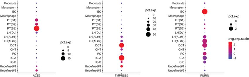
PERSPECTIVE    www.jasn.org

Figure 1. Single-cell RNA of SARS-COV-2 receptor (ACE2) and known priming proteases in the kidney. There is no clear correspon-
dence in single-cell RNA between ACE2 and TMPRSS2. EC, endothelial cell; PT, proximal tubule (S1, Segment one; S2 Segment two, S3
Segment three); LH(DL), loop of henle(descending thin limb); LH(AL), loop of henle(descending limb type one [#type 1] and type two [type
#2]); DCT, distal convoluted tubule; CNT, connecting tubule; PC, principal cell; IC-A, alpha intercalated cell; IC-B, beta intercalated cell;
pct. exp, percentexpressed. Data were extracted with permission from human kidney single-cell RNA-sequencing data.12,13

COVID-19 could, in some cases, foster           deposition in renal tubules of six pa-          doi:10.1101/2020.03.04.20031120). Su
the evolution of acute tubular necrosis         tients with SARS-CoV-2 infection, sug-          et al.3 similarly observed the presence
to cortical necrosis and, therefore, irre-      gesting activation of the complement            of virus-like particles in podocytes and
versible kidney failure.                        pathway. An interaction between angio-          renal tubular epithelial cells by electron
    Innate immunity and coagulation             tensin II (AngII) overactivity, innate/         microscopy, and SARS-CoV-2 nucleo-
pathways are intricately linked. 6              adaptive immune and complement                  protein antibody stained renal tubular
COVID-19–associated macrophage acti-            pathways, and the coagulation system            epithelia positive, but the specificity of
vation, hyperferritinemia, cytokine             could influence AKI severity and out-            the antibody used needs to be estab-
storm, and release of pathogen-                 comes. Inflammation-induced erythro-             lished. Although, as far as we know,
associated molecular patterns and               cyte aggregation (reflected as elevated          SARS-CoV-2 RNA, has not been de-
damage-associated molecular proteins            erythrocyte sedimentation rate) and             tected in the kidney, these results indi-
can result in release of tissue factor and      heme-mediated pathology may worsen              cate that SARS-CoV-2 could directly in-
activation of coagulation factors that          oxidative stress, inflammation, and              fect human kidney tubules and induce
create a predisposition to hypercoagula-        complement activation, to aggravate mi-         cytoplasmic renal tubular inclusions, a
bility. 6 Severe acute respiratory syn-         crovascular injury. 8 Further, organ            feature observed in other virus-
drome coronavirus 2 (SARS-CoV-2)                crosstalk between the injured lung, the         associated nephropathies. Thus, al-
may also target lymphocytes as they ex-         heart, and the kidney can worsen pathol-        though AKI may be attributable to hy-
press angiotensin-converting enzyme 2           ogy. Detailed studies to decipher the na-       potension and decreased kidney perfu-
(ACE2), leading to lymphocyte activa-           ture of coagulation dysfunction, micro-         sion secondary to hemodynamic or
tion and, consequently, activation-             angiopathy, and potential role for innate       hemostatic factors or associated sepsis,
induced cell death than can result in           immune and complement pathways are              one needs to consider that viral infection
lymphopenia of both CD41 and CD81               required to gain further insights regard-       of the kidneys with viral replication di-
T cells.7 Further, procoagulation path-         ing kidney pathology in COVID-19.               rectly in kidney parenchyma also plays
ways and complement systems can acti-              Also of interest is the finding that          a role.
vate each other. In support of this inter-      SARS-CoV-2 nucleocapsid protein was                 The main binding site for SARS-CoV-
action in COVID-19, Diao and                    observed in tubular structures in the kid-      2, like SARS-CoV, is the ACE2 protein,
colleagues (B. Diao, C.H. Wang, R.S.            neys from the six patients examined, and        which is expressed in the kidney much
Wang, Z.Q. Feng, Y.J. Tan, H.M. Wang,           nucleocapsid protein–positive inclusion         more than the lungs. 9,10 ACE2 is ex-
et al.: Human kidney is a target for novel      bodies were also observed in the cyto-          pressed on the brush border apical
severe acute respiratory syndrome coro-         plasm (B. Diao, C.H. Wang, R.S. Wang,           membrane of the proximal tubule,
navirus 2 [SARS-CoV-2] infection [pre-          Z.Q. Feng, Y.J. Tan, H.M. Wang, et al.:         where it colocalizes with angiotensin-
print posted online April 10, 2020].            Human kidney is a target for novel severe       converting enzyme (ACE), and is also
medRxiv doi:10.1101/2020.03.04.                 acute respiratory syndrome coronavirus          present at lower levels in podocytes.10
20031120) observed strong complement            2 [SARS-CoV-2] infection [preprint              It is conceivable that the virus could en-
C5b-9 (membrane attack complex)                 posted online April 10, 2020]. medRxiv          ter the kidney by invading podocytes

2        JASN                                                                                                          JASN 31: ccc–ccc, 2020
www.jasn.org PERSPECTIVE
first, and thus gain access to the tubular furin cleavage site in the Spike protein protein expression are altered in mouse
fluid and subsequently bind to ACE2 in that is processed during biogenesis.14 models of DKD and in patients with
the proximal tubule. In primary human Any effect of proteinuria, hyperin- DKD.10,17 Thus, patients with CKD, es-
airway epithelia, ACE2 is expressed api- flammation, or tubular injury on proxi- pecially those with DKD, who develop
cally, and SARS-CoV-2 infection pre- mal tubular ACE2 expression or SARS- COVID-19 may be at higher risk of
dominantly occurs on the apical surface, CoV-2 viral entry is currently unknown. AKI because of baseline upregulation of
but infection can occur on the basolat- Viral replication in podocytes and the the ACE and downregulation of ACE2, a
eral surface at low efficiency.11 Corona- ensuing damage could in theory account combination that primes a proinflam-
virus entry into host target cells also re- for the proteinuria that has been report- matory (including complement activa-
quires fusion of the viral envelope with ed in patients with COVID-19.2 Further, tion) and profibrotic state in the kidneys.
cellular membranes. Fusion-activated COVID-19–associated hemophagocytic Interestingly, a recent study described
SARS-CoV peptides are created by spe- macrophage activation and microangi- single-cell transcriptome analysis in 15
cific proteolytic cleavage of the S pro- opathy could also cause AKI and podo- normal human kidney samples. 18 In
teins, in a step called “priming.” As a cyte damage. Of interest, cases of this study, the proportions of kidney
consequence, cell infectivity not only de- COVID-19–associated collapsing glo- cells expressing ACE2, the SARS-CoV-2
pends on ACE2 expression, but is also merulopathy have been described.15 binding site, and proteases of the
governed by types of proteases found in Regardless of direct viral infection of TMPRSS family were compared between
a given cell type. In the kidney, Trans- the kidney, AngII is likely increased in occidental and Asian individuals.18 In-
membrane protease, serine 2 the context of acute lung injury16 and terestingly, the expression of ACE2 and
(TMPRSS2) 1 2 –14 (Figure 1), which there is evidence that ACE2 is downre- kidney disease–related genes was higher
primes the SARS-CoV-2 S protein, is ro- gulated in AKI. This may lead to type 1 in occidental donors relative to Asian
bustly expressed in the distal nephron angiotensin receptor activation as well as donors. This would suggest that the sus-
rather than the proximal tubule. It re- decreased angiotensin (1–7) formation ceptibility to kidney injury from corona-
mains to be determined if other and subsequent worsening of AKI. This virus infection might be higher in indi-
TMPRSS in the proximal tubule can me- is particularly important in subpopula- viduals of occidental rather than Asian
diate the priming step, such as TMPRSS tions of patients who have CKD, espe- descent. We are not aware, however, of
4, 5, or 9. Alternatively, tropism of SARS- cially those with diabetic kidney disease data supporting this possibility.
CoV-2 might be expanded by the unique (DKD). ACE2 and ACE mRNA and Despite the very limited information
on kidney involvement in COVID-19,
AKI appears to involve a complex pro-
cess driven by virus-mediated injury, cy-
tokine storm, AngII pathway activation,
dysregulation of complement, hypercoa-
gulation, and microangiopathy interact-
ing with common and known risk fac-
tors for AKI (Figure 2). There is paucity
of data regarding clinical and laboratory
characteristics of AKI in patients with
COVID-19. We urge that further studies
describing and analyzing the clinical
course of patients with COVID-19 in-
clude appropriate indices of kidney
function and diagnosis of AKI in their
analyses, including kidney injury mark-
ers, urine microscopy, quantified urine
protein, urine output, and urine electro-
lytes. Markers of macrophage activation,
coagulation, microangiopathy, and
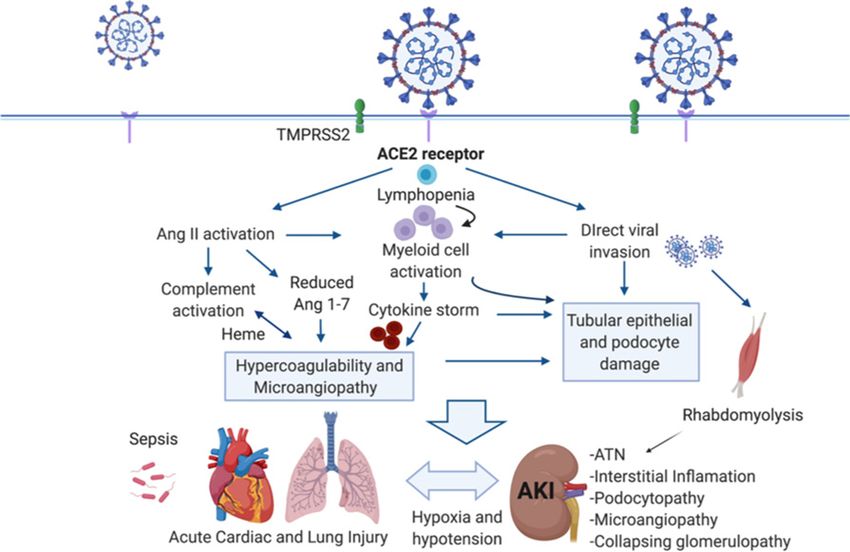
Figure 2. Targeting of ACE2 by SARS-CoV-2 results in angiotensin dysregulation, innate
complement activation, as well as kidney
and adaptive immune pathway activation, and hypercoagulation to result in organ injury
imaging and need for KRT (with relevant
and AKI associated with COVID-19. Organ crosstalk between the injured lungs, the heart,
and the kidney may further propagate injury. CD81 T-cells and natural killer cells can re- details), are important data needed to
strain macrophage activation and are potential targets for SARS-CoV-2. Ang 1–7, angio- further our understanding of AKI path-
tensin 1–7; ATN, acute tubular necrosis. ACE2, angiotensin converting enzyme 2; SARS- ophysiology associated with COVID-19.
CoV-2, severe acute respiratory syndrome coronavirus 2; TMPRSS2, transmembrane pro- Rates of reversibility of, or partial im-
tease, serine 2. provement in, kidney function and any
JASN 31: ccc–ccc, 2020 Pathophysiology of AKI in COVID-19 3PERSPECTIVE www.jasn.org
kidney biopsy results (including immu- Dr. Welling reports grants from NIDDK and the (angiotensin-converting enzyme 2)-independent.
Foudation LeDucq. Hypertension 75: 173–182, 2020
nofluorescence and electron micros-
10. Ye M, Wysocki J, William J, Soler MJ, Cokic I,
copy) should be reported. In the rush Batlle D: Glomerular localization and ex-
to report medical complications of REFERENCES pression of angiotensin-converting enzyme 2
COVID-19, we are missing valuable and angiotensin-converting enzyme: Impli-
clinical information. Speculation about 1. Bhatraju PK, Ghassemieh BJ, Nichols M, Kim
cations for albuminuria in diabetes. J Am Soc
specific interventions would not be ap- Nephrol 17: 3067–3075, 2006
R, Jerome KR, Nalla AK, et al.: Covid-19 in
11. Jia HP, Look DC, Shi L, Hickey M, Pewe L,
propriate until we obtain appropriate in- critically ill patients in the seattle region - case
Netland J, et al.: ACE2 receptor expression
formation. We advocate for a complete series [published online ahead of print Mar
and severe acute respiratory syndrome co-
30, 2020]. N Engl J Med doi:10.1056/
and standardized appraisal of the clinical ronavirus infection depend on differentiation
NEJMoa2004500
and laboratory picture so that preventa- 2. Cheng Y, Luo R, Wang K, Zhang M, Wang Z,
of human airway epithelia. J Virol 79:
tive and therapeutic strategies for AKI 14614–14621, 2005
Dong L, et al.: Kidney disease is associated
12. Wu H, Uchimura K, Donnelly EL, Kirita Y,
can be appropriately designed and with in-hospital death of patients with
Morris SA, Humphreys BD: Comparative
implemented. COVID-19 [published online ahead of print
analysis and refinement of human PSC-
Mar 20, 2020]. Kidney Int doi:10.1016/
derived kidney organoid differentiation with
j.kint.2020.03.005
3. Su H, Yang M, Wan C, Yi LX, Tang F, Zhu HY, single-cell transcriptomics. Cell Stem Cell
et al.: Renal histopathological analysis of 26 23: 869–881.e8, 2018
DISCLOSURES postmortem findings of patients with COVID- 13. Wilson PC, Wu H, Kirita Y, Uchimura K, Ledru
19 in China [published online ahead of print N, Rennke HG, et al.: The single-cell tran-
Apr 9, 2020]. Kidney International doi: scriptomic landscape of early human di-
Dr. Batlle reports nonfinancial support from An-
10.1016/j.kint.2020.04.003 abetic nephropathy. Proc Natl Acad Sci U S A
giotensin Therapeutics Inc., outside the submitted
116: 19619–19625, 2019
work. In addition, Dr. Batlle has a patent “Active 4. Tang N, Li D, Wang X, Sun Z: Abnormal co-
agulation parameters are associated with 14. Walls AC, Park YJ, Tortorici MA, Wall A,
low molecular weight variants of angiotensin con-
poor prognosis in patients with novel coro- McGuire AT, Veesler D: Structure, function,
verting enzyme 2” issued. Dr. Hiremath reports
navirus pneumonia. J Thromb Haemost 18: and antigenicity of the SARS-CoV-2 Spike
other from University of Ottawa, Department of
Medicine, outside the submitted work. Dr. Soler 844–847, 2020 glycoprotein. Cell 181: 281–292.e6, 2020
5. Zhou F, Yu T, Du R, Fan G, Liu Y, Liu Z, et al.: 15. Larsen CP, Bourne TD, Wilson JD, Saqqa O,
reports personal fees from AstraZeneca, nonfinan-
cial support from Boehringer, nonfinancial sup- Clinical course and risk factors for mortality of Sharshir MA: Collapsing glomerulopathy in a
port from Eli Lilly, nonfinancial support from Es- adult inpatients with COVID-19 in Wuhan, patient with coronavirus disease 2019
teve, personal fees from Janssen, personal fees from China: A retrospective cohort study. Lancet (COVID-19) [published online ahead of print
Novo Nordisk, outside the submitted work. Dr. 395: 1054–1062, 2020 Apr 9, 2020]. Kidney Int Rep doi:10.1016/
South reports other from National Institutes of 6. Delvaeye M, Conway EM: Coagulation and j.ekir.2020.04.002
Health, National Heart, Lung, and Blood Institute, innate immune responses: Can we view them 16. Reddy R, Asante I, Liu S, Parikh P, Liebler J,
and Loan Repayment Programs, during the con- separately? Blood 114: 2367–2374, 2009 Borok Z, et al.: Circulating angiotensin pep-
duct of the study. All remaining authors have noth- 7. Chen G, Wu D, Guo W, Cao Y, Huang D, tides levels in acute respiratory distress syn-
ing to disclose. Wang H, et al.: Clinical and immunological drome correlate with clinical outcomes: A
features of severe and moderate coronavirus pilot study. PLoS One 14: e0213096, 2019
disease 2019 [published online ahead of 17. Mizuiri S, Hemmi H, Arita M, Ohashi Y,
print Apr 13, 2020]. J Clin Invest doi:10.1172/ Tanaka Y, Miyagi M, et al.: Expression of ACE
JCI137244 and ACE2 in individuals with diabetic kidney
FUNDING 8. Frimat M, Tabarin F, Dimitrov JD, Poitou C, disease and healthy controls. Am J Kidney
Halbwachs-Mecarelli L, Fremeaux-Bacchi V, Dis 51: 613–623, 2008
Dr. Batlle reports grants from National Institute et al.: Complement activation by heme as a 18. Pan XW, Xu D, Zhang H, Zhou W, Wang LH,
of Diabetes and Digestive and Kidney Diseases secondary hit for atypical hemolytic uremic Cui XG: Identification of a potential mecha-
(NIDDK), during the conduct of the study. Dr. syndrome. Blood 122: 282–292, 2013 nism of acute kidney injury during the
Swaminathan reports grants from NIDDK, during 9. Serfozo P, Wysocki J, Gulua G, Schulze A, Ye M, COVID-19 outbreak: A study based on
the conduct of the study. Dr. South reports grants Liu P, et al.: Ang ii (angiotensin ii) conversion single-cell transcriptome analysis [published
from National Heart, Lung, and Blood Institute to angiotensin-(1-7) in the circulation is pop online ahead of print Mar 31, 2020]. Intensive
and NIDDK, during the conduct of the study. (prolyloligopeptidase)-dependent and ace2 Care Med doi:10.1007/s00134-020-06026-1
4 JASN JASN 31: ccc–ccc, 2020You can also read