Surgical stabilisation of hiatal hernia and gastroesophageal reflux associated with idiopathic inflammatory polymyopathy in a Wire Fox Terrier
←
→
Page content transcription
If your browser does not render page correctly, please read the page content below
Case Report                                                                        Veterinarni Medicina, 66, 2021

                                                                       https://doi.org/10.17221/47/2020-VETMED

Surgical stabilisation of hiatal hernia and
gastroesophageal reflux associated with idiopathic
inflammatory polymyopathy in a Wire Fox Terrier
Mu-Young Kim1, Ji-Hyeon Lee1, Hye-Mi Park2, Jung-Hyun Kim3,
Hun-Young Yoon1*
1
  Department of Veterinary Surgery, College of Veterinary Medicine, Konkuk University,
  Seoul, Republic of Korea
2
  Department of Veterinary Internal Medicine, Konkuk University Veterinary Medical
  Teaching Hospital, Seoul, Republic of Korea
3
  Department of Veterinary Internal Medicine, College of Veterinary Medicine,
  Konkuk University, Seoul, Republic of Korea
*Corresponding author: yoonh@konkuk.ac.kr

Citation: Kim MY, Lee JH, Park HM, Kim JH, Yoon HY (2021): Surgical stabilization of hiatal hernia and gasatroesophageal
reflux associated with idiopathic inflammatory polymyopathy in a Wire Fox Terrier. Vet Med-Czech 66.

Abstract: A one-and-a-half-year-old male Wire Fox Terrier weighing 3 kg presented with continuous drooling and
vomiting. Its body condition score was 3/9, and severe atrophy of the temporal/masticatory muscle, trismus,
and enophthalmos was observed on physical examination. The radiographic examination and fluoroscopic oesopha-
gography revealed a type 1 hiatal hernia with gastroesophageal reflux. The serology tests revealed increased muscle
enzyme activities. The antibody tests for acetylcholine receptor, type 2M fibre, and infectious agents were negative.
A conventional surgical treatment was performed, and a thickened, stretched, and flaccid diaphragmatic muscle
and an extended inelastic oesophageal hiatus were observed. On the histological examination of the diaphragmatic
muscle, a diffuse histiocytic myositis was confirmed. Although the postoperative gastroesophageal reflux totally
disappeared, the oesophageal motility and clinical signs did not improve significantly. Medical treatment with
immunosuppressive agents was attempted and was effective in alleviating the clinical signs and abnormal oesopha-
geal motility. The health condition of the dog was adequately maintained in the 12-month monitoring period.
These findings suggest that, although the medical and surgical treatment have different therapeutic effects, they
should be considered simultaneously for the management of a hiatal hernia associated with polymyopathy in dogs.

Keywords: muscular dystrophy; myositis; oesophageal hiatus; oesophageal motility

  A hiatal hernia describes the condition in which           stomach into the thorax (Sivacolundhu et al. 2002;
the abdominal contents bulge through the oesopha-            Katsianou et al. 2014). Other less frequent types in-
geal hiatus of the diaphragm into the thorax. Four           clude type 2, known as paraoesophageal hiatal her-
types of hiatal hernia have been described in dogs           nia, in which the gastroesophageal junction remains
and cats (Bright et al. 1990; Callan et al. 1993; Kat-       in its normal position, but a portion of the fundus
sianou et al. 2014). Type 1, also known as a slid-           bulges through the hiatus into the thorax; type 3,
ing hiatal hernia, is the most commonly reported             combines elements of both type 1 and type 2; and
type and is defined as the axial displacement of the         type 4, which is a type 3 hernia complicated with
distal oesophagus, gastroesophageal junction, and            a herniation of the organs other than the stomach

1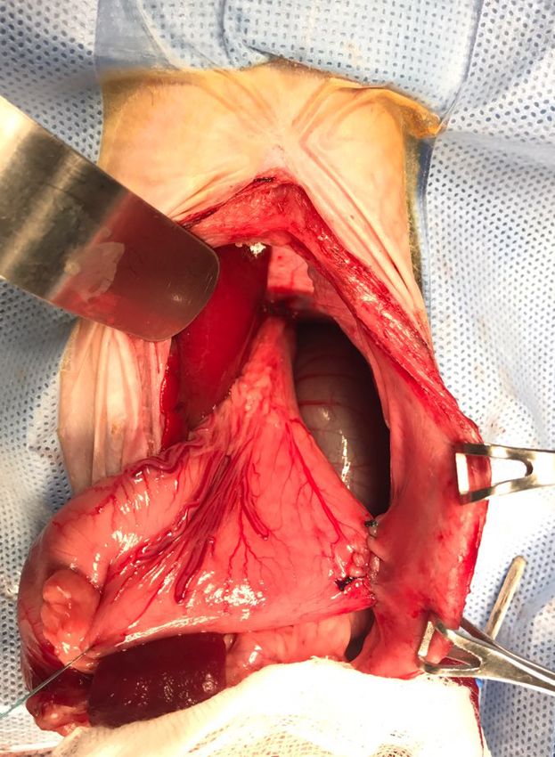
Case Report                                                                  Veterinarni Medicina, 66, 2021

                                                                  https://doi.org/10.17221/47/2020-VETMED

(Sivacolundhu et al. 2002; Katsianou et al. 2014).        myopathy; however, type 1 hiatal hernias related
It is essential to differentiate among the hiatal her-    to inflammatory polymyopathy have rarely been
nia types due to the differences in the pathophys-        reported. In particular, no report has been pub-
iology, which necessitate different treatment strate-     lished distinguishing the roles of the medical and
gies (Prymak et al. 1989; Sivacolundhu et al. 2002;       surgical treatment for this condition.
Katsianou et al. 2014). Type 1 hiatal hernias are            The present report describes the successful sur-
usually congenital and, thus, observed mainly             gical stabilisation of a type 1 hiatal hernia and
in young dogs (Keeley et al. 2008). The acquired          gastroesophageal reflux presumably related to the
form of a type 1 hiatal hernia occasionally occurs        idiopathic inflammatory polymyopathy in a Wire
as a result of a traumatic event or in combination        Fox Terrier, and the ensuing discussion addresses
with severe respiratory disease (Sivacolundhu et al.      the respective roles of the medical and surgical
2002). Clinical signs of a type 1 hiatal hernia include   treatment.
hypersalivation, regurgitation, vomiting, and dys-
phagia, which are usually secondary to reflux oe-
sophagitis or megaoesophagus (Sivacolundhu et al.         Case description
2002; Tauro et al. 2015). Occasionally, these clini-
cal signs may cause aspiration pneumonia (Ellison           A one-and-a-half-year-old male Wire Fox Terrier
et al. 1987; Lorinson and Bright 1998). The two main      was presented to the Veterinary Medical Teaching
treatment options for type 1 hiatal hernias are medi-     Hospital of Konkuk University (Seoul, Republic
cal therapy and surgery. Medical treatment aimed          of Korea) for evaluation of a hiatal hernia. The re-
at resolving the reflux oesophagitis and associated       ferring veterinarian reported perpetual vomiting,
megaoesophagus has been recommended as a first-           drooling, and acute weight loss on the initial obser-
line treatment by some authors (Prymak et al. 1989;       vation. On presentation, the patient was depressed,
Lorinson and Bright 1998; Sivacolundhu et al. 2002).      with a body condition score of 3/9. The patient
The most common surgical treatment for type 1 hia-        exhibited continuous vomiting and drooling, with
tal hernias is a combination of a diaphragmatic hi-       severe atrophy of the temporal/masticatory muscle,
atal plication, an oesophagopexy, and a left-sided        trismus, and enophthalmos. The serum biochemi-
gastropexy (Prymak et al. 1989; Callan et al. 1993;       cal abnormalities included elevated activities of as-
Lorinson and Bright 1998; Guiot et al. 2008). The         partate aminotransferase, 147 IU/l (reference range,
use of fundoplication techniques in dogs has result-      0–50 IU/l); alanine aminotransferase, 441 IU/l (ref-
ed in poor outcomes, despite modification of the          erence range, 10–100 IU/l); and creatine kinase,
original surgery (Ellison et al. 1987; Prymak et al.      1 661 IU/l (reference range, 10–200 IU/l). A plain
1989; Callan et al. 1993; Lorinson and Bright 1998).      thoracic radiography revealed findings compatible
   Inflammatory myopathies are characterised              with a hiatal hernia with a proximal stomach her-
by immunological responses in the skeletal mus-           niation into the thorax through the oesophageal
cles (Tauro et al. 2015). The underlying aetiology        hiatus (Figure 1). On the fluoroscopic oesophagog-
of these diseases in dogs includes an immune mal-         raphy, the esophagogastric junction and a portion
function and infection by protozoa, bacteria, rick-       of the stomach were displaced cranially into the
ettsiae, and/or parasites (Evans et al. 2004; Tauro       thoracic cavity, along with a severely decreased
et al. 2015). In human medicine, the most common          oesophageal motility and gastroesophageal reflux
inflammatory myopathies include polymyositis,             (Figure 2). The gastroesophageal junction sphincter
dermatomyositis, necrotising autoimmune myo-              did not appear to contract properly (Figure 2). The
sitis, and inclusion body myositis (Dalakas 2012).        contrast medium refluxed from the stomach to the
Polymyositis has been reported in various breeds,         oesophagus, and the diameter of the gastroesopha-
such as Boxers, German Shepherds, Retrievers, and         geal junction was almost equal to that of the influx.
the Hungarian Vizsla. Clinical, serological, electro-     The serum anti-acetylcholine receptor antibody,
myographic, and histological criteria are required        type 2M fibre antibody, and infectious agent an-
for the diagnosis of inflammatory polymyopathy            tibody tests were negative. A diagnosis of a type 1
(Podell 2002; Evans et al. 2004; Platt et al. 2006;       hiatal hernia was made. The dog was fed liquefied
Tauro et al. 2015). There have been a few reports         food in an elevated position to prevent food aspira-
describing hiatal hernias associated with poly-           tion. An empirical medical management was first

2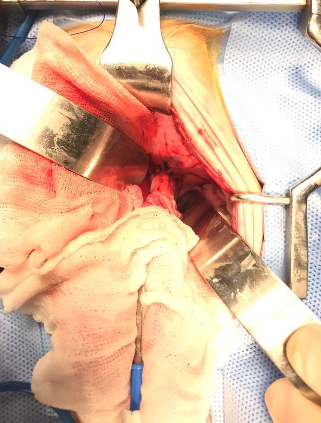
Case Report Veterinarni Medicina, 66, 2021
https://doi.org/10.17221/47/2020-VETMED
Figure 1 FIgure 2
Figure 1. Right lateral thorax radiograph with a soft
tissue opacity (white arrows), which represents the her-
niated cardia and the proximal part of the stomach Figure 2. Fluoroscopic oesophagography during the gas-
troesophageal reflux revealing the cranial displacement
initiated with metoclopramide [0.5 mg/kg, subcu- of the oesophagogastric junction and part of the stom-
taneous (s.c.), twice per day (b.i.d.)], metronida- ach into the thorax (white arrows), with a concurrent
zole [15 mg/kg, intravenous (i.v.), b.i.d.], cefazolin gastroesophageal junction dilation (blue arrow)
(30 mg/kg, s.c., b.i.d.), tramadol (4 mg/kg, s.c.,
b.i.d.), famotidine (1 mg/kg, i.v., b.i.d.), maropitant medical treatment, surgical stabilisation involving
[1 mg/kg, s.c., once per day (s.i.d.)], and aluminium a phrenoplasty, an oesophagopexy, and a left-sided
sucrose sulfate [Ulcerlmin, 5 ml, per os (p.o.), three gastropexy was performed (Figure 3). The dog was
times per day (t.i.d.)]. After failure of the empirical premedicated with cefazoline (30 mg/kg, i.v.), at-
Figure 3A Figure 3B
(A) (B)
Figure 3. Intraoperative images demonstrating the left-sided gastropexy (white arrow) (A) and phrenoplasty/oesopha-
gopexy (black arrow) (B)
3Case Report Veterinarni Medicina, 66, 2021
https://doi.org/10.17221/47/2020-VETMED
ropine sulfate (0.02 mg/kg, s.c.), and butorphanol Figure 4
(0.1 mg/kg, i.v.), followed by anaesthesia induction
with propofol (6 mg/kg, i.v.). The dog was intubat-
ed, and anaesthesia was maintained with isoflurane
in oxygen. Intravenous fluids were administered
at a rate of 5 ml/kg/h intraoperatively until com-
plete recovery from anaesthesia. A ventral midline
celiotomy incision was performed, and the sliding
hiatal hernia was reduced by manual traction on the
gastric fundus. The diameter of the oesophageal hi-
atus was approximately 4 cm. The phrenoplasty was
performed by approximating the left and right cru- Figure 4. Histopathological examination of the diaphrag-
ra of the diaphragm using two simple interrupted matic muscle showing an inflammatory cell infiltration
3-0 polypropylene sutures. The oesophageal hiatus and fragmented/degenerated myocytes
was reduced to a diameter through which only the
index finger could be inserted. The oesophagus was even after increasing the dose to 1 mg/kg, the clini-
then sutured to the diaphragm by placing two sim- cal signs were not alleviated. After administration
ple interrupted 3-0 polypropylene sutures on each of azathioprine (2 mg/kg, p.o., s.i.d.), the patient
side of the oesophageal hiatus between the tunica started to take food voluntarily and gained weight.
muscularis along the surface of the oesophagus and Azathioprine was slowly tapered off and replaced
the diaphragm. Finally, a left-sided gastropexy was by cyclosporin microemulsion (25 mg/dog, p.o.,
performed by incising the seromuscular layer of the s.i.d.) over a 1-month period. Although intermit-
gastric fundus and left ventrolateral abdominal tent vomiting (once per week) was observed by the
wall. The abdominal wall incision was extended owner, the dog was reported to be in good health
through the peritoneum and transversus abdominis during the 12-month monitoring period.
muscle. The gastric incision edges were sutured
to the corresponding abdominal wall incision edges
using 3-0 polydioxanone in a simple continuous su- DISCUSSION AND CONCLUSIONS
ture pattern. Copious lavage with sterile saline and
a routine closure completed the procedure. During Since the first report of a hiatal hernia in dogs
the surgery, there was evidence of a thickened, by Gaskell, numerous additional cases have been
stretched, and flaccid diaphragmatic muscle and reported in the veterinary literature (Gaskell et al.
an extended inelastic oesophageal hiatus. The post- 1974; Ellis 1980; Kirkby et al. 2005; Guiot et al. 2008).
operative management included intravenous fluid Although the exact aetiology and pathogenesis re-
therapy and maintenance of the preoperative medi- main uncertain, several interrelated factors have
cations. The postoperative analgesia was provided been postulated to be responsible for hiatal hernias,
by a continuous infusion of butorphanol (0.1 mg/ including: anatomical deformities of the hiatal ca-
kg/h, i.v.) for 24 h and intermittent doses of butor- nal and phrenoesophageal ligament; a neurological
phanol (0.1 mg/kg, i.v.) as needed for pain. Diffuse or muscular disorder; a severe respiratory disease;
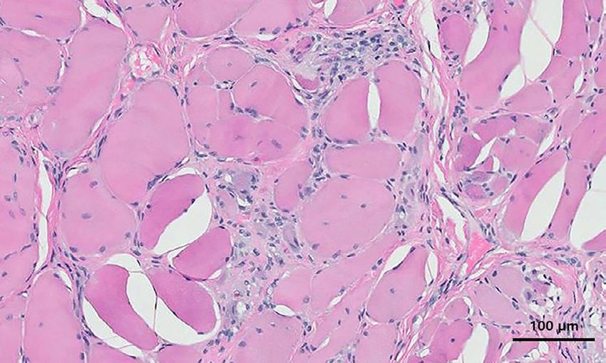
histiocytic myositis of the diaphragmatic muscle trauma; and an increasing gradient between the
was diagnosed based on the histopathologic exami- intraabdominal and intrathoracic pressure (Kahrilas
nation showing an inflammatory cell infiltration et al. 1999; Sivacolundhu et al. 2002). These factors
and fragmented/degenerated myocytes (Figure 4). compromise the function of the lower oesophageal
On the fluoroscopic oesophagography, 5 days after sphincter or separate the extrinsic anatomical
surgery, the gastroesophageal reflux virtually dis- structures (diaphragmatic crus, phrenoesophageal
appeared, and the gastroesophageal junction ap- ligament, fixation of the oesophagus to the liver)
peared to be in the normal position. In contrast, from the lower oesophageal sphincter, causing
the oesophageal motility remained decreased, and the dynamic axial motion of the gastroesophageal
vomiting was frequently observed. An acetylcho- junction into the thorax (Sivacolundhu et al. 2002;
linesterase inhibitor (pyridostigmine, 0.5 mg/kg, Mayhew et al. 2017). In the majority of the type 1
p.o., b.i.d.) was additionally administered; however, hiatal hernia cases reported to date, the clinical
4Case Report Veterinarni Medicina, 66, 2021
https://doi.org/10.17221/47/2020-VETMED
signs had been caused by gastroesophageal reflux cular atrophy and increased serum levels of cre-
(Katsianou et al. 2014). Mayhew et al. (2017) sug- atine kinase were observed together, serological
gested that the clinical signs are mediated by the testing for the determination of antibodies associ-
axial movement of the gastroesophageal junction ated with myopathies, including an acetylcholine
rather than the dysfunction of the lower oesoph- receptor antibody (myasthenia gravis), a type 2M
ageal sphincter. However, many dogs and cats fibre antibody (anti-masticatory muscle myosi-
have asymptomatic type 1 hiatal hernias (Bright tis), and an infectious agent antibody (ehrlichio-
et al. 1990). Why and how the gastroesophageal sis, toxoplasma, neosporosis, leishmaniosis), was
reflux occurs in some individuals and not others performed. However, all of these test results were
remains unclear. In human studies, a gastroesoph- negative. A marked elevation of creatine kinase lev-
ageal reflux disease by itself may further worsen els is an indication of skeletal muscle damage and,
the function of the lower oesophageal sphincter, in the case reported herein, was considered to be
thus initiating a self-perpetuating cycle (Ellis 1980; associated with the temporal/masseter/pterygoid
Dalakas 2012). muscle atrophy and diaphragm muscle myositis.
Muscular dysfunction caused by tetanus, myas- However, other skeletal muscles, such as the pha-
thenia gravis, masticatory muscle myositis, idio- ryngeal and oesophageal muscles, may also be as-
pathic polymyopathy, and/or muscular dystrophy sociated with serum creatine kinase activity, given
has been reported to be associated with hiatal that the myopathy of these muscles could contrib-
hernias (Ham and Bree 1992; Acke et al. 2004; ute to the dysphagia and gastroesophageal reflux.
Dalakas 2012; Tauro et al. 2015). However, the exact An association between the magnetic resonance
mechanism of a hiatal hernia and gastroesophageal imaging (MRI) and the histopathologically identi-
reflux secondary to the muscular dysfunction re- fied muscle inflammation has been reported (Platt
mains unclear. Tetanus appears to cause muscular et al. 2006). As such, an MRI may be a useful diag-
diaphragmatic spasms, which in turn cause the nostic method for differentiating abnormal from
stretching of the central tendon of the diaphragm unaffected muscles and, therefore, may facilitate
(Ham and Bree 1992; Acke et al. 2004). Concurrent a more accurate biopsy.
with this spasm, shortening of the oesophagus may The compromised function due to the diaphrag-
result in a hiatal hernia. Merieux et al. hypothesised matic myositis in the present case was regarded
a possible relationship between the polymyositis/ as the major cause of the hiatal hernia. A diaphrag-
dermatomyositis and oesophageal abnormalities matic muscle myopathy may cause the stretching
causing food reflux (Merieux et al. 1983). Muscular of the oesophageal hiatus, which may induce sepa-
dystrophy associated with oesophageal and dia- ration of the gastroesophageal junction from the
phragmatic muscular dysfunction has been stud- hiatus. Conventional surgical techniques, includ-
ied in various canine breeds (Merieux et al. 1983; ing phrenoplasty, oesophagopexy, and left-sided
Evans et al. 2004; Brumitt et al. 2006; Dalakas 2012). gastropexy, to correct the type 1 hiatal hernia were
In a report describing idiopathic inflammatory performed, and the postoperative fluoroscopic oe-
polymyopathy in the Hungarian Vizsla, the rela- sophagography demonstrated that these surgical
tionship between idiopathic inflammatory poly- techniques prevented the relapse of the hiatal hernia
myopathy and other idiopathic immune-mediated and gastroesophageal reflux. However, consider-
diseases, including atopic dermatitis, immune- ing that the clinical signs and oesophageal motility
mediated polyarthritis, inflammatory bowel dis- abnormalities were not appreciably alleviated, the
ease, keratoconjunctivitis sicca, sebaceous adenitis, surgical treatment did not appear to improve
and steroid responsive meningitis arteritis, have the muscular function of the oesophagus or other
been suggested (Tauro et al. 2015). musculature. Alleviation of the clinical signs after
An unusual aspect of the present case was that administration of the immunosuppressive agents
the hiatal hernia with the gastroesophageal reflux (azathioprine and cyclosporine) used to relieve the
occurred concurrently with the regional specific idiopathic inflammation of the associated muscular
severe atrophy of the temporal/masseter muscle, structures indirectly suggests that the abnormally
trismus, and enophthalmos. Enophthalmos has decreased oesophageal motility was responsive
been reported to be secondary to pterygoid muscle to the immunosuppressive agents. Consequently,
atrophy (Tauro et al. 2015). Given that the mus- the gastroesophageal reflux occurred not only
5Case Report Veterinarni Medicina, 66, 2021
https://doi.org/10.17221/47/2020-VETMED
because of the muscular dysfunction of the lower Callan M, Washabau R, Saunders H, Kerr L, Prymak C,
oesophageal sphincter and diaphragm, but also be- Holt D. Congenital esophageal hiatal hernia in the Chinese
cause of the malpositioning of the gastroesopha- Shar-Pei dog. J Vet Intern Med. 1993 Jul;7(4):210-5.
geal junction. Similar to other reports, abdominal Dalakas M. Pathogenesis and therapies of immune mediated
pressure on the oesophagus and the angle of the oe- myopathies. Autoimmun Rev. 2012 Jan;11(3):203-6.
sophagus into the cardia may act as part of the anti- Ellis H. Controversies regarding the management of hiatus
reflux mechanism. hernia. Am J Surg. 1980 Jun;139(6):782-8.
The results of this study demonstrated that medi- Ellison G, Lewis D, Phillips L, Tarvin B. Esophageal hiatal
cal management alone may not be sufficient for hernia in small animals: Literature review and a modified
a hiatal hernia caused by idiopathic inflammatory surgical technique. J Am Anim Hosp Assoc. 1987 Jan;23(4):
polymyopathy. Moreover, the concurrent surgical 391-9.
repositioning of the displaced gastroesophageal Evans J, Levesque D, Shelton GD. Canine inflammatory
junction into the normal position could effectively myopathies: A clinicopathologic review of 200 cases. J Vet
alleviate the clinical signs. The surgical techniques Intern Med. 2004 Sep-Oct;18(5):679-91.
used for this patient were not aimed at the func- Gaskell J, Gibbs C, Pearson H. Sliding hiatus hernia with
tional improvement of the respective muscular reflux oesophagitis in two dogs. J Small Anim Pract. 1974
structures, but for repositioning of the gastroe- Aug;15(8):503-10.
sophageal junction and extrinsic anatomical struc- Guiot L, Lansdowne G, Rouppert P, Stanley BJ. Hiatal her-
tures in a harmonised manner. nia in the dog: A clinical report of four Chinese shar peis.
In conclusion, the results of this study support the J Am Anim Hosp Assoc. 2008 Nov-Dec;44(6):335-41.
suspicion that other types of inflammatory poly- Ham L, Bree H. Conservative treatment of tetanus associ-
myopathy could be involved in a type 1 hiatal hernia ated with hiatus hernia and gastroesophageal reflux.
and gastroesophageal reflux disease. However, the J Small Anim Pract. 1992 Jun;33(6):289-94.
histological or immunohistochemical evaluation Kahrilas P, Lin S, Chen J, Manka M. The effect of hiatus
of the affected muscle tissues would be necessary hernia on gastro-oesophageal junction pressure. Gut.
to confirm this hypothesis. 1999 Apr;44(4):476-82.
Our data, along with those from other reports, Kirkby KA, Bright RM, Owen HD. Paraesophageal hiatal
demonstrate that a hiatal hernia with gastroesopha- hernia and megaesophagus in a three-week-old Alaskan
geal reflux caused by idiopathic inflammatory poly- Malamute. J Amall Anim Pract. 2005 Aug;46(8):402-5.
myopathy should be treated both surgically and Keeley B, Puggioni A, Pratschke K. Congenital oesophageal
medically. hiatal hernia in a pug. Ir Vet J. 2008 Jun;61(6):389-93.
Katsianou I, Svoronou M, Papazoglou L. Current views re-
garding hiatal hernia in dogs and cats. Hell J Companion
Conflict of interest Anim Med. 2014 Jan;3(2):31-9.
Lorinson D, Bright RM. Long-term outcome of medical and
The authors declare no conflict of interest. surgical treatment of hiatal hernias in dogs and cats:
27 cases (1978–1996). J Am Vet Med Assoc. 1998 Aug 1;
213(3):381-4.
REFERENCES Merieux P, Verity M, Clements P. Esophageal abnormalities
and dysphagia in polymyositis and dermatomyositis. Ar-
Acke E, Jones BR, Breathnach R, McAllister H, Mooney CT. thritis Rheumatol. 1983 Aug;26(8):961-8.
Tetanus in the dog: Review and a case report of concur- Mayhew P, Marks S, Pollard R, Culp W, Kass P. Prospective
rent tetanus with hiatal hernia. Ir Vet J. 2004 Oct 1;57(10): evaluation of surgical management of sliding hiatal hernia
593-7. and gastroesophageal reflux in dogs. Vet Surg. 2017 Nov;
Bright RM, Denovo C, Sackman J, Toal C. Hiatal hernia 46(8):1098-109.
in the dog and cat: A retrospective study of 16 cases. Prymak C, Saunders HM, Washabau RJ. Hiatal hernia repair
J Small Anim Pract. 1990 May;31(5):244-50. by restoration and stabilization of normal anatomy.
Brumitt J, Essman S, Kornegay J, Graham J, Weber W, An evaluation in four dogs and one cat. Vet Surg. 1989
Berry C. Radiographic features of golden retriever mus- Sep-Oct;18(5):386-91.
cular dystrophy. Vet Radiol Ultrasound. 2006 Oct-Nov; Podell M. Inflammatory myopathies. Vet Clin North Am
47(6):574-80. Small Anim Pract. 2002 Jan;32(1):147-67.
6Case Report Veterinarni Medicina, 66, 2021
https://doi.org/10.17221/47/2020-VETMED
Platt S, Fraser McConnel J, Garosi L, Ladlow J, De Stefani A, Tauro A, Addicott D, Foale RD, Bowman C, Hahn C, Long S,
Diane Shelton G. Magnetic resonance imaging in the di- Massey J, Haley AC, Knowler SP, Day MJ, Kennedy LJ,
agnosis of canine inflammatory myopathies in three dogs. Rusbridge C. Clinical features of idiopathic inflammatory
Vet Radiol Ultrasound. 2006 Nov 1;47(6):532-7. polymyopathy in the Hungarian Vizsla. BMC Vet Res. 2015
Sivacolundhu R, Read R, Marchevsky A. Hiatal hernia con- Apr 21;11(1): [13].
troversies – A review of pathophysiology and treatment
options. Aust Vet J. 2002 Mar 10;80(1):48-53. Received: February 20, 2020
Accepted: November 24, 2020
7You can also read