PODIATRIC TICKLERS FOR THE EM DOC - KATHLEEN COWLING MCEP WINTER SYMPOSIUM JANUARY 29TH 2021
←
→
Page content transcription
If your browser does not render page correctly, please read the page content below
KATHLEEN
COWLING
                    PODIATRIC
MCEP WINTER
SYMPOSIUM
                    TICKLERS FOR
JANUARY 29TH 2021
                    THE EM DOC
FUN FOOT FACTS

                 Toenails grow at an average of 1.62 mm per month,
                 much slower than fingernails.
                 May take over a year to completely grow back.
                 Biotin 2.5 mg daily can help them be stronger and
                 less prone to breakage.
                 Keep nails trimmed and cuticles pushed back for
                 optimum health.
PLANTAR PUNCTURE WOUND

Uncomplicated superficial puncture wounds should
have wound care
Tetanus prophylaxis
Plain films if any suspicion of foreign body, recognizing
that rubber from athletic shoes will likely not show up
Antibiotics for patients that are high risk e.g. Diabetics
Particularly covering Pseudomonas
PLANTAR PUNCTURE WOUND

Cellulitis usually will show up within 4 days
Should prompt imaging for retained foreign body
Begin 10 day course of anti strep and staph tx
Osteomyelitis occurs in 2% of all plantar wounds
Usually, forefoot wounds occurring in athletic shoes
Remember plain films are normal in early osteo
Follow closely elderly, DM, PVD, immunocompromised
TINEA PEDIS = ATHLETE’S FOOT
Most common dermatophytosis
     chronic intertriginous, caused by Trichophyton
     erythema and erosion of interdigital skin
     usually lateral 3 toes
Diagnose clinically, or with Potassium Hydroxide
Differentiate from dyshidrotic eczema, allergic dermatitis and
psoriasis
Treatment is moisture reduction and topical antifungal
PO therapy if recurrent with itraconazole 200mg QD month
Or Terbinafine 250 mg PO 2-6 weeks
 https://www.merckmanuals.com/professional/dermatologic-disorders/fungal-skin-infections/tinea-pedis-athletes-foot
TINEA PEDIS = ATHLETE’S FOOT = HYPERKERATOTIC
NAIL FUNGUS = ONYCHOMYCOSIS = TINEA UNGUIUM

                            Affects about 8-10 % of the general population
                            Causes discolored nails, toenails 10x more common than
                            fingers
                            Risk factors: tinea pedis, older, males, PVD, DM, exposure
                            to it (public bathing)
                            60-80% dermatophytes (Trichophyton) the rest are
                            (Aspergillus, Scopulariopsis, Fusarium)
                            Difficult to treat, high relapse rates, oral is better than
                            topical
                            Diagnose: clinical, PCR, Potassium Hydroxide wet mount,
                            must get sample from proximal portion of the nail
                            Terbinafine has higher efficacy 250mg QD x 12 weeks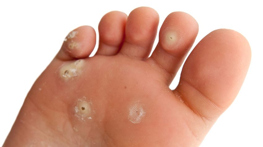
STINKY FEET = BROMHIDROSIS

Foul smelling odor from bacteria on the skin that eat your sweat

Feet have more sweat (eccrine) glands than any other part of the body

when the sweat is trapped by footwear, the bacteria produce isovaleric acid
STINKY FEET = BROMHIDROSIS

Cleanse feet daily, dry thoroughly

Don’t wear the same shoes day after day

Using foot powder may help

Avoid wearing shoes that are made from materials that don’t let the feet breathe

Coating the soles with aluminum chloride hexahydrate 20% can help keep the sweat minimized
BIOMECHANICS OF GAIT
RUNNING

https://youtu.be/QyiX0Fb-Lfw
Flat feet (also called pes planus or fallen arches) is a postural deformity in
which the arches of the foot collapse, with the entire sole of the foot coming
into complete or near-complete contact with the ground.

There is a functional relationship between the structure of the arch of the foot
and the biomechanics of the lower leg. The arch provides an elastic, springy
connection between the forefoot and the hind foot so that a majority of the
forces incurred during weight bearing on the foot can be dissipated before the
force reaches the long bones of the leg and thigh.
https://en.wikipedia.org/wiki/Flat_feet
PES PLANUS

True or False?

Children that go barefoot more have a greater likelihood of developing flat
feet.
POSTERIOR TIBIAL TENDON DYSFUNCTION = PTTD

The posterior tibial tendon is one of the major supports of the arch
When it becomes dysfunctional it leads to flattening of the arch
Often caused by overuse
Symptoms include pain, swelling and over pronation of the ankle
BIOMECHANICS OF FEET

Overpronators have too flexible feet
The feet of overpronators collapse too much and don’t get a good, rigid push-off when
they step because their foot is rolled in onto their arch

Supinators have too stiff feet
Supinators have arches that are raised too much, so they don’t absorb shock very well
when their feet first hit the ground
                          https://wexnermedical.osu.edu/blog/what-are-the-bottom-of-your-shoes-telling-you
BIOMECHANICS OF FEET
BIOMECHANICS OF FEET

According to the American Academy of Podiatric Sports Medicine, check
your athletic shoes after a total of 300 to 500 miles of running or walking,
or 45 to 60 hours of sports, such as basketball, dance, or tennis.

After that time, your shoes will have endured approximately one million
steps and may have lost their cushioning and support.

                                        http://www.aapsm.org/replace_shoes.html
OBESITY IS RELATED TO FOOT AND ANKLE INJURIES
Obesity (classified as BMI of 30 kg/m2 or greater) is becoming more prevalent in
America and so are musculoskeletal issues associated with it.
The healthy ankle joint allows for normal walking, and injuries to the joint, including
fractures, can have devastating effects if not properly addressed. The recent study
identified a correlation between more severe ankle fractures and obesity, especially for
obese men younger than 25, and obese women older than 50.
https://www.foothealthfacts.org/article/obesity-doubles-ankle-fracture-riskSPLAY FOOT = PES TRANSVERSOPLANUS Fan-like spreading of the metatarsal bones Mainly occurs from chronic obesity Places increased pressure on the medial metatarsophalangeal joint of the great toe This leads to re-distribution of loading on the heads of the other metatarsals, leading to callus formation, and hammertoes
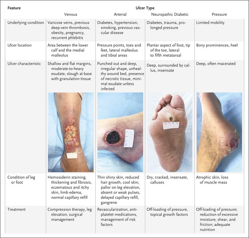
INGROWN TOENAILS = ONYCHOCRYPTOSIS

Usually blamed on poor trimming but not always the case

#1 risk is the shape of the nail, usually inherited trait

Trimming too far down, leaving a sharp corner can precipitate it

Pressure from tight shoes or trauma can increase risk

                                                            http://www.epodiatry.com/ingrown_nails.htm
INCOMPLETE MATRIXECTOMY Partial nail removal
MATRIXECTOMY

Digital block
Nail splitter
Remove entire lateral edge
Phenol applied for 30 seconds to wound base
PINCER NAIL = TRANSVERSE OVER-CURVATURE

Causes of pincer nails include
  Psoriasis
  Fungal infection
  Beta-blockers
  Arthritis and biomechanical changes
   Too narrow toe box
PINCER NAIL = TRANSVERSE OVER-CURVATURE

Treatment may require surgical resection of the widened matrix
Must include the entire length of the lateral edge of the matrix
May need to both sides
Treatment with 90% phenol is then applied to the base
LISFRANC JOINT INJURY

Traumatic injury of high force: MVA, falls
Disruption of the articulation of the medial cuneiform
   And the base of the second metatarsal
Injury usually operative repair
Important not to miss because of the risk for
   Compartment syndrome

                                                 https://www.orthobullets.com/foot-and-ankle/7030/lisfranc-injury
CALCANEAL FRACTURE
Most often caused from a fall
Look out for other related injuries, e.g. spine
Open fractures have a high risk for infectionCALCANEAL FRACTURE If there is any question on plain films, get the CT Watch out for compartment syndrome Place in a bulky Jones dressing with supportive posterior splint Non-weight bearing May require surgical fixation
5TH METATARSAL FRACTURE

90% are zone 1
Resulting from plantarflexion and hindfoot inversion

Zone 2 has vascular watershed supply which makes these prone to non-union

Zone 3 fracture is distal to the 4-5th metatarsal articulation, common site for stress fracture in athletes

                                        https://www.foothealthfacts.org/conditions/fractures-of-the-fifth-metatarsal
CRACKED HEELS = XEROSIS Skin around the heel is prone to being dry and developing thickening around the edges Prolonged standing on hard surfaces Obesity Medical conditions involving the skin e.g. eczema can lead to dryness, hypothyroidism This can all lead to developing fissures that can bleed
CRACKED HEELS = XEROSIS

Treatment includes
   good hygiene
   applying oil-based moisturizers daily
   Reducing the thickness gently using a pumice stone, regular maintenance
   Appropriate footwear
CORNS & CALLUS = HYPERKERATOSIS

Skin thickens due to a response to increase pressure
Usually “corns” on the toes and “callus” under the metatarsals
Biomechanical abnormalities caused by improper footwear, anatomy
When they become severe enough the body begins to reject it as a foreign body
Can be prone to infection if torn or cracked
CORNS & CALLUS = HYPERKERATOSIS

Proper foot hygiene, and appropriate footwear is critical
Avoid OTC topical acids and ”self” surgery
Surgical correction of bony prominence may ultimately be necessary
Adhesive cushions don’t fix the problem
CALCANEAL APOPHYSITIS = “SEVER’S” DISEASE

Typically seen in early adolescents
Calcaneal ossification center gets disturbed
Pain relieved with rest
May cause an antalgic gait
High impact sports and sudden activities can bring it on
Obesity and tight calve muscles make it worse
CALCANEAL APOPHYSITIS = “SEVER’S” DISEASE

Short period of rest
Ice, especially after activity
Stretch calve muscles
NSAIDS
Avoid going barefoot
Appropriate footwear, including a soft cushion heel raise
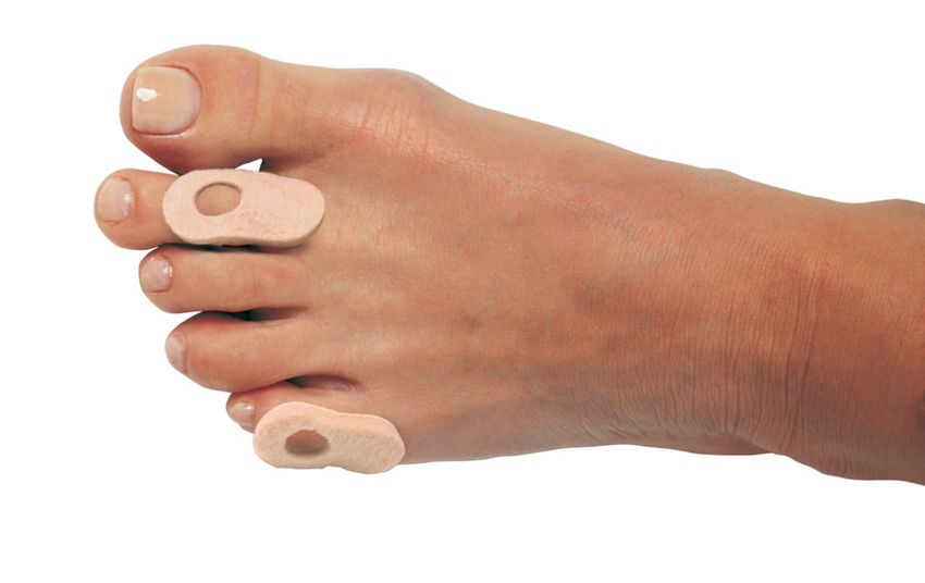
BUNIONS = HALLUX ABDUCTO VALGUS

1st metatarsophalangeal joint enlargement
Main contributing factor is footwear with tight toebox
Higher prevalence in women
Activities like ballet dancing can increase the proclivity of developing
Must have surgery if the problem becomes too severe
Proper footwear is key
Padding may help, especially if used between the toes
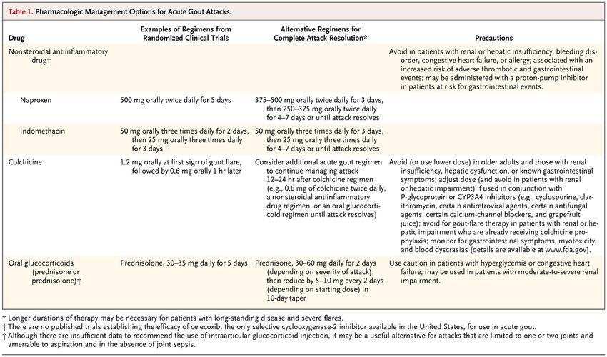
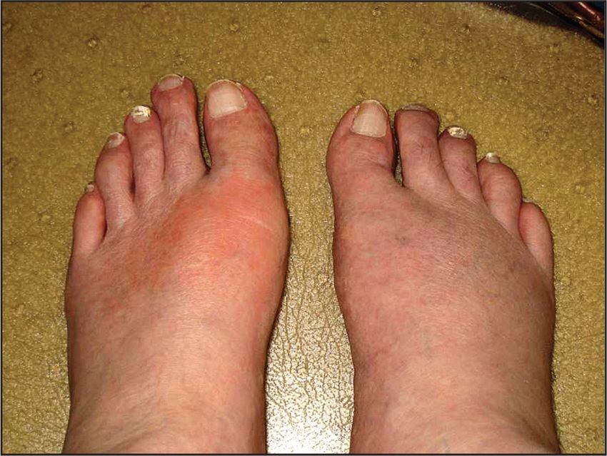
CRYSTAL ARTHROPATHY = GOUT

First metatarsal is the most common site
Affects men mostly until menopause and then women get it about the same
Swollen, and VERY painful to touch
Uric acid crystals precipitate out from hyperuricemia
Obesity, alcohol, high purine intake, renal disease all increase risk
CRYSTAL ARTHROPATHY = GOUT

Monosodium urate crystals in synovial fluid
Remember that you do not need an elevated serum uric acid level to have
Acute gout
Negatively birefringent urate crystals are seen on polarizing examination

                          https://journals.lww.com/em-news/Fulltext/2010/03000/Diagnosing_Gout__The_Basics.5.aspx
N Engl J Med 2011; 364:443-452
COMPARTMENT SYNDROME

                       Traumatic crush injury
                       Swelling increases pressure inside the fascial
                       compartment
                       Can cause vascular occlusion and ischemia
COMPARTMENT SYNDROME
Pain out of proportion
Pain with passive dorsiflexion
Decreased sensation
Loss of pulses
Tense swollen foot
https://www.orthobullets.com/trauma/1065/foot-compartment-syndromeCUTANEOUS LARVA MIGRANS Hookworm infection, usually self limited, migrating larvae die by 5 wks Severe pruritus usually leads patients to want immediate treatment Single dose of albendazole or ivermectin will work
PLANTAR FASCIITIS

Really a degenerative condition from wear and tear on the fascia, more a fasciopathy
Prolonged standing
Obesity
Unsupportive footwear
Age >40
High impact exercise
Tight calf muscles
Abnormal biomechanics, pes planus or cavus
PLANTAR FASCIITIS- WORK UP AND DIAGNOSIS

Pain most severe upon first stance
Plain radiographs, more to rule out other causes
Ultrasound
MRI
PLANTAR FASCIITIS- TREATMENT

Rest from high impact activities
Footwear, orthotics
Night splints
Stretching – this is the biggest preventative factor
NSAIDS
Physical therapy
ESWT- extracorporeal shockwave therapy-create microtears
Injections- AVOID steroids, cause fat pad atrophy, which is permanent
Proximal medial Gastrocnemius release
MORTONS NEUROMA

Not really a neuroma, but fibrosis around the interdigital nerve

9x more common in women, middle age, wearing tight shoes

Most common site is between the 3rd and 4th metatarsal heads

Sharp shooting pain most common symptom
MORTONS NEUROMA

                  Avoid tight shoes, wider toe box
                  Steroid injection may help
                  NSAIDS
                  Surgery if conservative management unsuccessful
                  Likely will have permanent numbness
N Engl J Med 2017; 377:1559-1567
https://stanfordmedicine25.stanford.edu/the25/ankle-brachial-index.html
PLANTAR WARTS = VERRUCA PLANTARIS

                             https://www.cfaortho.com/plantar-warts-plantarwarts
PLANTAR WARTS = VERRUCA PLANTARIS Warts involve the epithelium of the skin and are caused by infection with the human papillomavirus (HPV). Warts are the most common viral infection of the skin, affecting 7 to 10 percent of the general population. HPV thrives in warm, moist environments, such as public swimming pools and locker rooms, and transmits by direct contact, possibly through small cuts or abrasions in the stratum corneum layer of the skin Conventional treatment of warts frequently involves several ablative modalities including debridement, topical keratolytics (salicylic acid) and cantharone, with occlusive dressing. take care not to enter the dermis on dissection due to the potential to create painful scar tissue with healing.
INTRACTABLE PLANTAR KERATOSIS = IPK

                         Accumulation of dead skin cells that harden and thicken
                         Normally under the metatarsal head, heel or medial
                         aspect of great toe

                    https://www.semanticscholar.org/paper/Er%3AYAG-Laser-Treatment-of-
                    Intractable-Plantar-(IPK)-Koltaj/797859d52f093e287a5fe88c93ad4c8c8bf4f897
INTRACTABLE PLANTAR KERATOSIS - IPK
Painful lesion
Plantar surface
Typically, under metatarsal head
Clinical diagnosis, discern from verruca and epidermal inclusion cyst
Can cause antalgic gait, feels like walking on a marble/stone
Usually treated non-operatively, redistributing pressure, orthotics
Surgical treatment with YAG laserSUPERFICIAL CALCANEAL BURSITIS
https://www.lfaclinic.co.uk/conditions/superficial-calcaneal-bursitis/WORK UP Plain radiographs Ultrasound, looking for retrocalcaneal bursitis MRI
TREATMENT

Start with short rest period, avoiding sports/activity that trigger pain
Modify footwear, wearing sandals
ICE
NSAIDS/analgesics
Physical therapy focused on flexibility, biomechanical correction
Referral to DPM/ORTHO if not improving
RETROCALCANEAL BURSITIS

          Ultrasound-guided Diagnostic and Therapeutic Approach to Retrocalcaneal Bursitis
          The Journal of Rheumatology February 2011, 38 (2) 391-392; DOI:
HAGLUND’S DEFORMITY
Anatomical variant
Wearing tight shoes, high heels, overuse activities
https://www.lfaclinic.co.uk/conditions/haglunds-deformity/WORK UP Good examination, tenderness is above the calcaneus and anterior to the Achilles tendon Plain films Ultrasound MRI
TREATMENT

Rest period
ICE
NSAIDS/analgesics
Physical therapy
Stretching, correcting biomechanics
Operative

                   Calcaneus-Exostectomy-and-Achilles-Tendon-Reattachment-for-the-Treatment-of-Haglund-Deformity
CRPS = COMPLEX REGIONAL PAIN SYNDROME
Complex Regional Pain Syndrome
Sustained sympathetic activity
Pain out of proportion
Trauma with an exaggerated response, e.g. crush injury
Prolonged immobilization
ACE inhibitors on board at time of trauma
Smoking
Fibromyalgia
Females:males 4:1
40% occur in lower extremitiesCRPS = COMPLEX REGIONAL PAIN SYNDROME TREATMENT
Physical therapy
Nerve stimulation
Surgical sympathectomy
NSAIDS
Alpha and beta blockers
Antidepressants, GABA agonists
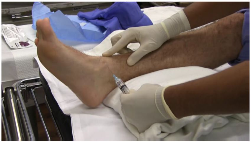
ketamineSAPHENOUS NERVE BLOCK
http://www.tamingthesru.com/ankle-and-foot-nerve-blocksSURAL NERVE BLOCK
http://www.tamingthesru.com/ankle-and-foot-nerve-blocksSUPERFICIAL PERONEAL NERVE BLOCK
http://www.tamingthesru.com/ankle-and-foot-nerve-blocksPOSTERIOR TIBIAL NERVE BLOCK
http://www.tamingthesru.com/ankle-and-foot-nerve-blocksREGIONAL FOOT BLOCKS
BLOCKSREFERENCES Evaluation and Management of Lower-Extremity Ulcers, October 19, 2017 Gout, February 3, 2011 N Engl J Med 2011; 364:443-452 https://www.podiatrytoday.com/how-treat-recalcitrant-plantar-warts https://www.lfaclinic.co.uk/conditions/superficial-calcaneal-bursitis/ https://www.lfaclinic.co.uk/conditions/haglunds-deformity/ https://www.jrheum.org/content/38/2/391 https://www.researchgate.net/profile/Phinit_Phisitkul/publication/317758943_Calcaneus_Exostectomy_and_Achilles_Tendon_Reattachment_ for_the_Treatment_of_Haglund_Deformity/links/5b2d3a28a6fdcc8506c2abfc/Calcaneus-Exostectomy-and-Achilles-Tendon-Reattachment https://www.ipfh.org/images/research_materials/2012_National_Foot_Health_Assessment_June_2012.pdf https://www.semanticscholar.org/paper/Er%3AYAG-Laser-Treatment-of-Intractable-Plantar-(IPK)- Koltaj/797859d52f093e287a5fe88c93ad4c8c8bf4f897 https://www.jospt.org/doi/pdf/10.2519/jospt.1985.7.3.91
REFERENCES

https://pubmed.ncbi.nlm.nih.gov/23107625/
http://www.epodiatry.com/ingrown_nails.htm
https://wexnermedical.osu.edu/blog/what-are-the-bottom-of-your-shoes-telling-you
http://www.aapsm.org/replace_shoes.html
https://www.jospt.org/doi/pdf/10.2519/jospt.1985.7.3.96
https://podiatrym.com/cme/August2000Levitz.pdf
https://www.foothealthfacts.org/conditions/obesity-and-your-feet
https://journals.lww.com/em-news/Fulltext/2010/03000/Diagnosing_Gout__The_Basics.5.aspx
https://www.orthobullets.com/trauma/1065/foot-compartment-syndrome
https://pubmed.ncbi.nlm.nih.gov/17403258/
https://www.merckmanuals.com/professional/dermatologic-disorders/fungal-skin-infections/tinea-pedis-athletes-foot
http://www.tamingthesru.com/ankle-and-foot-nerve-blocks
https://en.wikipedia.org/wiki/Flat_feet
https://stanfordmedicine25.stanford.edu/the25/ankle-brachial-index.html
You can also read