The Development of a Phytopathogenic Fungi Control Trial: Aspergillus flavus and Aspergillus niger Infection in Jojoba Tissue Culture as a Model ...
←
→
Page content transcription
If your browser does not render page correctly, please read the page content below
Hindawi
e Scientific World Journal
Volume 2021, Article ID 6639850, 8 pages
https://doi.org/10.1155/2021/6639850
Research Article
The Development of a Phytopathogenic Fungi Control Trial:
Aspergillus flavus and Aspergillus niger Infection in Jojoba Tissue
Culture as a Model
Nawal Abd El-Baky ,1 Raoufa Ahmed Abdel Rahman,2 Mona Mohammed Sharaf ,1
and Amro Abd Al Fattah Amara 1
1
Protein Research Department, Genetic Engineering and Biotechnology Research Institute,
City of Scientific Research and Technological Applications, Alexandria, Egypt
2
Pharmaceutical Bio-Products Research Department, Genetic Engineering and Biotechnology Research Institute,
City of Scientific Research and Technological Applications, Alexandria, Egypt
Correspondence should be addressed to Amro Abd Al Fattah Amara; amroamara@web.de
Received 31 October 2020; Accepted 7 January 2021; Published 18 January 2021
Academic Editor: Gerardo Caruso
Copyright © 2021 Nawal Abd El-Baky et al. This is an open access article distributed under the Creative Commons Attribution
License, which permits unrestricted use, distribution, and reproduction in any medium, provided the original work is
properly cited.
After introducing the idea of using concentrations equal to or less than the minimum inhibition concentration (MIC) of some
active chemical compounds for evacuating microbial cells, different types of microbes were evacuated. The original protocol was
given the name sponge-like protocol and then was reduced and modified from a microorganism to another to prepare microbial
ghosts for various applications such as immunological applications, drug delivery, and isolation of DNA and protein. Fungal
pathogens that infect plants critically affect cost effectiveness, quality, and quantity of their production. They kill plant cells and/or
cause plant stress. Plant fungal infections can originate from many sources such as infected soil, seeds, or crop debris causing
diseases and quality losses around the world with billions of US dollars annually as costs of the associated productivity loss. This
study focused on the application of the sponge-like protocol in protecting in vitro tissue cultures of plants against fungal
pathogens. This can be useful for research purposes or may be developed to be introduced in field applications. Aspergillus flavus
and Aspergillus niger infection in tissue culture of jojoba (Simmondsia chinensis (Link) Schn.) was used as a model to establish the
employment of this protocol to control plant fungal diseases. The best conditions for A. flavus and A. niger ghosts production
previously mapped by randomization experimental design (reduced Plackett–Burman experimental design) were used to prepare
fungal ghosts. SDS, NaOH, NaHCO3, and H2O2 were used in their MIC (+1 level) or minimum growth concentration (MGC, −1
level) according to the determined optimal experimental design. The release of both of DNA and protein from the fungal cells was
evaluated spectrophotometrically at 260nm and 280nm, respectively, as an indicator for cell loss of their cytoplasm. Fungal ghost
cells were also examined by transmission electron microscopy. After confirming the preparation of high-quality fungal ghost cells,
the same conditions were mimicked to control plant fungal infection. Jojoba grown in tissue culture was sprayed with fungal cells
(about 103 CFU) as a control experiment or fungal cells followed by treatment with solution (a) represents the fungal ghost cells
formation calculated critical concentration (FGCCC) of SDS, NaOH, and NaHCO3 and then treatment with solution (b)
represents H2O2 FGCCC. The plant was examined on day 0 (plant grown before any infection or infection followed by treatment),
day 5 (plant at day 5 after infection or infection followed by treatment), and day 10 (plant at day 10 after infection or infection
followed by treatment). We observed fungal growth in case of control experiments at days 5 and 10 on the tissue culture medium,
as well as plant, and the absence of any fungal growth in case of plant treated with FGCCC even after day 10. We recommend using
this FGCCC in the form of chemical spraying formulation to treat the plants aiming to control different plant fungal infections in
in vitro tissue culture systems or applied in field.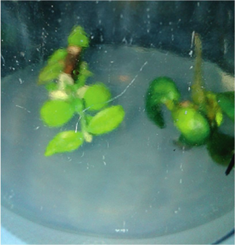
2                                                                                                 The Scientific World Journal

1. Introduction                                                       In this study, as an approach to develop the benefits of
                                                                  the sponge-like chemical protocol, it was applied to protect
Phytopathogenic fungi cause most of the diseases arising in       in vitro tissue cultures of plants against fungal pathogens. As
agricultural and horticultural setups [1]. Generally, phyto-      a model, tissue culture of jojoba (Simmondsia chinensis
pathogens developed infection strategies to attack any plant      (Link) Schn.) was infected with A. flavus and A. niger
[2], searching for entry and sourcing nutrients forcefully to     representing the phytopathogenic fungi. The infected plant
achieve microbial growth and development [3]. These               tissue culture was sprayed with FGCCC for each fungus. The
pathogens can overcome plant immune defences [4, 5]. This         fungal growth was observed after 5 and 10 days of infection
negatively affects the health, homeostasis, and physiology of      followed by treatment. The success of the protocol to prevent
plants and may result in systemic damage [6]. Plant fungal        plant fungal infection can be useful for research purposes or
pathogens can be responsible for a complexity of problems         may be developed to be introduced in field applications.
for farmers in plant production as listed by Dean et al. [7]
who reviewed the top ten plant fungal pathogens. To
overcome the problem of persistent attack of phytopatho-          2. Materials and Methods
genic fungi on agricultural products, numerous agro-              2.1. Fungal Strains. A. flavus and A. niger strains used in this
chemicals were used, some of which are toxic to humans and        study were kindly identified and provided by the Al-Azhar
a withdrawal period is essential between the last dosage and      University Mycology Center (Cairo, Egypt).
crops harvesting. Besides, they may also have negative en-
vironmental effects on soil organisms, insects, and plant
pollinators [8].                                                  2.2. Cultivation Conditions. A. flavus and A. niger strains
    Aspergillus niger (black mold) is one of the most             were cultivated in one-litre shake flasks containing 500 ml
common fungal species of the genus Aspergillus. It is             Sabouraud’s dextrose broth at 28°C for 7 days at 150 rpm.
abundant in soil and common in indoor environments [9]. It
shows phytopathogenic effects [10] causing black mold
disease on certain fruits and vegetables and is a common          2.3. FGCCC. The MIC and MGC values for NaOH, SDS,
contaminant of food. Aspergillus niger is responsible for         NaHCO3, and H2O2 against A. flavus and A. niger were
sooty mold of onions and ornamental plants. Aspergillus           determined as previously reported [25, 26]. The best con-
flavus (yellow mold) exists worldwide as a soil saprobe and        ditions for A. flavus and A. niger ghosts production were
infects various important agriculture crops. Common hosts         formerly mapped by randomization experimental design
of the pathogen are legumes, cereal grains, and tree nuts [11].   (reduced Plackett–Burman experimental design) [25, 26]
This pathogen can produce the polyketide-derived carci-           and then used to prepare fungal ghosts. The different var-
nogenic and mutagenic secondary metabolite, which is              iables were four chemical compounds: NaOH, SDS,
called aflatoxin [12, 13]. Aflatoxin causes aflatoxicosis due to     NaHCO3, and H2O2. The four variables were adjusted
inhaling or ingesting food and feed contaminated with high        according to the optimal experimental design for each
levels of aflatoxin.                                               fungus as described in Table 1.
    Amara et al. [14] were the first to introduce and de-              Each variable of the four used chemical compounds was
velop the MIC-MGC chemical technique to gently induce             represented at two levels (high and low), which are donated
pores in the microbial cells. They named the new tech-            by +1 (MIC) and −1 (MGC), as in Table 1 (except that SDS
nique as sponge-like protocol for preparing bacterial             was used in only the +1 level and NaOH was used in only the
ghosts. The protocol is based on using MIC-MGC of a               −1 level).
combination of some cheap, safe, and active chemical                  The fungal biomass of the cultivated fungi at 25°C for 7
compounds (SDS, NaOH, NaHCO3, and H2O2) for                       days was collected, washed gently by 0.5% saline, and then,
evacuating microbial cells. The Plackett–Burman opti-             recentrifuged at 6000 rpm for 10 min. The supernatant was
mization and randomization experimental design (full              discarded afterwards. 5X stock solution for each of NaOH,
and reduced) was applied to map the best chemical and             SDS, and NaHCO3 and 2X stock solution for H2O2 were
physical conditions to produce bacterial ghosts [14–16].          prepared from both +1 (MIC) and −1 (MGC) levels
The protocol enables evacuation of microbial cells while          according to Table 1 (except SDS and NaOH were used in
keeping their 3D structure. This turns live cells to dead         only the +1 and −1 level, respectively). All stock solutions
ghost cells with retained immunogenicity that could be            were filter sterilized by 0.22 μm TPP syringe filter (St. Louis,
used in immunization [17, 18]. Microbial ghosts prepared          Mo., USA).
by this protocol can also be applied as a drug delivery               Fungal ghost cells preparation was conducted in three
system [19]. Even the cytoplasm itself released from the          steps. The first step includes addition of 1 ml from NaOH,
evacuated ghost cells contains many important bio-                SDS, and NaHCO3 at concentration of 5X (+1 or −1) to 1 ml
macromolecules and biological structures other than               water and 1 ml of the fungal suspension (0.5 gm of the fungal
DNA and protein. The sponge-like chemical protocol was            mat/ml) to get a final concentration of 1X of each used
evolved to produce microbial ghosts from Gram-negative            chemical compound. Then, mixture was incubated for
[14–17, 20–22] and Gram-positive [23, 24] bacteria,               30 min at room temperature. The second step includes H2O2
Saccharomyces cerevisiae, and Candida, filamentous fungi           addition at concentration of 2X (+1 or −1) to 1 ml of the
[25, 26].                                                         treated fungal suspension from step 1 to reach final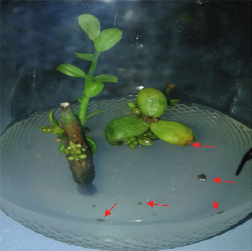
The Scientific World Journal                                                                                                              3

Table 1: The optimal experimental design for ghost cells preparation from each fungus including four chemicals variables in +1 or −1 level.
Fungal strain                    NaOH                            SDS                         NaHCO3                           H2O2
A. flavus                     −1 (0.001 g/ml)               +1 (0.0001 g/ml)                −1 (0.01 g/ml)                +1 (0.15 ml/ml)
A. niger                     −1 (0.001 g/ml)               +1 (0.0001 g/ml)                +1 (0.3 g/ml)                 −1 (0.001 ml/ml)

concentration of 1X of H2O2. The mixture was then incu-                50–60% relative humidity in a Tissue Culture Unit (Phar-
bated for 1 min at room temperature.                                   maceutical Bio-Products Research Department, Genetic
    After each abovementioned treatment step, the super-               Engineering and Biotechnology Research Institute, City of
natant was collected by centrifuging the treated fungal                Scientific Research and Technological Applications). Jojoba
suspension at 6000 rpm. Subsequently, the fungal pellet was            culture jars used in this study were incubated after infection
washed using 1X phosphate buffered saline (PBS) (alter-                 or infection followed by treatment separately from our usual
natively common saline solution can be used). In the third             culture establishment for the in vitro plant tissue culture to
step, the cell pellets were washed using 60% ethanol and left          avoid spread of fungal contamination throughout the
at room temperature for 20 min. After each abovementioned              system.
washing step and centrifugation, the supernatant was pre-
served to determine the amount of the released protein and
DNA.                                                                   2.10. Infection of Jojoba Tissue Culture with A. flavus or A. niger
                                                                       and Development of a Fungal Control Trial. Tissue culture of
                                                                       jojoba was infected by spraying with A. flavus or A. niger
2.4. Fungal Ghost Cells Evaluation Using a Light Microscope.           (103 CFU) representing the phytopathogenic fungi. After
The fungal ghost cells sample from each fungus was ex-                 24 hr, the plant tissue culture was sprayed with different
amined by using a light microscope. The quality of the                 FGCCC of SDS, NaOH, and NaHCO3 for each of A. niger
prepared ghost cells has been determined based on the                  and A. flavus for 20 min followed by treatment with (b)
cellular structure as being either intact or deformed, and             H2O2 FGCCC solution for 20 min. Then, the treated plants
then, the overall fungal ghost quality is given as %.                  were sprayed with sterile distilled water. Infected jojoba
                                                                       tissue culture without treatment was used as negative control
2.5. Determination of DNA Concentration. The concentra-                experiment. The fungal growth was observed after 5 and 10
tion of DNA in the supernatant after each ghost cells                  days of infection or infection followed by treatment.
preparation step for each fungus was determined by mea-
suring the absorbance at 260 nm. A quartz cuvette was used.            3. Results and Discussion
An extinction 260 � 1 corresponds to 50 μg dsDNA ml−1
[27].                                                                  It is obvious that phytopathogenic fungi result in several and
                                                                       diverse problems associated with a potential vast impact on
                                                                       plant production. Phytopathogenic fungi, thus, have a huge
2.6. Determination of Protein Concentration. Concentration
                                                                       negative part in providing food efforts for the ever-growing
of released protein from each ghost cells preparation step for
                                                                       world population. The diseases they cause in plants, as well
each fungus (the different supernatants) was determined by
                                                                       as the mycotoxins they produce, severely threaten agricul-
spectrophotometry at 280 nm. A quartz cuvette was used.
                                                                       tural production. Much effort has been made to minimize
The protein concentration was derived from the bovine
                                                                       damage caused by phytopathogenic fungi in agricultural
serum albumin (BSA) standard curve [14].
                                                                       setups including various management/control approaches
                                                                       and innovations. Fungal plant pathogens have been con-
2.7. Detection of Fungal Viability. Fungal ghost preparations          trolled by several approaches such as inducing plant natural
of both A. flavus and A. niger were investigated for the                resistance (using plant defence molecules in agricultural
possibility of the presence of any viable cells by subjecting          production) against invading fungi [29], fungicides [30],
them to growth on Sabouraud’s dextrose agar plates at 28°C             biological pesticides [31], and using plant extracts [8].
for 7 days.                                                                 The sponge-like protocol was primarily designed to use
                                                                       cheap and safe chemical compounds (NaOH, SDS,
                                                                       NaHCO3, and H2O2) for microbial ghost cells preparation
2.8. Transmission Electron Microscopy for Examination of
                                                                       [14]. Ghosts from various microbes were produced by this
A. flavus and A. niger Ghost Cells. A transmission electron
                                                                       gentle chemical protocol that was applied to induce evac-
microscope (JEOL TEM 100 CX) was used for the exami-
                                                                       uation-pores at the microbial cell wall [14–17, 19–26]. The
nation of A. flavus and A. niger ghost cells.
                                                                       main feature that distinguishes this protocol from other
                                                                       chemical evacuation methods was the combined use of MIC
2.9. In Vitro Tissue Culture of Jojoba (Simmondsia chinensis           and MGC of the used chemicals responsible for killing
(Link) Schn.). In vitro tissue culture of jojoba was prepared          microbes according to the optimal experimental design to
as previously described by Mohasseb et al. [28]. The plant             prepare microbial ghosts mapped by full or reduced
cultures were cultivated in jars and maintained in diffuse              Plackett–Burman experimental design. MIC of a used
light (1000–2000 lx), 16 h photoperiod at 25 ± 2°C, and                chemical will kill microbial cell while MGC allows cells to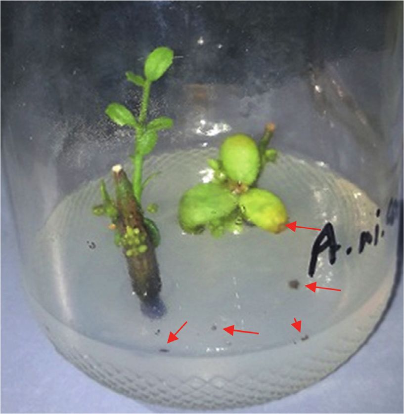
4                                                                                                             The Scientific World Journal

      Table 2: Spectrophotometry evaluation of released DNA and protein from ghost cells and percentage of ghost cell quality.
                  NaOH/SDS/NaHCO3 step              H2O2 step                   Ethanol step
Fungal strain                                                                                       Cell quality (%)
                DNA (μg/ml) Protein (mg/ml) DNA (μg/ml) Protein (mg/ml) DNA (μg/ml) Protein (mg/ml)
A. flavus            31.4         0.542        15.65           0.161          1             0.016          80%
A. niger           29.45         0.664         0.15           0.001        5.53            0.007          80%

                                    (a)                              (b)                                (c)

Figure 1: Transmission electron microscope images indicating evacuation and morphological changes in appearance of formed A. flavus
and A. niger ghost cells (magnification at 2000X). (a) A. flavus control cells; (b) A. flavus ghost and dead cells; and (c) A. niger ghost and dead
cells.

escape and live in the presence of a chemical compound, but                of fungal ghost cells preparation, the release of both of DNA
still it has an effect on the cell wall. The MIC of NaOH gently             and protein from the fungal cells was evaluated spectro-
induces the pore (s) in the microbial cell wall, while SDS is              photometrically at 260nm and 280nm, respectively, as pre-
essential to wash the “sponge-like” cells content, as well as to           sented in Table 2.
perturb and destabilize the cell wall/plasma membrane of                       The quality of the prepared ghost cells has been deter-
yeasts and fungi [32], and NaHCO3 increases the cell per-                  mined using a light microscope based on the cellular
meability. Finally, H2O2 is applied to hydrolyse the                       structure as being either intact or deformed, and then, the
remaining nucleic acids in the ghosts, and ethanol is used to              overall fungal ghost quality was given as % (Table 2).
inactivate the remaining nonlysed cells [14].                                  No growth was obtained on Sabouraud’s dextrose agar
     Our scientific group is working extensively to establish               plates after ghost cells cultivation from either A. flavus or
the sponge-like protocol and expanding its applications in                 A. niger, which proves the loss of the fungal cytoplasmic
humans and economically important animal and plant                         content during the process and the death of these fungi.
healthcare.                                                                Fungal cells were killed and well evacuated as proved by the
     Microbial ghosts prepared by this protocol can be ap-                 transmission electron microscopy (Figure 1).
plied in immunization [17, 18], as a drug delivery system                      After confirmation of preparing high-quality ghost cells,
[19], and even DNA and protein isolation [25, 26]. In this                 the same conditions were mimicked to control plant fungal
study, the protocol was applied to protect in vitro tissue                 infection.
cultures of plants against fungal pathogens. As a model,                       Jojoba grown in tissue culture and sprayed with fungal
tissue culture of jojoba (Simmondsia chinensis (Link) Schn.)               cells with or without treatment were examined on day 0
was infected with A. flavus and A. niger representing the                   (plant grown before any infection or infection followed by
phytopathogenic fungi. The success of the protocol to                      treatment), day 5 (plant at day 5 after infection or infection
prevent plant fungal infection can be developed to be in-                  followed by treatment), and day 10 (plant at day 10 after
troduced in field applications or research purposes (via                    infection or infection followed by treatment). We observed
spraying or immersing the plants in field or tissue cultures                the presence of fungal growth in case of control experiments
with chemical compounds involved in the study at con-                      at days 5 and 10 on the tissue culture medium, as well as
centrations achieving the best conditions for fungal cells                 plant parts (Figure 2), and the absence of any fungal growth
killing and ghosts production).                                            in case of plant infected with fungal cells and treated with
     The best conditions for A. flavus and A. niger ghosts                  FGCCC even at day 10 (Figure 3).
production previously mapped [25, 26] by randomization                         This study succeeded in preventing plant fungal infec-
experimental design (reduced Plackett–Burman experi-                       tion via fungal cell evacuation. The used FGCCC inhibits any
mental design) were used to prepare fungal ghosts. SDS,                    form of fungal growth. The spraying of fungal cells during
NaOH, NaHCO3, and H2O2 were used in their MIC (+1                          infection of plant tissue culture may cause some spores to
level) or MGC (−1 level) according to this determined                      contaminate the culture medium as seen in negative control
optimal experimental design for each fungus. After each step               experiments, while no sign of fungal growth was observed on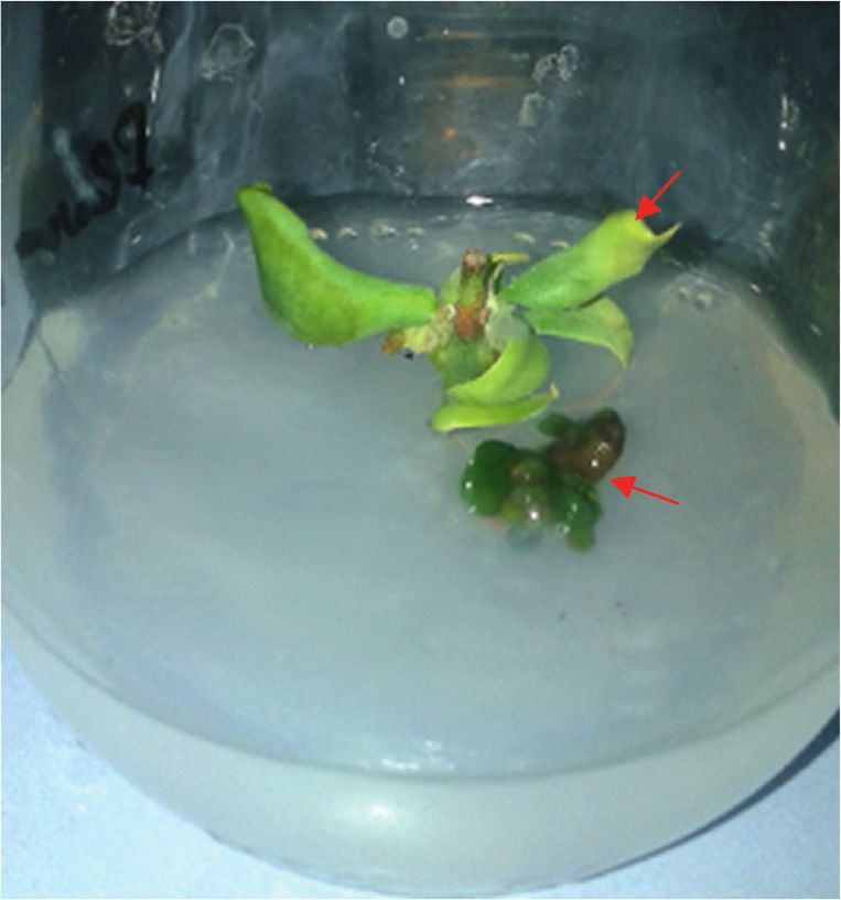
The Scientific World Journal                                                                                                                            5

                                                 (a)                                                  (b)

                                                 (c)                                                  (d)

                                                 (e)                                                  (f )

Figure 2: Jojoba grown in tissue culture and infected with either A. niger or A. flavus cells without any further treatments. (a), (c), and (e)
Plant at days 0, 5, and 10, respectively, after spraying with A. niger cells. (b), (d), and (f ) Plant at days 0, 5, and 10, respectively, after spraying
with A. flavus cells. Red arrows refer to fungal growth. In case of A. flavus, plant nearly stopped growth.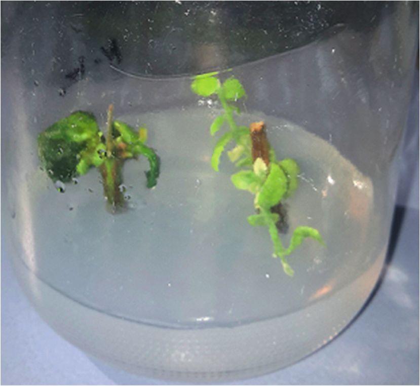
6 The Scientific World Journal
(a) (b)
(c) (d)
(e) (f )
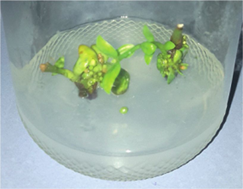
Figure 3: Jojoba grown in tissue culture, infected with either A. niger or A. flavus cells, and sprayed afterwards with FGCCC.(a), (c), and (e)
Plant at days 0, 5, and 10, respectively, after treatment with A. niger cells then sprayed with FGCCC. (b), (d), and (f ) Plant at days 0, 5, and 10,
respectively, after treatment with A. flavus cells and then sprayed with FGCCC. Plant continues to grow without any sign of fungal growth
on the tissue culture medium or plant parts.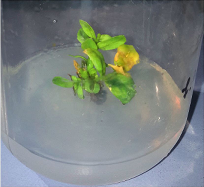
The Scientific World Journal                                                                                                                 7

the culture medium of treated plants even with multiple                  [5] A. Zvereva and M. Pooggin, “Silencing and innate immunity
spraying steps with fungal cells and FGCCC solutions. These                  in plant defense against viral and non-viral pathogens,” Vi-
results can be applied through spraying the plants in field or                ruses, vol. 4, no. 11, pp. 2578–2597, 2012.
tissue cultures with chemical compounds involved in the                  [6] G. N. Agrios, Plant Pathology, Elsevier Acad Press, Amster-
study at concentrations achieving the best conditions for                    dam, Netherlands, Fifth edition, 2005.
                                                                         [7] R. Dean, J. A. L. Van Kan, Z. A. Pretorius et al., “The Top 10
fungal cells killing and ghosts production. However, further
                                                                             fungal pathogens in molecular plant pathology,” Molecular
studies are recommended to screen the optimal incubation                     Plant Pathology, vol. 13, no. 4, pp. 414–430, 2012.
time for applying these chemicals to the plant to prevent any            [8] D. S. S. Shuping and J. N. Eloff, “The use of plants to protect
negative effect they may cause on plant growth and quality.                   plants and food against fungal pathogens: a review,” African
                                                                             Journal of Traditional, Complementary and Alternative
4. Conclusions                                                               Medicines, vol. 14, no. 4, pp. 120–127, 2017.
                                                                         [9] R. A. Samson, J. Houbraken, R. C. Summerbell, B. Flannigan,
This preliminary study aimed to use sponge-like protocol for                 and J. D. Miller, “Common and important species of fungi and
preparing FGCCC spray able to protect plants against fungal                  actinomycetes in indoor environments,” Microorganisms in
                                                                             Home and Indoor Work Environments, pp. 287–292, CRC,
pathogens. A. flavus and A. niger FGCCC were calculated for
                                                                             Boca Raton, FL, USA, 2001.
each, and ghost cells were confirmed by measuring the
                                                                        [10] N. V. Pawar, V. B. Patil, S. S. Kamble, and G. B. Dixit, “First
release of each of protein and DNA, as well as by the use of                 report of Aspergillus Niger as a plant pathogen on Zingiber
the light and electron microscope. The FGCCCs were ex-                       officinale from India,” Plant Disease, vol. 92, no. 9, p. 1368,
amined for their ability to suppress the fungal infection on                 2008.
jojoba tissue culture. No fungal growth was observed in the             [11] R. J. St. Leger, S. E. Screen, and B. Shams-Pirzadeh, “Lack of
treated experiments compared with the control. Apparently,                   host specialization in Aspergillus flavus,” Applied and Envi-
the used chemical compounds at the calculated concen-                        ronmental Microbiology, vol. 66, pp. 320–324, 2000.
trations did not affect the plant growth. This study might be a          [12] B. W. Horn and J. I. Pitt, Edited by N. Kokalis Burelle,
step towards a new approach to control phytopathogenic                       D. M. Porter, and R. Rodriguez-Kabana, Eds., “Yellow mold
fungi. Further studies are in need to validate and to optimize               and aflatoxin,” in Compendium of Peanut Diseases,
our results.                                                                 D. H. Smith Subrahmanyam, Ed., Vol. 44–49, Am. Phyto-
                                                                             pathol. Soc, St. Paul, MN, USA, 1997.
                                                                        [13] M. A. Klich, “Aspergillus flavus: the major producer of af-
Data Availability                                                            latoxin,” Molecular Plant Pathology, vol. 8, no. 6, pp. 713–722,
                                                                             2007.
All the data are given in the original file.                             [14] A. A. Amara, M. M. Salem-Bekhit, and F. K. Alanazi, “Sponge-
                                                                             like: a new protocol for preparing bacterial ghosts,” The
                                                                             Scientific World Journal, vol. 7, 2013.
Conflicts of Interest                                                   [15] A. A. Amara, M. M. Salem-Bekh, and F. K. Alanazi, “Prep-
                                                                             arationof bacterial ghosts for E. coli JM109 using “Spongei-
The authors declare that they have no conflicts of interest.
                                                                             kereduced protocol”,” Asian Journal of Biological Sciences,
                                                                             vol. 6, no. 8, pp. 363–369, 2013.
Authors’ Contributions                                                  [16] S. A. Sheweita, A. M. Batah, A. A. Ghazy, A. Hussein, and
                                                                             A. A. Amara, “A new strain of Acinetobacter baumannii and
Nawal Abd El-Baky and Raoufa Ahmed Abdel Rahman                              characterization of its ghost as a candidate vaccine,” Journal of
contributed equally to this study.                                           Infection and Public Health, vol. 12, no. 6, pp. 831–842, 2019.
                                                                        [17] A. A Amara, A. J. Neama, A. Hussein, E. A. Hashish, and
                                                                             S. A. Sheweita, “Evaluation the surface antigen of the Sal-
Acknowledgments                                                              monella typhimurium ATCC 14028 ghosts prepared by
                                                                             “SLRP”” The Scientific World Journal, vol. 2014, p. 6 pages,
This study recieved institutional instrumental and chemical                  2014.
support.                                                                [18] A. M. Batah and T. A. Ahmad, “The development of ghost
                                                                             vaccines trials,” Expert Review of Vaccines, vol. 19, no. 6,
References                                                                   p. 549, 2020.
                                                                        [19] A. A. Amara, “Bacterial and Yeast Ghosts: E. coli and Sac-
 [1] G. N. Agrios, Plant Pathogens and Disease: General Intro-               charomyces cerevsiae preparation as drug delivery model,”
     duction, Elsevier Inc., University of Florida, Gainesville, FL,         International Science and Investigation Journal, vol. 4,
     USA, 2009.                                                              pp. 11–22, 2015.
 [2] W. Knogge, “Fungal infection of plants,” The Plant Cell, vol. 8,   [20] M. Menisy, A. Hussein, A. A. Ghazy, S. Sheweita, and
     no. 10, pp. 1711–1722, 1996.                                            A. A. Amara, “Klebsiella pneumoniae Ghosts as vaccine using
 [3] R. Horbach, A. R. Navarro-Quesada, W. Knogge, and                       sponge like reduced protocol,” Journal of Cellular and Mo-
     H. B. Deising, “When and how to kill a plant cell: infection            lecular Medicine, vol. 3, pp. 1–8, 2017.
     strategies of plant pathogenic fungi,” Journal of Plant Phys-      [21] H. Park, S. Oh, N. Vinod et al., “Characterization of chem-
     iology, vol. 168, no. 1, pp. 51–62, 2011.                               ically-induced bacterial ghosts (BGs) using sodium hydrox-
 [4] B. P. H. J. Thomma, T. Nürnberger, and M. H. A. J. Joosten,            ide-induced Vibrio parahaemolyticus ghosts (VPGs),”
     “Of PAMPs and effectors: the blurred PTI-ETI dichotomy,”                 International Journal of Molecular Sciences, vol. 17, no. 11,
     The Plant Cell, vol. 23, no. 1, pp. 4–15, 2011.                         Article ID 1904, 2016.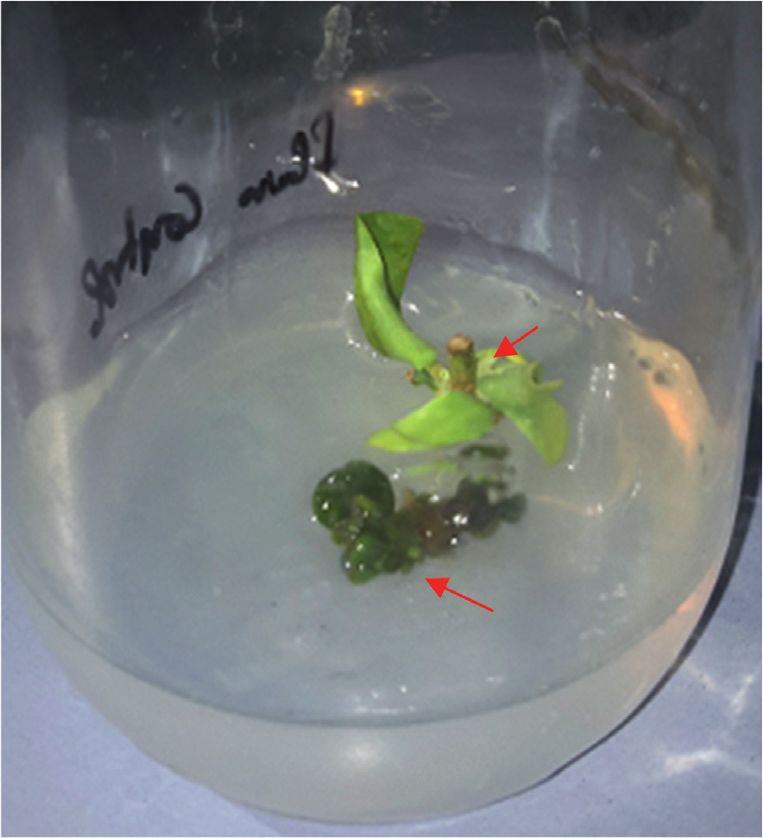
8                                                                       The Scientific World Journal

[22] N. Vinod, S. Oh, S. Kim, C. W. Choi, S. C. Kim, and
     C. H. Jung, “Chemically induced Salmonella enteritidis ghosts
     as a novel vaccine candidate against virulent challenge in a rat
     model,” Vaccine, vol. 32, no. 26, pp. 3249–3255, 2014.
[23] A. A. Amara, “The critical activity for the cell all degrading
     enzymes: could the use of the lysozyme for microbial ghosts
     preparation establish emergence oral vacccination protocol,”
     International Science and Invastigation Journal, vol. 5,
     pp. 351–369, 2016.
[24] X. Wu, X. Ju, L. Du et al., “Production of bacterial ghosts from
     gram-positive PathogenListeria monocytogenes,” Foodborne
     Pathogens and Disease, vol. 14, no. 1, pp. 1–7, 2017.
[25] N. A. El-Baky, M. M. Sharaf, E. Amer, H. R. Kholef,
     M. Z. Hussain, and A. A. Amara, “Protein and DNA isolation
     from Aspergillus Niger as well as ghost cells formation,” SOJ
     Biochemistry, vol. 4, pp. 1–7, 2018.
[26] N. A. El-Baky, M. M. Sharaf, E. Amer et al., “The minimum
     inhibition and growth concentration for controlling fungal
     infection as well as for ghost cells preparation: aspargillus
     flavus as a model,” Biomedical Journal of Scientific and
     Technical Research, vol. 10, pp. 1–5, 2018.
[27] J. Sambrook, E. F. Fritsch, and T. Mainiatis, Molecular Cloning
     a Laboratory Manual, Cold Spring Harbor Laboratory, Cold
     Spring Harbor, New YorkNY, USA, 2nd edition, 1989.
[28] H. A. A. Mohasseb, M. K. El-Bahr, Z. M. Adam, H. A. Moursy,
     and M. E. D. Solliman, “In vitro clonal propagation of Jojoba
     (Simmondsia chinensis (link) Schn.),” Australian Journal of
     Basic and Applied Sciences, vol. 3, pp. 3128–3136, 2009.
[29] A. Nega, “Review on concepts in biological control of plant
     pathogens,” Journal of Biology, Agriculture and Healthcare,
     vol. 4, pp. 33–35, 2014.
[30] N. Patel, P. Desai, N. Patel, and A. Jha, H. K. Gautam,
     Agronanotechnology for plant fungal disease management: a
     review,” International Journal of Current Microbiology and
     Applied Sciences, vol. 3, pp. 71–84, 2014.
[31] M.-Y. Yoon, B. Cha, and J.-C. Kim, “Recent trends in studies
     on botanical fungicides in agriculture,” The Plant Pathology
     Journal, vol. 29, no. 1, pp. 1–9, 2013.
[32] P.-A. Delley and M. N. Hall, “Cell wall stress depolarizes cell
     growth via hyperactivation of RHO1,” Journal of Cell Biology,
     vol. 147, no. 1, pp. 163–174, 1999.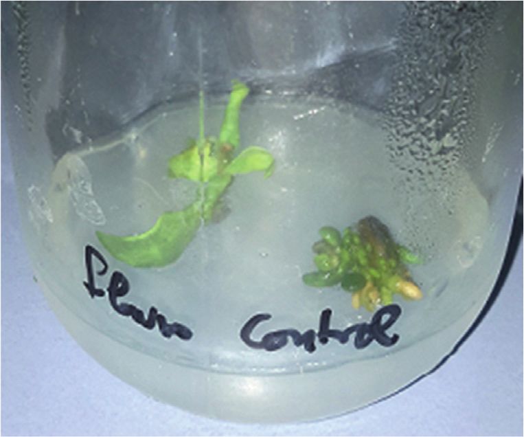
You can also read