Gastrointestinal manifestations of Talaromyces marneffei infection in an HIV-infected patient rapidly verified by metagenomic next-generation ...
←
→
Page content transcription
If your browser does not render page correctly, please read the page content below
Zhou et al. BMC Infectious Diseases   (2021) 21:376
https://doi.org/10.1186/s12879-021-06063-1

 CASE REPORT                                                                                                                                         Open Access

Gastrointestinal manifestations of
Talaromyces marneffei infection in an HIV-
infected patient rapidly verified by
metagenomic next-generation sequencing:
a case report
Ying Zhou1, Yongfeng Liu2 and Ying Wen1*

  Abstract
  Background: The manifestation of Talaromyces marneffei infection in some HIV-infected patients may be atypical.
  Cases with gastrointestinal involvement have rarely been reported. It is hard to make a diagnosis when patients are
  lacking the characteristic rash and positive blood culture.
  Case presentation: Here, we described a patient living with HIV who complained of fever and abdominal pain,
  and was rapidly diagnosed with Talaromyces marneffei infection by metagenomic next-generation sequencing
  (mNGS) using formalin-fixation and paraffin-embedded (FFPE) samples of omentum majus tissue. We also reviewed
  reported related cases.
  Conclusions: Talaromyces marneffei is an unusual cause of clinical presentations involving obvious abdominal pain
  and lower gastrointestinal bleeding, but can be included in the differential diagnosis. As an important diagnostic
  tool, the significance of mNGS using FFPE samples of lesions provides a more targeted diagnosis.
  Keywords: Human immunodeficiency virus, Talaromyces marneffei, Gastrointestinal involvement, Metagenomic
  next-generation sequencing

Background                                                                              Talaromyces marneffei infection considered when they
The common manifestations of Talaromyces marneffei                                      first present with gastrointestinal manifestations.
infection in human immunodeficiency virus (HIV)-in-
fected individuals consist of fever, anemia, weight loss,
characteristic skin papules, respiratory signs, lymphade-                               Case presentation
nosis, hepatosplenomegaly, and other organ involve-                                     A 33-year-old Chinese man presented with continuous
ment. In China, Talaromyces marneffei is mainly found                                   fever for one month from July 15th, 2019, followed by
in southern China. Therefore, HIV-infected patients with                                20 days of abdominal pain. The initial highest
a travel history to southern China should have                                          temperature was 38.9 °C, accompanied by night sweats,
                                                                                        anorexia, fatigue, weight loss and diarrhea (watery stool,
* Correspondence: wenying666466@163.com                                                 4–5 times per day). On July 25th, 2019, the patient pre-
1
 Department of Infectious Diseases, The First Affiliated Hospital of China              sented with intolerable abdominal pain and body
Medical University, No. 155, Nanjing North Street, Heping District, Shenyang
110001, Liaoning Province, China                                                        temperature had increased to 40 °C. HIV infection was
Full list of author information is available at the end of the article                  confirmed and the patient’s CD4+ T-cell count was
                                         © The Author(s). 2021 Open Access This article is licensed under a Creative Commons Attribution 4.0 International License,
                                         which permits use, sharing, adaptation, distribution and reproduction in any medium or format, as long as you give
                                         appropriate credit to the original author(s) and the source, provide a link to the Creative Commons licence, and indicate if
                                         changes were made. The images or other third party material in this article are included in the article's Creative Commons
                                         licence, unless indicated otherwise in a credit line to the material. If material is not included in the article's Creative Commons
                                         licence and your intended use is not permitted by statutory regulation or exceeds the permitted use, you will need to obtain
                                         permission directly from the copyright holder. To view a copy of this licence, visit http://creativecommons.org/licenses/by/4.0/.
                                         The Creative Commons Public Domain Dedication waiver (http://creativecommons.org/publicdomain/zero/1.0/) applies to the
                                         data made available in this article, unless otherwise stated in a credit line to the data.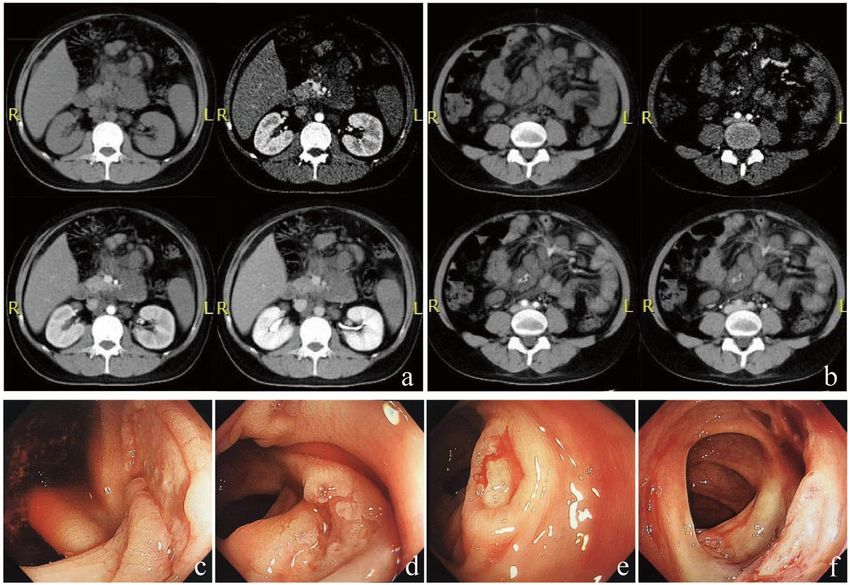
Zhou et al. BMC Infectious Diseases    (2021) 21:376                                                                                   Page 2 of 6

7cells/μL. The patient was born in Shenyang, located in                    and fungi cultures were repeated three times and all
the north of China. However, since July 2018, he had                       tests were negative. We did not observe parasite eggs in
worked and travelled a lot in Guangdong province, lo-                      the stool specimens. Giemsa stain of bone marrow aspir-
cated in the south of China. The patient had eaten                         ate did not find any pathogens. A bone marrow sample
roasted bamboo rat in December 2018.                                       culture was not carried out due to insufficient bone mar-
   The patient experienced abdominal tenderness and re-                    row samples obtained.
bound pain. Brain contrast magnetic resonance imaging                        For this kind of patient with obvious peritonitis despite
(MRI) and chest computed tomography (CT) scans were                        a negative T-SPOT result, empirical anti-tuberculosis
relatively normal. Abdominal CT scans showed severe                        treatment with a regimen of isoniazid, rifampin, etham-
fatty liver, thickened and swollen small intestinal wall,                  butol, and pyrazinamide was prescribed at the day 4 of
pelvic cavity effusion, and thickened mesentery accom-                     admission. A week later, fever and abdominal pain had
panied by multiple enlarged intra-abdominal lymph                          worsened. The patient complained of diffuse abdominal
nodes (Fig. 1a, b). The patient had normal leucocyte and                   pain and sustained fever. The patient displayed abdom-
platelet counts and mild anemia. The patient displayed                     inal rigidity. At the day 12 of admission, fungal infection
elevated C-reactive protein (135.30 mg/L) and galacto-                     was suspected and omentum majus biopsy was per-
mannan levels (4.39 μg/L). Serum ferritin was above                        formed. Hematoxylin and eosin (H&E) staining showed
2000.00 μg/L. Serum cryptococcal antigen was negative.                     granuloma with central necrosis and a large number of
Anti-neutrophil cytoplasmic antibodies were negative.                      foamy macrophages, lymphocytes, and neutrophil infil-
Toxoplasma gondii IgM and IgG antibodies were nega-                        tration. Periodic acid–Schiff (PAS) and Gomori’s methe-
tive. Cytomegalovirus (CMV) IgG antibody and herpes                        namine silver nitrate (GMS) staining showed clustered
simplex virus IgG antibody were positive, while IgM                        yeast in macrophages (Fig. 2). Acid-fast bacilli staining
antibodies were negative. Epstein-Barr virus (EBV) IgM                     (using Ziehl Neelsen), CMV-antigen, TB-DNA, and
antibody was negative while viral capsid antigen IgG and                   EBV-DNA in paraffin-embedded tissue sections were all
nuclear antigen IgG antibodies were positive. Serum                        negative. The patient began antifungal treatment with
CMV-DNA was undetectable. Whole blood EBV-DNA                              amphotericin B. In order to identify the specific fungal
was undetectable. The HIV RNA load was 5.1 × 10⁵ cop-                      species, particularly to differentiate between Talaro-
ies/mL. Blood bacteria and fungi cultures were repeated                    myces marneffei, histoplasma, and other deep fungal in-
three times and all tests were negative. Fecal bacteria                    fections, FFPE samples were sent to BGI PathoGenesis

 Fig. 1 Presentation of abdominal CT scan and colonoscopy. Abdominal CT scan showed severe fatty liver, pelvic cavity effusion (a), thickened
 and swollen small intestinal wall and thickened mesentery, accompanied by multiple enlarged intra-abdominal lymph nodes (a, b).
 Gastrointestinal endoscopy found multiple small shallow ulcers scattered in the cecum (c), ascending colon (d), transverse colon (e), and
 descending colon (f), partly accompanied by white exudates and active bleeding
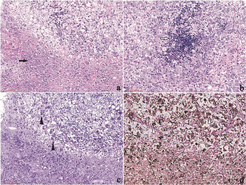
Zhou et al. BMC Infectious Diseases    (2021) 21:376                                                                                  Page 3 of 6

 Fig. 2 Histopathology of biopsy samples. H&E staining showing granuloma with central necrosis and concentrated inflammatory cell infiltrations
 involving foamy macrophages (containing a large number of yeasts), neutrophils (a) (200 × magnification), and lymphocytes (b) (200 ×
 magnification); Yeasts with positive PAS staining (c) (200 × magnification) and GMS staining (d) (400 × magnification) in macrophages

Pharmaceutical Technology (BGI-Shenzhen) for metage-                      achieved spontaneous remission. The patient’s serum
nomic next-generation sequencing (mNGS), which indi-                      creatinine increased to 120 μmol/L during amphotericin
cated Talaromyces marneffei infection 3 days later                        B treatment, but tenofovir alafenamide fumarate was not
(Fig. 3). In brief, the experimental procedure was per-                   available in China at that time, therefore, we suggested
formed as follows: DNA from the patient’s FFPE samples                    to use abacavir, and spent weeks to detect HLA-B5701
was extracted using the MagPure FFPE DNA LQ Kit fol-                      and applied for abacavir. After eight weeks of anti-fungal
lowing the manufacturer’s instructions. The DNA library                   treatment, ART was initiated with a regimen of lamivu-
was constructed and sequenced, human sequences were                       dine, abacavir, and dolutegravir. After 12 weeks of anti-
excluded, and low-complexity reads were removed, the                      fungal treatment, abdominal CT indicated that the thick-
remaining data were classified by simultaneously align-                   ened mesentery and small intestine had recovered, the
ing to four microbial genome databases, consisting of                     retroperitoneal lymph node had shrunk, and colonos-
4061 whole genome sequences of viral taxa, 2473 bacter-                   copy showed that the colon lesions had recovered. A 12-
ial genomes or scaffolds, 199 fungi connected to human                    month follow-up revealed that the patient’s CD4+ T-cell
infection, and 135 parasites associated with human dis-                   count had increased to 85cells/μL and HIV RNA was
eases [1].                                                                undetectable. The patient continues to take 200 mg itra-
   Fever and abdominal pain rapidly resolved after the                    conazole per day as secondary prevention until CD4 + T
initial days of starting amphotericin B treatment, while                  cells count reach 100 cells/μL for at least 6 months.
gastrointestinal bleeding occurred with a total bloody
stool volume of 1000 mL. Gastrointestinal endoscopy re-                   Discussion and conclusions
vealed multiple small shallow ulcers scattered in the                     This is a case of gastrointestinal Talaromyces marneffei
cecum, ascending colon, transverse colon, and descend-                    infection with negative blood culture, and the absence of
ing colon, partly accompanied by white exudates and ac-                   any respiratory involvement or rash. The mNGS rapidly
tive bleeding (Fig. 1c-f). After endoscopic hemostasis                    aided in identifying Talaromyces marneffei nucleotide
therapy with 1:1 epinephrine solution injected around                     sequences in omentum majus FFPE samples from our
the lesion, the bleeding temporarily stopped. Amphoteri-                  patient, which had never been previously reported; In 3
cin B treatment was continued followed by oral itracona-                  previous published papers, mNGS has been reported to
zole. Intestinal bleeding had another two relapses and                    help to diagnose Talaromyces marneffei infection in the
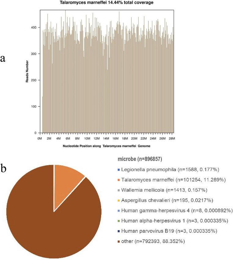
Zhou et al. BMC Infectious Diseases    (2021) 21:376                                                                                Page 4 of 6

 Fig. 3 Pathogen identification from paraffin-embedded tissue samples using the metagenomic next-generation sequencing method. The number
 of sequencing reads identified that corresponded to Talaromyces marneffei was 101,254 (b) with 14.44% genome coverage (a). Reads distribution
 of the total DNA sequence in the sample was without human host

bronchoalveolar lavage fluid [2, 3], bone marrow [2],                    symptoms (e.g., diarrhea) are relatively common with a
cerebrospinal fluid [2, 4], and skin lesion [2] specimens.               prevalence of approximately 25% [6], the prevalence of
  Talaromyces marneffei is a common opportunistic in-                    colonic involvement caused by Talaromyces marneffei
fection among HIV-infected patients in southeast Asia,                   infection is only 1.9% [7]. Including the present case,
southern China, and northeastern India, which are en-                    prominent abdominal involvement from Talaromyces
demic areas for Talaromyces marneffei. Possible epi-                     marneffei infection has been reported in a total of 14 pa-
demiological risk factors are as follows: (1) a history of               tients (Table 1) [4–13]. The main macroscopic patho-
travel or living in endemic areas and soil exposure, espe-               logical changes include multiple gastrointestinal ulcers
cially during the rainy season, has been suggested to be                 and mesenteric lymphadenitis. Common distribution of
a critical risk factor; (2) people living with HIV infection,            colonic infections include the cecum, ascending colon,
especially CD4+ T-cell counts below 200cells/μL, con-                    appendix, transverse colon, descending colon, or sigmoid
tributes to an increased risk of Talaromyces marneffei                   colon, small intestine, and duodenum. Common clinical
infection. Common manifestations of disseminated                         manifestations are fever, diarrhea, abdominal pain, lower
Talaromyces marneffei include fever, anemia, weight                      gastrointestinal bleeding, and intestinal obstruction.
loss, skin lesions, respiratory signs, lymphadenopathy,                  Most patients survive with anti-fungal treatment. Wild
and hepatosplenomegaly. Characteristic cutaneous le-                     bamboo rats exhibit a 100% prevalence of Talaromyces
sions aids to diagnosis and Talaromyces marneffei infec-                 marneffei infection [14]. It is important to note that the
tion can be confirmed by positive culture from blood,                    bamboo rat is a common species of rodent bred for meat
skin lesion, and bone marrow samples [5]. Inhalation of                  and wool in southern China. The potential for bamboo
conidia is the primary route of infection, which then dis-               rats to transmit pathogens to humans remains unclear
seminates to the reticuloendothelial system, skin, and                   because most patients with Talaromyces marneffei infec-
gastrointestinal organs. Although gastrointestinal                       tion in Guangdong did not have a history of contact
Zhou et al. BMC Infectious Diseases          (2021) 21:376                                                                                        Page 5 of 6

Table 1 Summary of clinical characteristics for 14 HIV-infected cases with intestinal Talaromyces marneffei
Case    Age    Area and Abdominal                Other clinical      Skin and             Involved organ or           Treatment maintenance        Outcome
No.     (yr)/  year of  symptoms                 presentations       mucous               tissue/diagnostic
        gender report                                                membrane             methods
                                                                     appearance
1       72/M      Hong         GI bleeding       anorexia,           jejunal ulcer(S)     small intestine(B + C),     NM                           Died
                  Kong                           dysphagia,                               mesenteric lymph
                  China                          weight loss                              node, liver(A)
                  1992 [4]
2       32/M      Hong         diarrhea          fever, night        multiple solitary    cecum, transverse and       Amphotericin B/Itraconazole survived
                  Kong                           sweats, dry         ulcers(E)            descending colon(B +
                  China                          cough                                    C)
                  1996 [5]
3、4     NM        Thai 1998 abdominal            fever               NM                   mesenteric lymph node Amphotericin B                     survived
                  [8]       pain                                                          (B), blood and bone
                                                                                          marrow (C)
5       52/M      Taiwan       diarrhea,         fever, erupted      shallow ulcers(E)    skin, bone marrow(B +       Amphotericin B/Itraconazole survived
                  China        abdominal         papule, anomia,                          C), colons(B)
                  1999 [6]     pain
6       30/M      Taiwan       diarrhea,         dyspepsia, fever,   shallow ulcers(E)    cecum, ascending and Amphotericin B/Itraconazole survived
                  China        abdominal         anomia, weight                           transverse colons(B + C)
                  1999 [6]     pain, bloody      loss
                               stool
7       33/M      India        abdominal         fever, loss of      duodenum             duodenum(B + C), bone Amphotericin B/Itraconazole survived
                  2008 [7]     pain              appetite, weight    narrowing(E)         marrow(C)
                                                 loss, vomiting
8       39/M      Hong         Abdominal         fever, weight       perioral             neck and                    Amphotericin B/Itraconazole Survived
                  Kong         pain              loss                umbilicated          retroperitoneal
                  China                                              lesions              lymph nodes (H + C),
                  2010 [9]                                                                blood (C)
9       28/M      India        non-colicky       fever, weight       perioral             neck nodes and              Amphotericin B/Itraconazole survived
                  2014 [10]    abdominal         loss                umbilicated          retroperitoneal lymph
                               pain                                  lesions              nodes(B + C), blood(C)
10      52/M      China        pain in the       anorexia, weight    multiple solitary transverse colon (B + H) Itraconazole                       survived
                  2017 [11]    lower left        loss                shallow ulcers (E)
                               abdomen
11      38/F      India        colicky           loss of appetite,   skin lesions,        skin, jejunal ulcers(B +    Amphotericin B/Itraconazole survived
                  2020 [12]    abdominal         weight loss         jejunal ulcers(E)    C),
                               pain
12      37/M      China        Abdominal         NM                  multiple ulcers      colon (B), blood (C)        Amphotericin B/Itraconazole Survived
                  2020 [13]    pain                                  (E)
13      50/M      China        Abdominal         weight loss         multiple ulcers      colon (B)                   Voriconazole+Amphotericin    Survived
                  2020 [13]    pain                                  (E)                                              B/Itraconazole
14      33/M      China        colicky           fever, weight       colon ulcers(E)      Mesenteric lymph            Amphotericin B/Itraconazole survived
                  [PR]         abdominal         loss, night                              node(B + N)
                               pain, bloody      sweats
                               stool
ND Not done, NM not mentioned, PR present report
Diagnostic methods to demonstrate P marneffei were autopsy (A), biopsy (B), culture (C), histopathology (H), surgery(S), Endoscope(E), NGS(N)

with bamboo rats [15]. Although the patient’s history of                           ulcers identified during the endoscopic examination
bamboo rat consumption is very suggestive, the link be-                            due to intolerance of the patient and the risk of
tween bamboo rat ingestion history in this case and pre-                           hemorrhage. Although the lack of an intestinal patho-
dominantly gastrointestinal presentation requires further                          logical confirmation from non-specific shallow ulcers
study.                                                                             infiltrated by lymphocytes and histiocytes distended
  This case report has several limitations. The limited                            with yeast [6], intestinal Talaromyces marneffei infec-
size of the omentum majus biopsy tissue was insuffi-                               tion was considered based on the patient’s abdominal
cient for tissue culture. Another limitation is a lack of                          symptoms of diarrhea, abdominal pain, and bloody
microbial cultivation of bone marrow aspirate. It is                               stool, which showed total improvement following
unfortunate that we did not perform biopsies of the                                anti-fungal treatment.
Zhou et al. BMC Infectious Diseases (2021) 21:376 Page 6 of 6
In conclusion, as a type of culture-independent 4. Tsui WMS, Ma KF, Tsang DNC. Disseminated Penicillium marneffei infection
method, mNGS provides a rapid etiological diagnosis, in HIV-infected subject. Histopathology. 1992;20(4):287–93. https://doi.org/1
0.1111/j.1365-2559.1992.tb00985.x.
especially in patients with an uncommon presentation of 5. Leung R, Sung JY, Chow J, Lai CKW. Unusual cause of fever and diarrhea in
Talaromyces marneffei infection. FFPE samples of le- a patient with AIDS: Penicillium marneffei infection. Dig Dis Sci. 1996;41(6):
sions and fresh biopsy specimens may represent suitable 1212–5. https://doi.org/10.1007/BF02088239.
6. Ko CI, Hung CC, Chen MY, Hsueh PR, Hsiao CH, Wong JM. Endoscopic
specimens for mNGS, which may be convincing for diagnosis of intestinal penicilliosis marneffei: report of three cases and
obtaining a targeted diagnosis and treatment. review of the literature. Gastrointest Endosc. 1999;50(1):111–4. https://doi.
org/10.1016/S0016-5107(99)70359-7.
Abbreviations 7. George IA, Sudarsanam TD, Pulimood AB, Mathews MS. Acute abdomen: an
HIV: Human immunodeficiency virus; mNGS: Metagenomic next-generation unusual presentation of disseminated Penicillium marneffei infection. Indian
sequencing; MRI: Magnetic resonance imaging; CT: Computed tomography; J Med Microbiol. 2008;26(2):180–2. https://doi.org/10.4103/0255-0857.40538.
H&E: Hematoxylin and eosin; PAS: Periodic acid–Schiff; GMS: Gomori’s 8. Ukarapol N, Sirisanthana V, Wongsawasdi L. Penicillium marneffei
methanamine silver nitrate staining; FFPE: Formalin-fixation and paraffin- mesenteric lymphadenitis in human immunodeficiency virus-infected
embedded children. J Med Assoc Thail. 1998;81(8):637–40.
9. Hung HG, Lok KH. Intestinal Penicillium marneffei: an unusual cause of
Acknowledgements chronic diarrhea in an AIDS patient. J Dig Dis. 2010;11(3):189–91. https://doi.
We thank the patient for agreeing to submit his case. We also thank Xu org/10.1111/j.1751-2980.2010.00435.x.
Wang (Department of endoscopy of the First Affiliated Hospital of China 10. Ghalige HS, Sahoo B, Sharma S, Devi KR, Singh Th SC. Acute abdomen due
Medical University) for their professional assistance. to Penicillium marneffei: an Indicator of HIV infection in Manipur state. J
Clin Diagn Res. 2014;8(9):ND05–6.
11. Feng S, Wang X, Zhang X, Yang H, Wang Z. Pathological diagnosis of a rare
Authors’ contributions
intestinal penicillium marneffei infection in an acquired immunodeficiency
YW made the conception and design of the work. YZ helped to collect the
syndrome patient: a case report and literature review. Int J Clin Exp Pathol.
data of the case. YW, YZ,YFL wrote the manuscript. All authors carried out
2017;10(3):3710–5.
final approval of the version to be published.
12. Philip Sridhar R, Coelho VV, Roopavathana B, Chase S. Opportunistic
penicilliosis infection causing intestinal obstruction in people living with HIV
Funding complicating antiretroviral therapy. BMJ Case Rep. 2020;13(2):e230121.
This work was supported by the science and technology foundation of 13. Pan M, Huang J, Qiu Y, Zeng W, Li Z, Tang S, Wei X, Zhang J. Assessment of
Shenyang (China) of Ying Wen (18–014–4-30) in the First Affiliated Hospital Talaromyces Marneffei Infection of the Intestine in Three Patients and a
of China Medical University. And Ying Wen made the conception and design Systematic Review of Case Reports. Open Forum Infect Dis. 2020;7(6):
of the work. ofaa128.
14. Cao C, Liang L, Wang W, Luo H, Huang S, Liu D, et al. Common reservoirs
Availability of data and materials for Penicillium marneffei infection in humans and rodents. China Emerg
Not applicable (no datasets were generated or analyzed during the current Infect Dis. 2011;17(2):209–14. https://doi.org/10.3201/eid1702.100718.
report). 15. Li X, Yang Y, Zhang X, Zhou X, Lu S, Ma L, et al. Isolation of Penicillium
marneffei from soil and wild rodents in Guangdong. SE China
Declarations Mycopathologia. 2011;172(6):447–51. https://doi.org/10.1007/s11046-011-
9443-5.
Ethics approval and consent to participate
Not applicable.
Publisher’s Note
Springer Nature remains neutral with regard to jurisdictional claims in
Consent for publication published maps and institutional affiliations.
Written informed consent for patient information to be published was
provided by the patient.
Competing interests
The authors declared no potential conflicts of interest with respect to the
research, authorship, and/or publication of this article.
Author details
1
Department of Infectious Diseases, The First Affiliated Hospital of China
Medical University, No. 155, Nanjing North Street, Heping District, Shenyang
110001, Liaoning Province, China. 2BGI PathoGenesis Pharmaceutical
Technology, BGI-Shenzhen, Shenzhen, Guangdong Province, China.
Received: 1 June 2020 Accepted: 9 April 2021
References
1. Zhu YM, Ai JW, Xu B, Cui P, Cheng Q, Wu H, et al. Rapid and precise
diagnosis of disseminated T.marneffei infection assisted by high-throughput
sequencing of multifarious specimens in a HIV-negative patient: a case
report. BMC Infect Dis. 2018;18(1):379.
2. Pongpech N, Rotjanapan P. Absence of cutaneous involvement in
disseminated Talaromyces marneffei infection in an AIDS patient: a case
report and literature review. Infect Drug Resist. 2019;12:1493–9. https://doi.
org/10.2147/IDR.S207819.
3. Praneenararat S. Fungal infection of the colon. Clin Exp astroenterol. 2014;7:
415–26.You can also read