A PROGRESSIVE AND COMPLEX CLINICAL COURSE IN TWO FAMILY MEMBERS WITH ERF-RELATED CRANIOSYNOSTOSIS: A CASE REPORT - DIVA-PORTAL
←
→
Page content transcription
If your browser does not render page correctly, please read the page content below
Körberg et al. BMC Medical Genetics     (2020) 21:90
https://doi.org/10.1186/s12881-020-01015-z

 CASE REPORT                                                                                                                                      Open Access

A progressive and complex clinical course
in two family members with ERF-related
craniosynostosis: a case report
Izabella Körberg1*, Daniel Nowinski2, Marie-Louise Bondeson1, Malin Melin1, Lars Kölby3 and Eva-Lena Stattin1

  Abstract
  Background: ERF-related craniosynostosis are a rare, complex, premature trisutural fusion associated with a broad
  spectrum of clinical features and heterogeneous aetiology. Here we describe two cases with the same pathogenic
  variant and a detailed description of their clinical course.
  Case presentation: Two subjects; a boy with a BLSS requiring repeated skull expansions and his mother who had
  been operated once for sagittal synostosis. Both developed intracranial hypertension at some point during the
  course, which was for both verified by formal invasive intracranial pressure monitoring. Exome sequencing revealed
  a pathogenic truncating frame shift variant in the ERF gene.
  Conclusions: Here we describe a boy and his mother with different craniosynostosis patterns, but both with
  verified intracranial hypertension and heterozygosity for a truncating variant of ERF c.1201_1202delAA
  (p.Lys401Glufs*10). Our work provides supplementary evidence in support of previous phenotypic descriptions of
  ERF-related craniosynostosis, particularly late presentation, an evolving synostotic pattern and variable expressivity
  even among affected family members.
  Keywords: ERF, Craniosynostosis, Intracranial hypertension

Background                                                                           pressure and intellectual disability. It is usually classified
Craniosynostosis (CS) is clinically and genetically a                                based on suture fusion type: sagittal, metopic, bi−/unicor-
heterogenous congenital anomaly with an incidence of 1                               onal, bi−/unilambdoid and complex, or multisutural.
in 2500 live births [1]. In most patients, CS presents as                               Bilateral lambdoid and sagittal synostosis (BLSS) was
an isolated anomaly (70–75%; non-syndromic). In 25–                                  first described 1976 by Neuhauser et al,. as a syndrome
30% of all patients, CS is manifested with additional                                called “craniofacial dyssynostosis”, [OMIM 218350].
anomalies and/or developmental delay (syndromic).                                    Seven patients, including two siblings, presenting with
  The most common clinical feature in CS is abnormal                                 BLSS where described, which also presented with short
head shape. This is a consequence of growth of the under-                            stature, Chiari type 1, intellectual disability and sus-
lying brain and restriction of skull growth due to prema-                            pected autosomal recessive inheritance [2]. BLSS was
ture ossification of skull sutures [1]. CS can result in                             later called “Mercedes Benz pattern” of CS due to the
numerous morphologic and functional abnormalities,                                   characteristic symbol of the three fused sutures on CT
including craniofacial malformation, increased intracranial                          imaging [3]. This is a rare complex trisuture synostosis
                                                                                     with a distinct clinical picture, including severe frontal
* Correspondence: izabella.baranowska.korberg@akademiska.se                          bossing, biparietal narrowing, turribrachycephaly, occipi-
1
 Department of Immunology, Genetics and Pathology, Uppsala University,
Science for Life Laboratory, Uppsala, Sweden
                                                                                     tal bulging or flatness, and low set ears [4]. Chiari I
Full list of author information is available at the end of the article

                                      © The Author(s). 2020 Open Access This article is licensed under a Creative Commons Attribution 4.0 International License,
                                      which permits use, sharing, adaptation, distribution and reproduction in any medium or format, as long as you give
                                      appropriate credit to the original author(s) and the source, provide a link to the Creative Commons licence, and indicate if
                                      changes were made. The images or other third party material in this article are included in the article's Creative Commons
                                      licence, unless indicated otherwise in a credit line to the material. If material is not included in the article's Creative Commons
                                      licence and your intended use is not permitted by statutory regulation or exceeds the permitted use, you will need to obtain
                                      permission directly from the copyright holder. To view a copy of this licence, visit http://creativecommons.org/licenses/by/4.0/.
                                      The Creative Commons Public Domain Dedication waiver (http://creativecommons.org/publicdomain/zero/1.0/) applies to the
                                      data made available in this article, unless otherwise stated in a credit line to the data.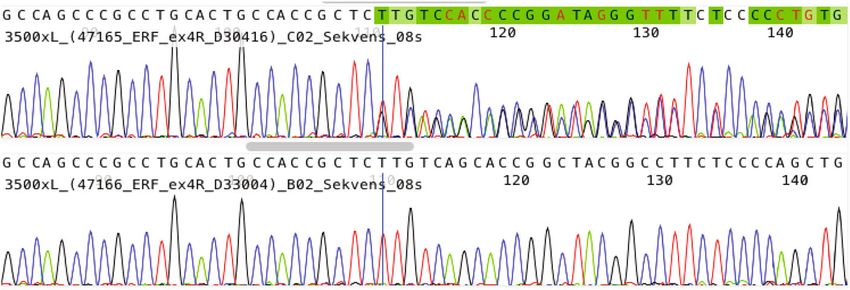
Körberg et al. BMC Medical Genetics (2020) 21:90 Page 2 of 6
malformation is common and may require neurosurgical months of age. During follow-up, at 1 year of age, a decline
decompression [5]. in head circumference was noted, the head shape turned
The aetiology of CS is heterogeneous, and several brachycephalic, there was a mild exorbitism, and
chromosomal hotspot loci and candidate genes have repeated ophthalmological examinations revealed papil-
been associated with CS [6–8]. One of the most recent ledema (Fig. 1). A repeat MRI showed development of a
addition is ETS2 repressor factor (ERF), in which patho- Chiari type 1 malformation and CT showed patho-
genic variants have been identified in patients presenting logical gyral impression and restenosis with complete
with CS [9–12]. pansynostosis. At this stage a formal invasive monitor-
Here we describe the clinical and molecular aspects of ing of intracranial pressure was performed with an
CS in a patient and his mother with a pathogenic variant intraparenchymatous catheter, confirming intracranial
in the ERF gene. hypertension (30–40 mmHg), which in turn resulted in
performing a secondary posterior calvarial expansion
Case presentation with internal distractors. The operation and postopera-
Clinical genetic investigation tive care were performed with the intracranial pressure
All patients with craniosynostosis and craniofacial syn- catheter in place and a return to normal intracranial
dromes, referred to the Craniofacial centre in Uppsala, pressure dynamics was seen during the first days after
are assessed by a team clinical geneticist and formally surgery. At 3 years follow-up the boy was in good
included in genetic research with approval from the health, papilledema had resolved and head shape was
ethical committee (Dnr 2013/294). The Craniofacial normal (Fig. 1). The family move to the south of
team in Uppsala is one of two national reference centres Sweden and the patient was therefore referred to the
for paediatric craniofacial surgery and has a close collab- craniofacial team in Gothenburg for further follow-up.
oration with the other licensed team in Gothenburg. At 3.5 years of age, headaches and papilledema reap-
Data from the care of the patients is entered in a quality peared. Intracranial pressure was high (above 40
registry with 100% coverage ratio. Blood samples are mmHg), and a third transcranial skull expanding pro-
obtained from the patients and parents in connection to cedure was performed including frontal remodelling
surgery or outpatient visits. and expansion and posterior expansion with springs
Patients with coronal, complex or atypical craniosyn- over osteotomy lines. The boy was identified as a
ostosis are routinely investigated for chromosomal patient with a positive family history, complex cranio-
abnormalities and/or genetic variants with numerous synostosis and negative findings on, at that time, imple-
methods including microarray analysis and a craniosyn- mented genetic CS panel. This warranted further
ostosis gene panel. Selected patients with negative investigation with clinical WES.
findings on routine genetic work-up are investigated The mother of the boy had been referred to the cranio-
further with whole-exome sequencing. facial team in Gothenburg at the age of 4 years due to
exophthalmos, left sided blindness and abnormal head
Subjects shape (Fig. 1). There was no family history of craniofacial
A baby-boy was referred at 2 months of age to the malformations. She had intermittent nocturnal headache
Craniofacial Centre, Uppsala University Hospital, due to and vomiting indicating raised intracranial pressure.
craniosynostosis. He was born after an uneventful preg- Imaging with CT and MRI showed shallow orbits, marked
nancy in gestational week 38 + 3. The birth weight was gyral impressions and increased CSF around optic nerves.
2.586 kg, and the length 46 cm. He was the first child of The anterior part of the sagittal suture was closed prema-
non-consanguineous parents and had been hospitalized turely. Ophthalmological examination revealed left sided
due to stagnation in weight gain. The mother had optic nerve atrophy. A formal 48-h ICP monitoring with
noticed an abnormal head shape of the boy and a com- an epidural sensor indicated intracranial hypertension.
puted tomography (CT) performed at the local hospital She was operated at the age of five with frontoorbital
confirmed complete fusion of the sagittal suture and remodelling and biparietal expansion. After the operation
bilateral partial fusions of both lambdoid sutures. headaches resolved and previous attention, and concentra-
Clinically, there was a constriction of the posterior skull, tion difficulties were improved.
compensatory forehead expansion, slight hypertelorism and
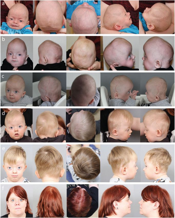
slightly increased tension on the anterior fontanelle (Fig. 1). Whole-exome sequencing
There were no other obvious malformations and no Clinical whole-exome sequencing (WES) and analysis
papilledema on ophthalmological examination. Magnetic protocols were developed by the Clinical genomics
Resonance Imaging (MRI) was performed and there was no facility, Uppsala and were adapted as a clinical WES
Chiari type malformation or other signs of intracranial test at Dept. of Clinical Genetics, Uppsala University
pathology. A craniectomy procedure was performed at 4 Hospital, Sweden, as described by Gudmundsson andKörberg et al. BMC Medical Genetics (2020) 21:90 Page 3 of 6 Fig. 1 Craniofacial features of the index patient presenting with bilateral lambdoid and sagittal synostosis (a-e). Mother of the index patient (f). (a) First visit at the age of seven weeks and two days. Constriction of the posterior skull, with a compensatory forehead expansion, and slight hypertelorism (b) The forehead expansion was more pronounced before operation at the age of 5 months. (c) After operation at the age of 6 months. Note brachycephalic head shape, and mild exorbitism (d) After operation at the age of 13 months. Note brachycephalic head shape, and more pronounced exorbitism (e) Index patient at the age of 3 years and 6 months. Note a normal head shape, and even more pronounced exorbitism (f) Mother of the index patient at the age of 26 years, operated for a sagittal synostosis at the age of 5 years. Note a scaphocephalic head shape, and exorbitism collegues, with minor modifications [13]. Venous was sequenced on an Illumina HiSeq2500 with 100 bp blood was collected from the boy and his parents and paired-end reads. Alignment of raw data to (GRch37/ DNA was extracted using an automated system UCSChg19) and variant calling was performed using (QuickGene, QIAGEN Inc.) according to standard the Bcbio Nextgen v 0.8.9 pipeline tool (https:// protocols. Whole exome sequencing was performed github.com/chapmanb/bcbio-nextgen). Briefly, align- using a trio-based approach (patient, mother and ment was performed using BWA 0.7.12, variant call- father). In brief, 250 ng genomic DNA was used for ing using GATK haplotype caller (GATK framework library preparation with Clinical Research Exome and 3.2.4, Genomie Analysis TK 3.2.2), and quality control Sure SelectQXT Target Enrichment System (Agilent parameters were calculated using FastQC 0.11.3, Technologies, Santa Clara, CA, USA). Enriched DNA Picard HsMetrics 1.96 (broadinstitute.github.io/picard)
Körberg et al. BMC Medical Genetics       (2020) 21:90                                                                                   Page 4 of 6

and GATK Depth of Coverage (GATK framework                                  variant in the index patient and his affected mother while
3.2.4, Genomie Analysis TK 3.2.2). For filtering of                         the father was not a carrier (Fig. 2b,c).
variants BENCHlab NGS (Agilent Technologics Inc.)
was used and allelic variants identified were classified                    Discussion and conclusions
according to the American College of Medical Genet-                         We report a boy and his mother carrying a heterozygous
ics and Genomics and the Association for Molecular                          frameshift variant c.1201_1202delAA (p.Lys401Glufs*10),
Pathology [14]. The pathogenic variant ERF (NM_                             in ERF gene causing sagittal, and BLSS also called “Mer-
006494.3) c.1201_1202delAA (p.Lys401Glufs*10) was                           cedes Benz pattern “of CS, and isolated sagittal CS
confirmed by Sanger sequencing of DNA from the                              respectively. Interestingly, both mother and son devel-
trio. Primers and PCR conditions are available upon                         oped intracranial hypertension which was verified by
request. PCR products were sequenced in both for-                           invasive monitoring. Phenotypically there were several
ward and reverse directions using BigDye terminator                         similarities, but also some differences. Both had sagittal
v3.1 cycle sequencing kit followed by automated                             synostosis, shallow orbits and mild hypertelorism (Fig.
sequencing on 3500 XL Genetic Analyzer (Applied                             1). Only the son, however, had lambdoid suture involve-
Biosystems, Foster City, CA). Sequencing data was                           ment, which probably explained why only he developed
analysed using SequencePilot v3.5.1 software (JSI                           a Chiari 1 malformation.
Medical Systems, GmbH).                                                       An interesting observation is the pansynostosis
                                                                            (including coronal sutures) that developed over time in
Identification of a pathogenic variant c.1201_1202delAA                     the index patient. ERF-related pansynostosis has been
(p.Lys401Glufs*10) in ERF                                                   described in previously reported cases [9, 12]. Thus, the
Whole-exome sequencing of the index patient (the boy)                       postoperative pansynostosis observed here could be a
generated 95 M reads where 91% of the reads mapped to                       postnatal progression of the sutural fusion, similarly to
the reference genome. The average read depth was 94X                        what is seen in Crouzon syndrome. Alternatively, the
and 97% of the exome was covered >10x. Filtering of vari-                   coronal fusion could be a phenomenon secondary to the
ants revealed a heterozygous variant c.1201_1202delAA                       early craniectomy procedure. Indeed, it is well known
(p.Lys401Glufs*10) in ERF (NM_006494.3) (Fig. 2a). The                      that secondary, postsurgical coronal synostosis develops
variant was classified as likely pathogenic since it is                     in a proportion of patients with non-syndromic sagittal
predicted to result in a truncated protein; it is absent in                 synostosis [15, 16]. However, the progressive evolution
1000 genomes and has been reported in one out of 236,                       of slight exorbitism in the index patient rather indicates
032 individuals in gnomAD. Sanger sequencing confirmed                      a primary and progressive sutural fusion causing growth
the presence of the c.1201_1202delAA (p.Lys401Glufs*10)                     restriction of the anterior skull base and orbits.

 Fig. 2 Structure of ERF gene and variant identified. (a) Main ERF transcript consist of 4 exons, ERF c.1201_1202delAA (p.Lys401Glufs*10) marked
 with red line. (b) Confirmation of truncating frameshift variant illustrated with sequencing. (c) pedigree
Körberg et al. BMC Medical Genetics (2020) 21:90 Page 5 of 6
Secondary coronal synostosis after surgery for sagittal and variable expressivity seen in autosomal dominant
synostosis, which is believed to be caused by the loss of disorders, between families and within families and
growth promoting forces across the coronal sutures after even among patients carrying the same variant in the
the decompressive widening of the skull, has not been ERF gene.
seen to cause progressive supraorbital retrusion and Other heterozygous variants causing CS has previously
exorbitism. been reported in ERF gene. Twigg et al. sequenced ERF
The ERF sequence variant has been reported previ- in 411 patients and detected mutations in ERF in 12
ously in four patients (family 12, and patient 19, and families of which most were truncating variants [9].
25), family 12 presenting with pansynostosis, hyperte- There were nine patients (9/12) with multiple synostosis,
lorism, delayed development, poor attention span and or pansynostosis, and among them two with BLSS. In
problems with writing (Table 1) [9]. Furthermore, the 2015, Chaudhry et al. sequenced 40 patients, and hetero-
patient presented with brachydactyly of hands and zygous mutations in ERF were detected in 2 patients
feet, broad halluces and dysplastic ears, thus (5%), one patient with bicoronal and metopic synostosis,
syndromic CS [9]. Patient 19 presented with pansy- and the other with pansynostosis [10]. Glass et al.
nostosis and Chari-1 malformation, he had orbital reported 16 unrelated probands and 20 family members
hypertelorism, malar hypoplasia, frontal bossing, long with multi sutural synostosis, predominantly BLSS [12].
philtrum, high palate, low set ears, inverted nipples In total 13 different heterozygous ERF-variants were
and clinodactyly [12]. reported (Table 1). These studies suggest a wide
In patient 25, the variant was paternally inherited spectrum of pathogenic variants causing ERF-related CS
and she presented with unicoronal synostosis, long [9, 10, 12]. The phenotype in the mother and son
philtrum, and short turned up nose. Interestingly, her presented here is consistent with the phenotype previ-
father did not present with synostosis, but had orbital ously described.
hypertelorism, mild malar hypoplasia and prognath- Hence, our work further supports that ERF is a candi-
ism. Both patients had raised ICP, in patient 19 at date gene that should be considered, particularly in
the age of 28 months, and < 1 month of age in patient patients presenting with multiple suture or BLSS of CS
25. This further supports the decreased penetrance and in patients with a progressive course.
Table 1 Overview of the clinical features of ERF variant c.1201_1202delAA p.Lys401Glufs*10 as reported by Twigg et al. 2013, Glass
et al. 2019 and the present study
Family/Patient Sex Age at Phenotype, CSa Chari-1 Facial dysmorphism Other phenotypic Reference
assessment malfomation traits
(years)
family 12 Mb 4 pansynostosis unknown hypertelorism, brachydactyly of Twigg et al. 2013 [9]
dysplastic ears hands and feet,
broad halluces,
delayed
development,
poor attention
span, problems
with writing
patient 19 M 28 pansynostosis yes hypertelorism, malar inverted nipples, Glass et al. 2019 [12]
hypoplasia, frontal clinodactyly
bossing, long philtrum,
high palate, lowset ears
patient 25 Fc < 1 unicoronal synostosis no long philtrum, short joint hypermobility Glass et al. 2019 [12]
turned up nose
father of patient 25 M – – – hypertelorism, mild – Glass et al. 2019 [12]
malar hypoplasia,
prognathism
patient 1 M 0,16 sagittal, bilateral yes Constriction of the – Baranowska
(son/index) lambdoid posterior skull, Körberg et al.
compensatory
forehead expansion,
hypertelorism,
exorbitism
patient 2 (mother) F 4 sagittal unknown Hypertelorism, poor attention span? Baranowska
exorbitism Körberg et al.
Abbreviations: acraniocynostosis (CS), bmale (M), cfemale (F)Körberg et al. BMC Medical Genetics (2020) 21:90 Page 6 of 6
Abbreviations 6. Shiihara T, Kato M, Kimura T, Hayasaka K, Yamamori S, Ogata T.
BLSS: Bilateral lambdoid and sagittal synostosis; CS: Craniosynostosis; Craniosynostosis with extra copy of MSX2 in a patient with partial 5q-
ERF: ETS2 repressor factor; CT: Computed tomography; MRI: Magnetic trisomy. Am J Med Genet A. 2004;128A(2):214–6.
resonance imaging; WES: Whole exome sequencing; ICP: Intracranial pressure 7. Tagariello A, Heller R, Greven A, Kalscheuer VM, Molter T, Rauch A, et al.
Balanced translocation in a patient with craniosynostosis disrupts the SOX6
Acknowledgements gene and an evolutionarily conserved non-transcribed region. J Med Genet.
We sincerely thank the family members who participated in this study. The 2006;43(6):534–40.
WES was performed by Clinical Genomics Uppsala, Science for Life 8. Cohen MM Jr. Craniofacial disorders caused by mutations in homeobox
Laboratory, Uppsala on instrument at the SNP&SEQ technology platform, genes MSX1 and MSX2. J Craniofac Genet Dev Biol. 2000;20(1):19–25.
Science for Life Laboratory, Uppsala. 9. Twigg SR, Vorgia E, McGowan SJ, Peraki I, Fenwick AL, Sharma VP, et al.
Reduced dosage of ERF causes complex craniosynostosis in humans and
Authors´ contributions mice and links ERK1/2 signaling to regulation of osteogenesis. Nat Genet.
IBK compiled all data and was the major contributor in writing the 2013;45(3):308–13.
manuscript. DN contributed with the clinical data, including phenotypic 10. Chaudhry A, Sabatini P, Han L, Ray PN, Forrest C, Bowdin S. Heterozygous
description of the cases, from Uppsala and wrote the manuscript. MLB mutations in ERF cause syndromic craniosynostosis with multiple suture
identified and classified the variant. MM provided access to WES and the involvement. Am J Med Genet A. 2015;167A(11):2544–7.
SNP&SEQ facility. LK contributed with the clinical data, including phenotypic 11. Balasubramanian M, Lord H, Levesque S, Guturu H, Thuriot F, Sillon G, et al.
description of the cases, from Gothenburg, ELS recruited the patients, Chitayat syndrome: hyperphalangism, characteristic facies, hallux valgus and
described them phenotypically, coordinated the study and wrote the bronchomalacia results from a recurrent c.266A>G p.(Tyr89Cys) variant in
manuscript. The authors read and approved the final manuscript. the ERF gene. J Med Genet. 2017;54(3):157–65.
12. Glass GE, O'Hara J, Canham N, Cilliers D, Dunaway D, Fenwick AL, et al. ERF-
Funding related craniosynostosis: the phenotypic and developmental profile of a
This study was supported by grants from the Medical faculty at Uppsala new craniosynostosis syndrome. Am J Med Genet A. 2019;179(4):615–27.
University, Sweden, and Stiftelsen Olle Engkvist Byggmästare. The role of the 13. Gudmundsson S, Anneren G, Marcos-Alcalde I, Wilbe M, Melin M, Gomez-
funders was to enable research and provide relevant equipment. Open Puertas P, et al. A novel RAD21 p.(Gln592del) variant expands the clinical
access funding provided by Uppsala University. description of Cornelia de Lange syndrome type 4 - Review of the literature.
Eur J Med Genet. 2018;62:103526.
Availability of data and materials 14. Richards S, Aziz N, Bale S, Bick D, Das S, Gastier-Foster J, et al. Standards and
The data of the current study are available from the corresponding author guidelines for the interpretation of sequence variants: a joint consensus
on reasonable request. recommendation of the American College of Medical Genetics and
Genomics and the Association for Molecular Pathology. Genet Med. 2015;
Ethics approval and consent to participate 17(5):405–24.
The study was approved by the ethical committee (Dnr 2013/294). A written 15. Arnaud E, Capon-Degardin N, Michienzi J, Di Rocco F, Renier D.
consent was obtained from the patient and patients’ parents for Scaphocephaly part II: secondary coronal synostosis after scaphocephalic
participation. surgical correction. J Craniofac Surg. 2009;20(Suppl 2):1843–50.
16. van Veelen ML, Eelkman Rooda OH, de Jong T, Dammers R, van Adrichem
LN, Mathijssen IM. Results of early surgery for sagittal suture synostosis:
Consent for publication
long-term follow-up and the occurrence of raised intracranial pressure.
An informed written consent was obtained from the patient and patients’
Childs Nerv Syst. 2013;29(6):997–1005.
parents for publication of this case report, including any accompanying
images.
Publisher’s Note
Competing interests Springer Nature remains neutral with regard to jurisdictional claims in
The authors declare that they have no competing interests. published maps and institutional affiliations.
Author details
1
Department of Immunology, Genetics and Pathology, Uppsala University,
Science for Life Laboratory, Uppsala, Sweden. 2Department of Surgical
Sciences, Plastic Surgery, Uppsala University, Uppsala, Sweden. 3Department
of Plastic Surgery, Institute for Clinical Sciences, Sahlgrenska Academy,
University of Gothenburg, Sahlgrenska University Hospital, Gothenburg,
Sweden.
Received: 18 September 2019 Accepted: 29 March 2020
References
1. Lattanzi W, Barba M, Di Pietro L, Boyadjiev SA. Genetic advances in
craniosynostosis. Am J Med Genet A. 2017;173(5):1406–29.
2. Neuhauser G, Kaveggia EG, Opitz JM. Studies of malformation syndromes of
man XXXIX: a craniosynostosis-craniofacial dysostosis syndrome with mental
retardation and other malformations: "craniofacial dyssynostosis". Eur J
Pediatr. 1976;123(1):15–28.
3. Moore MH, Abbott AH, Netherway DJ, Menard R, Hanieh A. Bilambdoid and
posterior sagittal synostosis: the Mercedes Benz syndrome. J Craniofac Surg.
1998;9(5):417–22.
4. Hing AV, Click ES, Holder U, Seto ML, Vessey K, Gruss J, et al. Bilateral
lambdoid and sagittal synostosis (BLSS): a unique craniosynostosis
syndrome or predictable craniofacial phenotype? Am J Med Genet A. 2009;
149A(5):1024–32.
5. Rhodes JL, Kolar JC, Fearon JA. Mercedes Benz pattern craniosynostosis.
Plast Reconstr Surg. 2010;125(1):299–304.You can also read