Medical Image Synthesis for Data Augmentation and Anonymization using Generative Adversarial Networks
←
→
Page content transcription
If your browser does not render page correctly, please read the page content below

Medical Image Synthesis for Data Augmentation and
                                          Anonymization using Generative Adversarial Networks

                                         Hoo-Chang Shin1 , Neil A Tenenholtz2 , Jameson K Rogers2 , Christopher G Schwarz3 ,
                                          Matthew L Senjem3 , Jeffrey L Gunter3 , Katherine Andriole2 , and Mark Michalski2
                                                                             1
                                                                       NVIDIA Corporation
arXiv:1807.10225v2 [cs.CV] 13 Sep 2018

                                                     2MGH & BWH Center for Clinical Data Science, Boston, MA, USA
                                                                3 Mayo Clinic, Rochester, MN, USA
                                              hshin@nvidia.com, jrogers24@partners.org, gunter.jeffrey@mayo.edu

                                                Abstract. Data diversity is critical to success when training deep learning mod-
                                                els. Medical imaging data sets are often imbalanced as pathologic findings are
                                                generally rare, which introduces significant challenges when training deep learn-
                                                ing models. In this work, we propose a method to generate synthetic abnormal
                                                MRI images with brain tumors by training a generative adversarial network us-
                                                ing two publicly available data sets of brain MRI. We demonstrate two unique
                                                benefits that the synthetic images provide. First, we illustrate improved perfor-
                                                mance on tumor segmentation by leveraging the synthetic images as a form of
                                                data augmentation. Second, we demonstrate the value of generative models as
                                                an anonymization tool, achieving comparable tumor segmentation results when
                                                trained on the synthetic data versus when trained on real subject data. Together,
                                                these results offer a potential solution to two of the largest challenges facing ma-
                                                chine learning in medical imaging, namely the small incidence of pathological
                                                findings, and the restrictions around sharing of patient data.

                                                Keywords: Generative Models, GAN, Image Synthesis, Deep Learning, Brain
                                                Tumor, Magnetic Resonance Imaging, MRI, Segmentation

                                         1   Introduction
                                         It is widely known that sufficient data volume is necessary for training a successful ma-
                                         chine learning algorithm [6] for medical image analysis. Data with high class imbalance
                                         or of insufficient variability [18] leads to poor classification performance. This often
                                         proves to be problematic in the field of medical imaging where abnormal findings are
                                         by definition uncommon. Moreover, in the case of image segmentation tasks, the time
                                         required to manually annotate volumetric data only exacerbates this disparity; manually
                                         segmenting an abnormality in three dimensions can require upwards of fifteen minutes
                                         per study making it impractical in a busy radiology practice. The result is a paucity of
                                         annotated data and considerable challenges when attempting to train an accurate algo-
                                         rithm. While traditional data augmentation techniques (e.g., crops, translation, rotation)
                                         can mitigate some of these issues, they fundamentally produce highly correlated image
                                         training data.
                                              In this paper we demonstrate one potential solution to this problem by generating
                                         synthetic images using a generative adversarial network (GAN) [9], which provides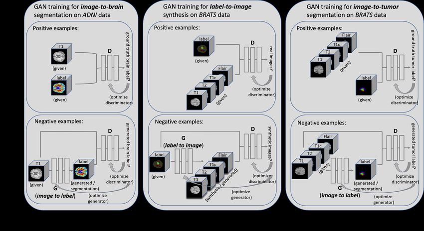
an additional form of data augmentation and also serves as a effective method of data
anonymization. Multi-parametric magnetic resonance images (MRIs) of abnormal brains
(with tumor) are generated from segmentation masks of brain anatomy and tumor. This
offers an automatable, low-cost source of diverse data that can be used to supplement
the training set. For example, we can alter the tumor’s size, change its location, or
place a tumor in an otherwise healthy brain, to systematically have the image and the
corresponding annotation. Furthermore, GAN trained on a hospital data to generate
synthetic images can be used to share the data outside of the institution, to be used as
an anonymization tool.
    Medical image simulation and synthesis have been studied for a while and are
increasingly getting traction in medical imaging community [7]. It is partly due to
the exponential growth in data availability, and partly due to the availability of better
machine learning models and supporting systems. Twelve recent research on medical
image synthesis and simulation were presented in the special issue of Simulation and
Synthesis in Medical Imaging [7].
    This work falls into the synthesis category, and most related works are those of
Chartsias et al [3] and Costa et al [4]. We use the publicly available data set (ADNI and
BRATS) to demonstrate multi-parametric MRI image synthesis and Chartsias et al [3]
use BRATS and ISLES (Ischemic Stroke Lesion Segmentation (ISLES) 2015 challenge)
data set. Nonetheless, evaluation criteria for synthetic images were demonstrated on
MSE, SSIM, and PSNR, but not directly on diagnostic quality. Costa et al [4] used
GAN to generate synthetic retinal images with labels, but the ability to represent more
diverse pathological pattern was limited compared to this work. Also, both previous
works were demonstrated on 2D images or slices/views of 3D images, whereas in this
work we directly process 3D input/output. The input/output dimension is 4D when it
is multi-parametric (T1/T2/T1c/Flair). We believe processing data as 3D/4D in nature
better reflects the reality of data and their associated problems.
    Reflecting the general trend of the machine learning community, the use of GANs
in medical imaging has increased dramatically in the last year. GANs have been used
to generate a motion model from a single preoperative MRI [10], upsample a low-
resolution fundus image [13], create a synthetic head CT from a brain MRI [16], and
synthesizing T2-weight MRI from T1-weighted ones (and vice-versa) [5]. Segmentation
using GANs was demonstrated in [22,21]. Finally, Frid-Adar et al. leveraged a GAN for
data augmentation, in the context of liver lesion classification [8]. To the best of our
knowledge, there is no existing literature on the generation of synthetic medical images
as form of anonymization and data augmentation for tumor segmentation tasks.

2     Data
2.1   Dataset
We use two publicly available data set of brain MRI:

Alzheimer’s Disease Neuroimaging Initiative (ADNI) data set
The ADNI was launched in 2003 as a public-private partnership, led by principal inves-
tigator Michael W. Weiner, MD. The primary goal of ADNI has been to test whether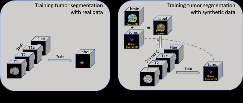
serial magnetic resonance imaging (MRI), positron emission tomography (PET), other
biological markers, and clinical and neuropsychological assessment can be combined to
measure the progression of mild cognitive impairment (MCI) and early Alzheimer’s dis-
ease (AD). For up-to-date information on the ADNI study, see www.adni-info.org.
We follow the approach of [17] that is shown to be effective for segmenting the brain
atlas of ADNI data. The atlas of white matter, gray matter, and cerebrospinal fluid (CSF)
in the ADNI T1-weighted images are generated using the SPM12 [1] segmentation and
the ANTs SyN [19] non-linear registration algorithms. In total, there are 3,416 pairs of
T1-weighted MRI and their corresponding segmented tissue class images.

Multimodal Brain Tumor Image Segmentation Benchmark (BRATS) data set
BRATS utilizes multi-institutional pre-operative MRIs and focuses on the segmenta-
tion of intrinsically heterogeneous (in appearance, shape, and histology) brain tumors,
namely gliomas [14]. Each patient’s MRI image set includes a variety of series in-
cluding T1-weighted, T2-weighted, contrast-enhanced T1, and FLAIR, along with a
ground-truth voxel-wise annotation of edema, enhancing tumor, and non-enhancing tu-
mor. For more details about the BRATS data set, see braintumorsegmentation.org.
While the BRATS challenge is held annually, we used the BRATS 2015 training data
set which is publicly available.

2.2   Dataset Split and Pre-Processing

As a pre-processing step, we perform skull-stripping [11] on the ADNI data set as skulls
are not present in the BRATS data set. The BRATS 2015 training set provides 264
studies, of which we used the first 80% as a training set, and the remaining 20% as a test
set to assess final algorithm performance. Hyper-parameter optimization was performed
within the training set and the test set was evaluated only once for each algorithm
and settings assessed. Our GAN operates in 3D, and due to memory and compute
constraints, training images were cropped axially to include the central 108 slices,
discarding those above and below this central region, then resampled to 128 × 128 × 54
for model training and inference. For a fair evaluation of the segmentation performance
to the BRATS challenge we used the original images with a resolution of 256 × 256 × 108
for evaluation and comparison. However, it is possible that very small tumor may get
lost by the downsampling, thus affecting the final segmentation performance.

3     Methods

The image-to-image translation conditional GAN (pix2pix) model introduced in [12] is
adopted to translate label-to-MRI (synthetic image generation) and MRI-to-label (image
segmentation). For brain segmentation, the generator G is given a T1-weighted image of
ADNI as input and is trained to produce a brain mask with white matter, grey matter and
CSF. The discriminator D on the other hand, is trained to distinguish “real” labels versus
synthetically generated “fake” labels. During the procedure (depicted in Figure 1 (a)) the
generator G learns to segment brain labels from a T1-weighted MRI input. Since we did
not have an appropriate off-the-shelf segmentation method available for brain anatomy
Fig. 1. Illustration of training GAN for (a) MRI-to-brain segmentation; (b) label-to-MRI synthesis;
(c) MRI-to-tumor segmentation.

in the BRATS data set, and the ADNI data set does not contain tumor information, we
first train the pix2pix model to segment normal brain anatomy from the T1-weighted
images of the ADNI data set. We then use this model to perform inference on the T1
series of the BRATS data set. The segmentation of neural anatomy, in combination with
tumor segmentations provided by the BRATS data set, provide a complete segmentation
of the brain with tumor.
     The synthetic image generation is trained by reversing the inputs to the generator
and training the discriminator to perform the inverse task (i.e., “is this imaging data
acquired from a scanner or synthetically generated?” as opposed to “is this segmentation
the ground-truth annotation or synthetically generated?” – Figure 1 (b)). We generate
synthetic abnormal brain MRI from the labels and introduce variability by adjusting
those labels (e.g., changing tumor size, moving the tumor’s location, or placing tumor
on a otherwise tumor-free brain label). Then GAN segmentation module is used once
again, to segment tumor from the BRATS data set (input: multi-parametric MRI; output:
tumor label). We compare the segmentation performance 1) with and without additional
synthetic data, 2) using only the synthetic data and fine-tuning the model on 10% of
the real data; and compare their performance of GAN to a top-performing algorithm4
[20]from the BRATS 2017 challenge.

3.1   Data Augmentation with Synthetic Images

The GAN trained to generate synthetic images from labels allows for the generation
of arbitrary multi-series abnormal brain MRIs. Since we have the brain anatomy label
and tumor label separately, we can alter either the tumor label or the brain label to
 4 https://github.com/taigw/brats17
Fig. 2. Workflow of getting synthetic images with variation. On BRATS data set, MRI-to-label
image translation GAN is applied to T1-weighted images to get brain atlas. It is then merged
with the tumor label given in the BRATS data set, possibly with alterations (shift tumor location;
enlarge; shrink). The merged labels (with possibly alterations) are then used as an input to label-
to-MRI GAN, to generate synthetic multi-parametric MRI with brain tumor.
get synthetic images with the characteristics we desire. For instance, we can alter the
tumor characteristics such as size, location of the existing brain and tumor label set, or
place tumor label on an otherwise tumor-free brain label. Examples of this are shown in
Figure 3.
The effect of the brain segmentation algorithm’s performance has not been evaluated
in this study.
Since the GAN was first trained on 3,416 pairs of T1-weighted (T1) images from the
ADNI data set, generated T1 images are of the high quality, and, qualitatively difficult
to distinguish from their original counterparts. BRATS data was used to train the
generation of non-T1-weighted image series. Contrast-enhanced T1-weighted images
use the same image acquisition scheme as T1-weighted images. Consequently, the
synthesized contrast-enhanced T1 images appear reasonably realistic, although higher
contrast along the tumor boundary is observed in some of the generated images. T2-
weighted (T2) and FLAIR image acquisitions are fundamentally different from the T1-
weighted images, resulting in synthetic images that are less challenging to distinguish
from scanner-acquired images. However, given a sufficiently large training set on all
these modalities, this early evidence suggests that the generation of realistic synthetic
images on all the modalities may be possible.Fig. 3. Examples of generated images. The first row depicts the original (“real”) images on which
the synthetic tumors were based. Generated images without adjustment of the segmentation label
are shown in the second row. Examples of generated images with various adjustments to the tumor
segmentation label are shown in the third through fifth rows. The last row depicts examples of
synthetic images where a tumor label is placed on a tumor-free brain label from the ADNI data
set.
Other than increasing the image resolution and getting more data especially for the
sequences other than T1-weighted images, there are still a few important avenues to
explore to improve the overall image quality. For instance, more attention likely needs
to be paid for the tumor boundaries so it does not look superimposed and discrete when
synthetic tumor is placed. Also, performance of brain segmentation algorithm and its
ability to generalize across different data sets needs to be examined to obtain higher
quality synthetic images combining data sets from different patient population.
The augmentation using synthetic images can be used in addition to the usual
data augmentation methods such as random cropping, rotation, translation, or elastic
deformation [15]. Moreover, we have more control over the augmented images using the
GAN-based synthetic image generation approach, that we have more input-option (i.e.,
label) to perturb the given image than the usual data augmentation techniques. The usual
data augmentation methods rely mostly on random processes and operates on the whole
image level than specific to a location, such as tumor. Additionally, since we generate
image from the corresponding label, we get more images for training without needing to
go through the labor-intensive manual annotation process. Figure 4 shows the process
of training GAN with real and synthetic image and label pairs.Fig. 4. Training GAN for tumor segmentation with (a) real; and (b) synthetic image-label pairs.
Synthetic data generation can increase the training data set with desired characteristics (e.g., tumor
size, location, etc.) without the need of labor-intensive manual annotation.
3.2 Generating Anonymized Synthetic Images with Variation
Protection of personal health information (PHI) is a critical aspect of working with
patient data. Often times concern over dissemination of patient data restricts the data
availability to the research community, hindering development of the field. While remov-
ing all DICOM metadata and skull-stripping will often eliminate nearly all identifiable
information, demonstrably proving this to a hospital’s data sharing committee is near
impossible. Simply de-identifying the data is insufficient. Furthermore, models them-
selves are subject to caution when derived from sensitive patient data. It has been shown
[2] that private data can be extracted from a trained model.
Development of a GAN that generates synthetic, but realistic, data may address these
challenges. The first two rows of Figure 3 illustrate how, even with the same segmentation
mask, notable variations can be observed between the generated and original studies.
This indicates that the GAN produces images that do not reflect the underlying patients
as individuals, but rather draws individuals from the population in aggregate. It generates
new data that cannot be attributed to a single patient but rather an instantiation of the
training population conditioned upon the provided segmentation.
4 Experiments and Results
4.1 Data Augmentation using Synthetic Data
Dice score evaluation of the whole tumor segmentation produced by the GAN-based
model and the model of Wang et al. [20] (trained on real and synthetic data) are shown
in Table 1. The segmentation models are trained on 80% of the BRATS’15 training data
only, and the training data supplemented with synthetic data. Dice scores are evaluated
on the 20% held-out set from the BRATS’15 training data. All models are trained for
200 epochs on NVIDIA DGX systems.Table 1. Dice score evaluation (mean / standard deviation) of GAN-based segmentation algorithm
and BRATS’17 top-performing algorithm [20], trained on “real” data only; real + synthetic data;
and training on synthetic data only and fine-tuning the model on 10% of the real data. GAN-
based models were trained both with (with aug) and without (no aug) including the usual data
augmentation techniques (crop, rotation, translation, and elastic deformation) during training. All
models were trained for 200 epochs to convergence.
Synthetic only,
Method Real Real + Synthetic Synthetic only
fine-tune on 10% real
GAN-based (no aug) 0.64/0.14 0.80/0.07 0.25/0.14 0.80/0.18
GAN-based (with aug) 0.81/0.13 0.82/0.08 0.44/0.16 0.81/0.09
Wang et al. [20] 0.85/0.15 0.86/0.09 0.66/0.13 0.84/0.15
A much improved performance with the addition of synthetic data is observed
without usual data augmentation (crop, rotation, elastic deformation; GAN-based (no-
aug)). However, a small increase in performance is observed when added with usual
data augmentation (GAN-based (no-aug)), and it applies also to the model of Wang et
al. [20] that incorporates usual data augmentation techniques.
Wang et al. model operates in full resolution (256x256) combining three 2D models
for each axial/coronal/sagittal view, whereas our model and generator operates in half
the resolution (128x128x54) due to GPU memory limit. We up-sampled the GAN-
generated images twice the generated resolution for a fair comparison with BRATS
challenge, however it is possible that very small tumor may get lost during the down-
/up- sampling. A better performance may be observed using the GAN-based model with
an availability of GPU with more memory. Also, we believe that the generated synthetic
images having half the resolution, coupled with the lack of the image sequences for
training other than T1-weighted ones possibly led to the relatively small increase in
segmentation performance compared to using the usual data augmentation techniques.
We carefully hypothesize that with more T2/Flair images being available, better image
quality will be observed for these sequences and so better performance for more models
and tumor types.
4.2 Training on Anonymized Synthetic Data
We also evaluated the performance of the GAN-based segmentation on synthetic data
only, in amounts greater than or equal to the amount of real data but without including
any of the original data. The dice score evaluations are shown in Table 1. Sub-optimal
performance is achieved for both our GAN-based and the model of Wang et al. [20]
when training on an amount of synthetic data equal to the original 80% training set.
However, higher performance, comparable to training on real data, is achieved when
training the two models using more than five times as much synthetic data (only), and
fine-tuning using a 10% random selection of the “real” training data. In this case, the
synthetic data provides a form of pre-training, allowing for much less “real” data to be
used to achieve a comparable level of performance.5 Conclusion
In this paper, we propose a generative algorithm to produce synthetic abnormal brain
tumor multi-parametric MRI images from their corresponding segmentation masks
using an image-to-image translation GAN. High levels of variation can be introduced
when generating such synthetic images by altering the input label map. This results in
improvements in segmentation performance across multiple algorithms. Furthermore,
these same algorithms can be trained on completely anonymized data sets allowing for
sharing of training data. When combined with smaller, institution-specific data sets,
modestly sized organizations are provided the opportunity to train successful deep
learning models.
References
1. John Ashburner and Karl J Friston. Unified segmentation. Neuroimage, 26(3):839–851,
2005.
2. Nicholas Carlini, Chang Liu, Jernej Kos, Úlfar Erlingsson, and Dawn Song. The secret
sharer: Measuring unintended neural network memorization & extracting secrets. arXiv
preprint arXiv:1802.08232, 2018.
3. A. Chartsias, T. Joyce, M. V. Giuffrida, and S. A. Tsaftaris. Multimodal mr synthesis via
modality-invariant latent representation. IEEE Transactions on Medical Imaging, 37(3):803–
814, 2018.
4. P. Costa, A. Galdran, M. I. Meyer, M. Niemeijer, M. AbrÃămoff, A. M. MendonÃğa, and
A. Campilho. End-to-end adversarial retinal image synthesis. IEEE Transactions on Medical
Imaging, 37(3):781–791, 2018.
5. Salman Ul Hassan Dar, Mahmut Yurt, Levent Karacan, Aykut Erdem, Erkut Erdem, and
Tolga Çukur. Image synthesis in multi-contrast mri with conditional generative adversarial
networks. arXiv preprint arXiv:1802.01221, 2018.
6. Pedro Domingos. A few useful things to know about machine learning. Communications of
the ACM, 55(10):78–87, 2012.
7. A. F. Frangi, S. A. Tsaftaris, and J. L. Prince. Simulation and synthesis in medical imaging.
IEEE Transactions on Medical Imaging, 37(3):673–679, 2018.
8. Maayan Frid-Adar, Eyal Klang, Michal Amitai, Jacob Goldberger, and Hayit Greenspan.
Synthetic data augmentation using gan for improved liver lesion classification. In IEEE
International Symposium on Biomedical Imaging (ISBI), 2018.
9. Ian Goodfellow, Jean Pouget-Abadie, Mehdi Mirza, Bing Xu, David Warde-Farley, Sherjil
Ozair, Aaron Courville, and Yoshua Bengio. Generative adversarial nets. In Advances in
neural information processing systems, pages 2672–2680, 2014.
10. Yipeng Hu, Eli Gibson, Tom Vercauteren, Hashim U Ahmed, Mark Emberton, Caroline M
Moore, J Alison Noble, and Dean C Barratt. Intraoperative organ motion models with an
ensemble of conditional generative adversarial networks. In MICCAI, 2017.
11. Juan Eugenio Iglesias, Cheng-Yi Liu, Paul M Thompson, and Zhuowen Tu. Robust brain
extraction across datasets and comparison with publicly available methods. IEEE transactions
on medical imaging, 30(9):1617–1634, 2011.
12. Phillip Isola, Jun-Yan Zhu, Tinghui Zhou, and Alexei A. Efros. Image-to-image translation
with conditional adversarial networks. In The IEEE Conference on Computer Vision and
Pattern Recognition (CVPR), July 2017.13. Dwarikanath Mahapatra, Behzad Bozorgtabar, Sajini Hewavitharanage, and Rahil Garnavi.
Image super resolution using generative adversarial networks and local saliency maps for
retinal image analysis. In MICCAI, 2017.
14. Bjoern H Menze, Andras Jakab, Stefan Bauer, Jayashree Kalpathy-Cramer, Keyvan Farahani,
Justin Kirby, Yuliya Burren, Nicole Porz, Johannes Slotboom, Roland Wiest, et al. The mul-
timodal brain tumor image segmentation benchmark (brats). IEEE transactions on medical
imaging, 34(10):1993–2024, 2015.
15. Fausto Milletari, Nassir Navab, and Seyed-Ahmad Ahmadi. V-net: Fully convolutional neural
networks for volumetric medical image segmentation. In 3D Vision (3DV), 2016 Fourth
International Conference on, pages 565–571. IEEE, 2016.
16. Dong Nie, Roger Trullo, Jun Lian, Caroline Petitjean, Su Ruan, Qian Wang, and Dinggang
Shen. Medical image synthesis with context-aware generative adversarial networks. In
MICCAI, 2017.
17. Christopher G Schwarz, Jeffrey L Gunter, Heather J Wiste, Scott A Przybelski, Stephen D
Weigand, Chadwick P Ward, Matthew L Senjem, Prashanthi Vemuri, Melissa E Murray,
Dennis W Dickson, et al. A large-scale comparison of cortical thickness and volume methods
for measuring alzheimer’s disease severity. NeuroImage: Clinical, 11:802–812, 2016.
18. Hoo-Chang Shin, Holger R Roth, Mingchen Gao, Le Lu, Ziyue Xu, Isabella Nogues, Jianhua
Yao, Daniel Mollura, and Ronald M Summers. Deep convolutional neural networks for
computer-aided detection: Cnn architectures, dataset characteristics and transfer learning.
IEEE transactions on medical imaging, 35(5):1285–1298, 2016.
19. Nicholas J Tustison, Philip A Cook, Arno Klein, Gang Song, Sandhitsu R Das, Jeffrey T
Duda, Benjamin M Kandel, Niels van Strien, James R Stone, James C Gee, et al. Large-scale
evaluation of ants and freesurfer cortical thickness measurements. Neuroimage, 99:166–179,
2014.
20. Guotai Wang, Wenqi Li, Sebastien Ourselin, and Tom Vercauteren. Automatic brain tumor
segmentation using cascaded anisotropic convolutional neural networks. arXiv preprint
arXiv:1709.00382, 2017.
21. Dong Yang, Daguang Xu, S Kevin Zhou, Bogdan Georgescu, Mingqing Chen, Sasa Grbic,
Dimitris Metaxas, and Dorin Comaniciu. Automatic liver segmentation using an adversarial
image-to-image network. In MICCAI, 2017.
22. Yizhe Zhang, Lin Yang, Jianxu Chen, Maridel Fredericksen, David P Hughes, and Danny Z
Chen. Deep adversarial networks for biomedical image segmentation utilizing unannotated
images. In MICCAI, 2017.Fig. 5. More examples of generated images.
You can also read