Diagnostic accuracy of three-dimensional- rotational angiography and heavily T2- weighted volumetric magnetic resonance fusion imaging for the ...
←
→
Page content transcription
If your browser does not render page correctly, please read the page content below

Spine
J NeuroIntervent Surg: first published as 10.1136/neurintsurg-2020-017252 on 4 March 2021. Downloaded from http://jnis.bmj.com/ on March 15, 2021 by guest. Protected by copyright.
Original research
Diagnostic accuracy of three-dimensional-rotational
angiography and heavily T2-weighted volumetric
magnetic resonance fusion imaging for the diagnosis
of spinal arteriovenous shunts
Bikei Ryu ,1,2,3 Shinsuke Sato ,1,2,3 Masayuki Takase,4 Tatsuki Mochizuki,3
Shogo Shima,3 Tatsuya Inoue,3 Yoshikazu Okada,3 Yasunari Niimi1
1
Neuroendovascular Therapy, ABSTRACT neuroradiologists are likely to play a major role
St. Luke’s International Hospital, Background Spinal arteriovenous shunts (SAVSs) in the diagnosis and treatment strategy. Recently,
Chuo-ku, Tokyo, Japan
2
Neurosurgery, Tokyo Women’s are rare entities occurring in various areas, from the with the advancement of imaging modalities and
Medical University, Shinjuku-ku, craniocervical junction to the sacral level. Recently, better techniques, better understanding of SAVS angio-
Tokyo, Japan understanding of SAVS angioarchitecture and elucidation architecture and elucidation of its pathogenesis
3
Neurosurgery, St. Luke’s of its pathogenesis have become possible with the have become possible.2–6 In particular, three-
International Hospital, Chuo-ku,
advancement of imaging techniques. However, the utility dimensional-rotational angiography (3D-RA),
Tokyo, Japan
4
Radiology, St. Luke’s of fusing different image modalities for SAVS diagnostics including reconstruction with high spatial reso-
International Hospital, Chuo-ku, has not been determined. This study aimed to investigate lution of maximum intensity projection (MIP)-
Tokyo, Japan whether three-dimensional-rotational angiography (3D- multiplanar reconstruction (MPR) images, has
RA) and 3D-heavily T2-weighted volumetric MR (3D-MR) allowed investigators to better delineate the 3D
Correspondence to fusion imaging would improve the diagnostic accuracy relationship with the fine microvasculature and
Dr Bikei Ryu, Neuroendovascular
Therapy, St. Luke’s International
for SAVSs. surrounding bony structures2 5 7 8; such images have
Hospital, Chuo-ku, Tokyo, Japan; Methods We retrospectively reviewed 12 SAVSs in been shown to provide a more exact depiction of
ryu.bikei@twmu.a c.jp 12 patients. Assessment of 3D-RA and 3D-RA/3D- anatomical details that are important in selecting
MR fusion images for SAVS was performed by seven the treatment strategy, resulting in increased treat-
Received 29 December 2020 blinded reviewers. The final diagnosis was performed ment efficacy.2 5–7 However, while 3D-RA has the
Revised 10 February 2021
Accepted 11 February 2021 by two interventional neuroradiologists with extensive advantage of being able to simultaneously visu-
experience, and the interobserver agreement between alize bony structures and blood vessels, it cannot
the reviewers and the final diagnosis was calculated visualize the surrounding soft tissues such as the
using κ statistics. The comparison of the interobserver spinal cord and surrounding dura mater.2 9 The
agreement between 3D-RA and 3D-RA/3D-MR fusion development of imaging methods that can depict
images was performed for the diagnosis of SAVS both the neural structures and angioarchitecture
subtypes. We also statistically compared the image- would be valuable for the identification of the
quality gradings (on a 4-grade scale) to delineate the 3D SAVSs in relation to the surrounding soft tissues,
relationship between vascular malformations and the leading to diagnosis of the subtypes of SAVSs,
surrounding anatomical landmarks. which cannot always be easily discriminated.5
Results The interobserver agreement for the 3D- Recently, image fusion techniques between
RA/3D-MR fusion images was substantial (κ=0.7071) 3D-RA and high-resolution volumetric MRI have
and higher than that for the 3D-RA images (κ=0.3534). been widely used for diagnosis and pretreatment
Significantly better image quality grades were assigned planning in the field of intracranial disease. Their
to 3D-RA/3D-MR fusion images than to 3D-RA images utility to visualize the relationship between lesions
(p
Spine
J NeuroIntervent Surg: first published as 10.1136/neurintsurg-2020-017252 on 4 March 2021. Downloaded from http://jnis.bmj.com/ on March 15, 2021 by guest. Protected by copyright.
METHODS 3D-RA/3D-MR fusion image processing and data analysis
All procedures performed in this series involving human partic- Commercially available experimental reconstruction software
ipants were in accordance with the ethical standards of the (Syngo X workplace VD20B, Siemens) was used for the recon-
institution and with the 1964 Helsinki declaration and its later struction and fusion of the 3D-RA and 3D-MRI data in a 3D
amendments or comparable ethical standards. The requirement workstation (XLeonardo with Dyna3D, Siemens Medical Solu-
for written informed consent was waived because of the retro- tions). The fusion of both imaging modalities was performed
spective design. The STROBE guidelines were followed in this semi-automatically. After the automatic co-registration, manual
study. co-registration was performed to reach the optimal overlap of
the anatomical structures, including the vertebral bone and T2
flow voids, by experienced neuroradiology technologists with
Patients
the assistance of neurointerventionalists. The obtained 3D data
This retrospective study included 41 consecutive patients diag-
were reconstructed into 1.0–5.0 mm thin slab-MIP images and
nosed with SAVSs between January 2015 and December 2020.
MPR images. The contrast window level was separately adjusted
Of these, the data of 12 patients were analyzed for whom both
to achieve correct visualization. Appropriate quality fusion
preoperative 3D- heavily T2-
weighted volumetric MRI (3D-
images could be created in approximately 15 min.
MRI) and conventional digital subtraction angiography (DSA)
We retrospectively examined the images obtained from all
with 3D-RA data were available. Demographic data, baseline
patients, and the obtained images were simultaneously evalu-
clinical status, and imaging results were also collected from the
ated by two well experienced interventional neuroradiologists
medical records. 3D-RA/3D-MR fusion images were created for
(consulting specialists, board certified members of the Japanese
these 12 patients.
Society of Neuroendovascular Therapy (JSNET) with over 40
The diagnosis and detailed angioarchitecture of the SAVSs
and 20 years of clinical experience, respectively). The angioar-
were evaluated based on multimodal imaging including
chitecture, including shunt location, feeders, and drainers, in
3D- RA/3D- MR fusion images. SAVSs were further classified
relation to the surrounding anatomical structures was analyzed
based on previous studies according to the shunt location: spinal
in detail. At the end of each case review the two consulting
cord arteriovenous malformation (SAVM), perimedullary arte-
specialists made the final diagnosis of the SAVSs as a reference
riovenous fistula (PMAVF), spinal dural AVF (SDAVF), spinal
standard.
epidural AVF (SEDAVF), and radicular AVF (RAVF).2 5 6
For the purpose of assessing the diagnostic accuracy of each
reconstructed image, seven blinded reviewers (two specialists of
Imaging the JSNET with 20 and 12 years of clinical experience, respec-
A biplane angiography system (Artis zee Q, Siemens Healthcare tively, and five neurosurgery fellows) independently diagnosed
GmbH, Forchheim, Germany) was used for conventional spinal the subtype of SAVSs, including the shunt point, and scored
DSA. All patients underwent spinal angiography under general the quality of the obtained 3D images in all cases. In this study,
anesthesia. In addition to conventional DSA, 3D-RA of the main two review sessions were performed for the diagnostic accu-
feeding artery was subsequently performed using a 30×40 cm racy of the SAVSs. The reviewers assigned these ratings in two
flat panel detector. The parameters were: resolution of 960×960 steps. In the first session the reviewers independently evalu-
pixels (30×30 cm), image acquisition covering 100°, left anterior ated the angioarchitecture of each case with 3D-RA without
oblique to 100° right anterior oblique in a screw axis rotation of using 3D-RA/3D-MR fusion images. In the second session the
the C-arm with the X-ray tube moving under the patient, total reviewers evaluated the angioarchitecture with the addition of
acquisition time of 5–20 s for 133 projection images, 70 Kv using the 3D-RA/3D-MR fusion images and scored the image quality
the 360 nGy/frame dose mode, and angulation step of 1.50°/ of each case again. Both sessions were performed independently,
frame. and each review session included SAVSs subtype diagnosis,
The MIP and volume rendering (VR) technique were also used diagnosis of the shunt point, and evaluation of the quality of
for the 3D display. The obtained fusion 3D data were recon- the images. A 4-grade scale (1=Poor, 2=Moderate, 3=Good,
structed into thin 1.0–5.0 mm slab-MIP images. Conventional 4=Excellent) was used to score the image quality to elucidate
spinal DSA and reconstructed images obtained from 3D-RA with the relationship between the vessels and surrounding anatomical
adjustable transparency of the surrounding structures were used structures including the spinal cord, dura mater, subarachnoid
to analyze the detailed angioarchitecture and anatomical rela- space, and bone and soft tissues. Based on the review results, we
tionships of the SAVSs. statistically analyzed whether the 3D-RA/3D-MR fusion images
Preoperative 3D- heavily T2-weighted volumetric spinal contributed to the diagnosis of the SAVS subtypes and to the
MRI was performed using a 3.0 Tesla imaging system (CUBE: delineation of their anatomy.
Discovery MR750w, GE Healthcare, Milwaukee, Wisconsin,
USA and VISTA: Phillips Medical Systems, Eindhoven, The Statistical analysis
Netherlands). The T2 sagittal CUBE parameters (fast spin The final diagnoses and shunt points of the SAVSs judged by the
echo 3D) were: slice thickness, 1.6 mm; ACQ voxel size, two consulting specialists were compared with the answers of the
0.75×0.83×1.6 mm (freq×phase×thickness); REC voxel size, seven blinded reviewers. We statistically analyzed the interob-
0.47×0.47×0.8 mm (freq×phase×thickness); freq field of view server agreement using the κ statistic (Fleiss’ κ value) between
(FOV), 24; phase FOV, 0.7; repetition/echo time, 1500/90 ms; the seven blinded reviewers' results and the final diagnosis of
scan time, 5 min 10 s. A 26-channel body 36 AA coil was used. each case produced by the consulting specialists, and the interob-
The T2-coronal VISTA parameters (fast spin echo 3D) were: server agreement was determined by calculating the κ coefficient:
slice thickness, 0.8 mm; ACQ voxel size, 0.87×0.91×0.8 mm κ
Spine
J NeuroIntervent Surg: first published as 10.1136/neurintsurg-2020-017252 on 4 March 2021. Downloaded from http://jnis.bmj.com/ on March 15, 2021 by guest. Protected by copyright.
Figure 1 3D-RA/3D-MR fusion images of a SDAVF (case 1). Preoperative clinical imaging of a SDAVF in a patient in their 70s who presented with
progressive myelopathy. Thoracolumbar sagittal T2-weighted MRI (A) shows high intensity of the spinal cord and dilated tortuous vessels around
the spinal cord. Selective left Th12 segmental artery angiography (B) and selective microcatheter angiography of the feeding artery (C) show an
arteriovenous fistula with a single drainage vein into the radiculomedullary vein supplied by multiple fine feeders from the radiculomeningeal
artery. MIP coronal (D), axial (E), and volume rendering (F) images reconstructed from the 3D-RA of the left Th12 segmental artery show the detailed
angioarchitecture of the SDAVF. 3D-RA/3D-MR fusion images (slab MIP coronal images (G, H) and axial image (I)) show the clear 3D relationship with
differential contrast between the detailed angioarchitecture of the SDAVF and the surrounding tissue structure, suggesting that the SDAVF shunts into
the radiculomedullary vein on the dura mater of the spinal nerve root sleeve (G–I). The dura mater is clearly visualized by a black line in contrast to
the spinal fluid in the subarachnoid space. The asterisk indicates the shunt point. The yellow line indicates the dura mater of the spinal canal. 3D-MR,
three-dimensional-heavily T2-weighted volumetric magnetic resonance; 3D-RA, three-dimensional-rotational angiography; A-P, anterior-posterior; MIP,
maximum intensity projection; MRI, magnetic resonance imaging; SDAVF, spinal dural arteriovenous fistula.
was performed using Pearson’s χ2 test. The significance level was structures, and their 3D relationship with high spatial resolution,
set at p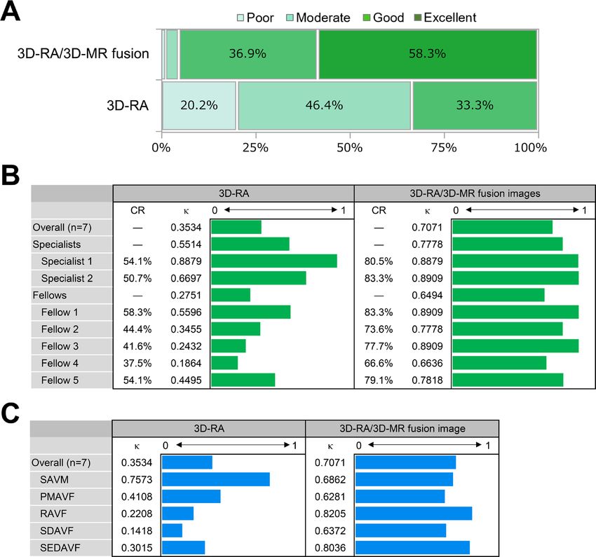
Spine
J NeuroIntervent Surg: first published as 10.1136/neurintsurg-2020-017252 on 4 March 2021. Downloaded from http://jnis.bmj.com/ on March 15, 2021 by guest. Protected by copyright.
Figure 2 3D-RA/3D-MR fusion images of a SEDAVF (case 2). Preoperative clinical imaging of a SEDAVF in a patient in their 60s who presented
with muscle weakness and sensory disturbance of the lower extremities. Thoracic sagittal T2-weighted MRI (A) shows low signal intensity of the
ventral spinal cord at the Th4 level. Subclavian artery angiography (B) and selective left Th4 segmental artery angiography (C) show a SEDAVF with
an epidural VP supplied by the left supreme intercostal artery and dorsal somatic branch. MIP axial (D), coronal (E), and volume rendering (F) images
reconstructed from the 3D-RA of the left subclavian artery show the detailed angioarchitecture of the SEDAVF. The arteriovenous fistula drains into the
intradural perimedullary vein through the epidural VP located in the lateral epidural space. 3D-RA/3D-MR fusion images (slab MIP axial image (G) and
coronal image (H), slab MIP image with scheme (I, J)) show a clear 3D relationship with differential contrast between the detailed angioarchitecture
of the SEDAVF and surrounding tissue structure, suggesting that the SEDAVF shunts into the lateral epidural VP. The dura mater is clearly visualized
by a black line in contrast to the spinal fluid in the subarachnoid space. An epidural vein draining from the VP penetrates the dura mater of the spinal
canal. The draining vein ascends the subarachnoid space and converges on the perimedullary vein. The asterisk indicates the shunt point. The yellow
line indicates the dura mater of the spinal canal. 3D-MR, three-dimensional-heavily T2-weighted volumetric magnetic resonance; 3D-RA, three-
dimensional-rotational angiography; A-P, anterior-posterior; MIP, maximum intensity projection; MRI, magnetic resonance imaging; SEDAVF, spinal
epidural arteriovenous fistula; VP, venous pouch.
DISCUSSION of contrast or the need for lumbar puncture. The advanced
In this study we evaluated whether using 3D-RA/3D-MR fusion 3D-RA-MR fusion technique enabled simultaneous visualization
images would improve the diagnostic performance for SAVSs. of the precise angioarchitecture of the SAVSs. 3D-RA/3D-MR
The interobserver agreement for the diagnosis, expressed with fusion imaging is a promising technique to precisely visualize
the kappa coefficient, markedly improved using fusion images. SAVSs, but further studies with larger samples are required to
In particular, the diagnostic accuracy of the fusion images in confirm the utility of this fusion technique.
the evaluation performed by the fellows was remarkable, and The complex vasculature of SAVSs occasionally renders
acceptable agreement was noted between the fellows and the surgical and endovascular treatment challenging. It is difficult
expert diagnoses. The fusion images allowed significantly better to delineate this complex vasculature with only conventional
recognition of the spatial relationship between the angioarchi- DSA because of its 2D acquisition and inherent vessel overlap.
tecture of the SAVSs and the surrounding anatomical landmarks, Thus, it is well known that 3D-RA provides accurate evaluation
including bony and soft structures, than 3D-RA with conven- of the shunt angioarchitecture with high spatial resolution and
tional DSA alone. The results of the present study show that more precisely shows the relationship between the shunt point
the addition of 3D-RA/3D-MR fusion images changed the diag- and the surrounding bony structures than conventional DSA.7 8
nosis of the lesions, suggesting that only using conventional DSA However, the spinal cord and surrounding soft tissues including
with 3D-RA may lead to misdiagnosis. There was no significant the dura mater are poorly distinguishable and recognizable with
difference between the 3D-RA and 3D-RA/3D-MR fusion images 3D-RA. For this reason, some SAVSs are often misdiagnosed as
for the accurate identification of the shunt points. This finding another type of SAVS at individual centers or there are diag-
indicates that 3D-RA with conventional DSA is valuable for the nostic disagreements even among experts.2 5
identification of shunt points. However, the categorical diag- Recently, the T2-weighted volumetric spinal MR sequence
nosis of the SAVS subtype is determined based on the anatomical was also shown to be well suited for the identification of vascular
landmarks where the shunt occurs. Thus, fusion imaging may flow voids with a high degree of accuracy of detection, localiza-
be essential for precise categorical shunt type diagnosis. To our tion, and characterization of SAVSs.17–20 Clear visualization of
knowledge, this is the first study to confirm the diagnostic accu- even subtle flow voids and neural structures could be achieved.20
racy of 3D-RA/3D-MR fusion images for the evaluation of SAVSs. However, fine arterial feeders and draining veins cannot be
Our fusion images provided a more detailed understanding of visualized and the actual fistula point cannot be seen with this
anatomy than 3D-RA without increasing the X-ray exposure of sequence alone. The type of spinal vascular malformation cannot
the patient. Unlike time-resolved imaging of contrast kinetics or be completely differentiated with MRI alone.18 21 Conversely,
computed tomography-myelography,14 15 the 3D-MRI used in this volumetric sequence provides high spatial resolution and
our fusion images can be obtained without the administration tissue interface contrast between the parenchyma, cerebral
4 Ryu B, et al. J NeuroIntervent Surg 2021;0:1–7. doi:10.1136/neurintsurg-2020-017252Spine
J NeuroIntervent Surg: first published as 10.1136/neurintsurg-2020-017252 on 4 March 2021. Downloaded from http://jnis.bmj.com/ on March 15, 2021 by guest. Protected by copyright.
Figure 3 3D-RA/3D-MR fusion images of a RAVF (case 3). Preoperative clinical imaging of a RAVF in a patient in their 70s who presented with
muscle weakness of the extremities. Cervical sagittal T2-weighted MRI (A) shows dilated tortuous vessels around the ventral cervical spinal cord.
Left vertebral artery angiography (B, C), selective left C6 segmental artery angiography (D), and selective left C5 segmental artery angiography
(E) show the spinal arteriovenous fistula with marked ascending intramedullary venous drainage supplied by multiple feeding arteries from the
radiculomeningeal arteries. The MIP axial image (F) reconstructed from the 3D-RA of the left vertebral artery shows the arteriovenous fistula
presumed to be located in the left C5 intervertebral foramen. 3D-RA/3D-MR fusion images (slab MIP axial image (G) and coronal image (H), slab
MIP coronal image with scheme (I)) show a clear 3D relationship with differential contrast between the detailed angioarchitecture of the AVF and
surrounding tissue structure. The dura mater of the spinal nerve root sleeve is clearly visualized by a black line in contrast to the spinal fluid in the
subarachnoid space. The shunt point is distinctly located in the intradural C5 nerve root sleeve, suggesting the location of the RAVF at the spinal nerve
root with intramedullary venous drainage. The asterisk indicates the shunt point. The yellow line indicates the dura mater of the spinal canal. 3D-MR,
three-dimensional-heavily T2-weighted volumetric magnetic resonance; 3D-RA, three-dimensional-rotational angiography; A-P, anterior-posterior; Lat,
lateral; MIP, maximum intensity projection; MRI, magnetic resonance imaging; RAVF, radicular arteriovenous fistula.
spinal fluid, ligaments, nerve roots, and dura mater of the spinal of their angioarchitecture is essential for the selection of an
canal.17–20 Owing to high spatial resolution and isotropic data optimal treatment strategy.3 5 The diagnostic significance of
acquisition, multiplanar reconstruction in any plane is feasible by the shunt lesion is that clinical characteristics and outcomes
applying detailed evaluation of the SAVSs and allowing favorable differ depending on the type of SAVS. In SAVSs at the cranio-
registration during image fusion with other modalities.18–20 22 cervical junction, hemorrhagic presentation is significantly
Therefore, fusion images combined with the features of 3D-RA, more frequent in RAVF, in which radical treatment is needed.2
which can reveal the fine angioarchitecture and the bony struc- However, it is difficult to distinguish between RAVF with a shunt
tures, and 3D-MRI, which can clearly reveal the surrounding on the intradural spinal nerve root and SDAVF with a shunt on
anatomical landmarks, is expected to increase the understanding the dura mater of the spinal nerve root sleeve based on 3D-RA
of SAVSs. During the creation of the fusion images, the target with conventional DSA because visualization of the true dura
vessels of interest can be color coded according to user prefer- mater, spinal nerves, and subarachnoid space is not possible
ence. Adjusting the threshold and thickness of the reconstructed with 3D-RA. Furthermore, other clinical differences have been
images can be performed at the workstation, which would lead reported; the preoperative neurological deficits of SEDAVFs are
to a better understanding of the 3D anatomy. In our present more severe than those of SDAVFs, the rate of gradual onset
results, the interobserver agreement for 3D-RA alone was not of PMAVFs was significantly higher than that of SAVMs, and
sufficient, especially for SDAVF, SEDAVF, and RAVF. This may the natural history of symptomatic spinal cord AVSs is poor.23 24
be because the relationship between the dura mater and shunt Therefore, there is a need to clearly delineate the shunt point of
point cannot be well delineated with 3D-RA alone. In fact, the SAVSs. Multimodal fusion images are promising for the evalu-
dura mater was clearly distinguishable from the arachnoid space ation and understanding of their angioarchitecture along with
on 3D-MRI, which improved the diagnostic interobserver agree- the surrounding anatomy,2 4 9 14 21 leading to the establishment
ment using the 3D-RA/3D-MR fusion images. Moreover, from of treatment strategies for both endovascular and surgical treat-
the fusion images, it is possible to speculate on the association ments.10–12 Thus, we recommend using this fusion technique for
between risky structures for vascular malformation, aneurysms, obtaining the optimal diagnosis of SAVS in the clinical setting.
varices and any accompanying complications of SAVSs such as In addition, proper diagnosis using fusion images would allow
hematoma or spinal cord focal localized edema. for the prediction of clinical outcomes and proper case manage-
The treatment strategy to obtain complete obliteration of ment. These fusion images could alter the management and
SAVSs is based on their angioarchitecture. Treatment outcomes treatment strategy for SAVS, leading to better treatment results.
differed among the types of SAVS.2 The differences in angioar- The present study has some limitations. This was a retrospec-
chitecture of spinal cord AVSs affect the complexity of treat- tive study with a relatively small sample size, given the rarity of
ment. Therefore, precise differentiation and identification the disease, and potentially involved selection biases. Therefore,
Ryu B, et al. J NeuroIntervent Surg 2021;0:1–7. doi:10.1136/neurintsurg-2020-017252 5Spine
J NeuroIntervent Surg: first published as 10.1136/neurintsurg-2020-017252 on 4 March 2021. Downloaded from http://jnis.bmj.com/ on March 15, 2021 by guest. Protected by copyright.
Figure 4 Interobserver agreement for the final diagnosis of SAVSs. (A) Assessment of image quality to delineate the relationship between the
vessels and surrounding anatomical structures: 4-grade scale (excellent, good, moderate, poor). The image quality grade was significantly higher
in the 3D-RA/3D-MR fusion image than in the 3D-RA image. (B) Interobserver agreements of the seven blinded reviewers for the SAVS diagnosis.
Comparison of the kappa coefficient between the 3D-RA and 3D-RA/3D-MR fusion images. (C) Interobserver agreements for each diagnostic category.
Comparison of the kappa coefficient between the 3D-RA and 3D-RA/3D-MR fusion images. 3D-MR, three-dimensional-heavily T2-weighted volumetric
magnetic resonance; 3D-RA, three-dimensional-rotational angiography; CR, concordance rate; PMAVF, perimedullary arteriovenous fistula; RAVF,
radicular arteriovenous fistula; SAVM, spinal arteriovenous malformation; SAVS, spinal arteriovenous shunts; SDAVF, spinal dural arteriovenous fistula;
SEDAVF, spinal epidural arteriovenous fistula; κ, kappa coefficient.
generalization of the conclusions of this study may be limited. of SAVS pathogenesis. It is expected to improve the overall diag-
There were also some methodological limitations. First, unlike nostic accuracy for SAVSs.
conventional DSA, 3D-RA/3D-MR fusion images do not provide
temporal information on blood flow. This disadvantage may be Acknowledgements We would like to thank our radiological technologist at St
eliminated with the use of a 4D-DSA dataset, instead of 3D-RA, Luke’s International Hospital for the technical assistance. We also thank Editage (
for the fusion images.13 Second, fusion of the different modality www.editage.jp) for English language editing.
images may not be precisely obtained because the bony land- Contributors BR and YN initiated the project. BR, SSa, MT, TM, SSh, TI, YO, and
marks may change based on patient posture, breathing, and YN were involved in the design of the experiments. BR conducted the experiments
motion during the examination.14 Diminution of the cerebro- and analyzed the data. BR and YN wrote the manuscript. All authors discussed the
results, commented on the paper, and approved the final version of the manuscript.
spinal fluid space in spinal canal stenosis or other pathologies
could also hamper the identification of a fistula.18 Therefore, Funding The authors have not declared a specific grant for this research from any
funding agency in the public, commercial or not-for-profit sectors.
fusion techniques require manual data corrections by well-
experienced radiological technologists and neurointerventional- Competing interests None declared.
ists. The possibility of misregistration should be considered as a Patient consent for publication Not required.
detriment of fusion imaging. Ethics approval All procedures performed in this series involving human
participants were in accordance with the ethical standards of the institutional
research committee (Saint Luke’s International Hospital, No. 19-R200) and the 1964
CONCLUSION Helsinki declaration and its later amendments or comparable ethical standards. The
We report the diagnostic accuracy of 3D- RA/3D- MR fusion need for written informed consent was waived because of the retrospective design
images for the evaluation of SAVSs. 3D- RA/3D- MR fusion of the study.
images provide high spatial resolution, allowing highly accurate Provenance and peer review Not commissioned; externally peer reviewed.
and detailed evaluation of the anatomical structure surrounding Data availability statement Data are available upon reasonable request.
the shunt. This fusion technique is promising for the evaluation All relevant data supporting the results of the present study are included within
6 Ryu B, et al. J NeuroIntervent Surg 2021;0:1–7. doi:10.1136/neurintsurg-2020-017252Spine
J NeuroIntervent Surg: first published as 10.1136/neurintsurg-2020-017252 on 4 March 2021. Downloaded from http://jnis.bmj.com/ on March 15, 2021 by guest. Protected by copyright.
the article and its Supplementary Information, and can be obtained from the 9 Tanoue S, Endo H, Hiramatsu M, et al. Delineability and anatomical variations
corresponding author on reasonable request. of perforating arteries from normal vertebral artery on 3D DSA: implications for
endovascular treatment of dissecting aneurysms. Neuroradiology 2020. doi:10.1007/
Supplemental material This content has been supplied by the author(s). It
s00234-020-02549-y. [Epub ahead of print: 21 Sep 2020].
has not been vetted by BMJ Publishing Group Limited (BMJ) and may not have
10 Shimizu S, Suzuki H, Maki H, et al. A novel image fusion visualizes the
been peer-reviewed. Any opinions or recommendations discussed are solely those
angioarchitecture of the perforating arteries in the brain. AJNR Am J Neuroradiol
of the author(s) and are not endorsed by BMJ. BMJ disclaims all liability and
2003;24:2011–4.
responsibility arising from any reliance placed on the content. Where the content
11 Sato M, Tateishi K, Murata H, et al. Three-dimensional multimodality fusion imaging
includes any translated material, BMJ does not warrant the accuracy and reliability
of the translations (including but not limited to local regulations, clinical guidelines, as an educational and planning tool for deep-seated meningiomas. Br J Neurosurg
terminology, drug names and drug dosages), and is not responsible for any error 2018;32:509–15.
and/or omissions arising from translation and adaptation or otherwise. 12 Ide S, Hirai T, Morioka M, et al. Usefulness of 3D DSA-MR fusion imaging in
the pretreatment evaluation of brain arteriovenous malformations. Acad Radiol
Open access This is an open access article distributed in accordance with the 2012;19:1345–52.
Creative Commons Attribution Non Commercial (CC BY-NC 4.0) license, which 13 Tritt S, Ommer B, Gehrisch S, et al. Optimization of the surgical approach in
permits others to distribute, remix, adapt, build upon this work non-commercially, AVMs using MRI and 4D DSA fusion technique: a technical note. Clin Neuroradiol
and license their derivative works on different terms, provided the original work is 2017;27:443–50.
properly cited, appropriate credit is given, any changes made indicated, and the use 14 Takai K, Kin T, Oyama H, et al. The use of 3D computer graphics in the
is non-commercial. See: http://creativecommons.org/licenses/by-nc/4.0/. diagnosis and treatment of spinal vascular malformations. J Neurosurg Spine
2011;15:654–9.
ORCID iDs 15 Mandalapu S, Kannath S, Kesavadas C. Fusion imaging of time resolved imaging of
Bikei Ryu http://orcid.org/0000-0003-0323-6628 contrast kinetics (TRICKS) and high resolution volumetric T2 MR sequences in the
Shinsuke Sato http://o rcid.org/0 000-0003-2 911-6755 evaluation of spinal vascular malformations. J Neuroradiol 2019;46:276–7.
16 Crewson PE. Reader agreement studies. AJR Am J Roentgenol 2005;184:1391–7.
REFERENCES 17 Baumert B, Wörtler K, Steffinger D, et al. Assessment of the internal craniocervical
1 Rodesch G, Lasjaunias P. Spinal cord arteriovenous shunts: from imaging to ligaments with a new magnetic resonance imaging sequence: three-dimensional
management. Eur J Radiol 2003;46:221–32. turbo spin echo with variable flip-angle distribution (SPACE). Magn Reson Imaging
2 Hiramatsu M, Sugiu K, Ishiguro T, et al. Angioarchitecture of arteriovenous fistulas 2009;27:954–60.
at the craniocervical junction: a multicenter cohort study of 54 patients. J Neurosurg 18 Kannath SK, Alampath P, Enakshy Rajan J, et al. Utility of 3D space T2-weighted
2018;128:1839–49. volumetric sequence in the localization of spinal dural arteriovenous fistula. J
3 Hiu T, Kitagawa N, Morikawa M, et al. Efficacy of DynaCT digital angiography in the Neurosurg Spine 2016;25:125–32.
detection of the fistulous point of dural arteriovenous fistulas. AJNR Am J Neuroradiol 19 Kannath SK, Mandapalu S, Thomas B, et al. Comparative analysis of volumetric
2009;30:487–91. high-resolution heavily T2-weighted MRI and time-resolved contrast-enhanced
4 Ryu B, Sato S, Mochizuki T, et al. Spinal arteriovenous fistula located in the filum MRA in the evaluation of spinal vascular malformations. AJNR Am J Neuroradiol
terminale externa: a case report and review of the literature. Interv Neuroradiol 2019;40:1601–6.
2020:159101992096836. 20 Kannath SK, Rajendran A, Thomas B, et al. Volumetric T2-weighted MRI improves the
5 Kiyosue H, Matsumaru Y, Niimi Y, et al. Angiographic and clinical characteristics diagnostic accuracy of spinal vascular malformations: comparative analysis with a
of thoracolumbar spinal epidural and dural arteriovenous fistulas. Stroke conventional MR study. J Neurointerv Surg 2019;11:1019–23.
2017;48:3215–22. 21 Krings T, Lasjaunias PL, Hans FJ, et al. Imaging in spinal vascular disease.
6 Kiyosue H, Tanoue S, Okahara M, et al. Spinal ventral epidural arteriovenous fistulas Neuroimaging Clin N Am 2007;17:57–72.
of the lumbar spine: angioarchitecture and endovascular treatment. Neuroradiology 22 Morris JM, Kaufmann TJ, Campeau NG, et al. Volumetric myelographic magnetic
2013;55:327–36. resonance imaging to localize difficult-to-find spinal dural arteriovenous fistulas. J
7 Prestigiacomo CJ, Niimi Y, Setton A, et al. Three-dimensional rotational spinal Neurosurg Spine 2011;14:398–404.
angiography in the evaluation and treatment of vascular malformations. AJNR Am J 23 Yu J-X, Hong T, Krings T, et al. Natural history of spinal cord arteriovenous shunts: an
Neuroradiol 2003;24:1429–35. observational study. Brain 2019;142:2265–75.
8 Li Z-F, Hong B, Xv Y, et al. Using DynaCT rotational angiography for angioarchitecture 24 Takai K, Endo T, Yasuhara T, et al. Microsurgical versus endovascular treatment of
evaluation and complication detection in spinal vascular diseases. Clin Neurol spinal epidural arteriovenous fistulas with intradural venous drainage: a multicenter
Neurosurg 2015;128:56–9. study of 81 patients. J Neurosurg Spine 2020:1–11.
Ryu B, et al. J NeuroIntervent Surg 2021;0:1–7. doi:10.1136/neurintsurg-2020-017252 7You can also read