Issue One 2018 - National Imaging Facility
←
→
Page content transcription
If your browser does not render page correctly, please read the page content below
Issue One 2018

Headshield Slug (Philine sp.) micro-CT scan shows internal shell and stomach of
probable new species found in Asia © Australian Museum

Imaging Data collected by Dr. Karine Mardon - Centre for Advanced Imaging, The
University of Queensland
In This Issue
    CEO’s Message

    Research Project
    •   Dicom2Cloud - Developing a Graphical
        User Interface for Anonymizing and
        Uploading Clinical Brain Scans to an
        Image Processing Cloud Instance

    National Cross-Capability
    Project
    •   National Network of Trusted Data
        Repositories

    International Collaboration
    •   MRI Studies of Freezing in Cold Hardy
        Plants

    News
    •   Imaging Uncovers Internal Structure
        of Australian Treasures
    •   Impact

2
CEO's Message

                                                       often not exciting, but essential to delivering
                                                       quality, reproducible outcomes, and greatest
                                                       impact. So NIF invests heavily in developing
                                                       tools, platforms and analysis workflows, to relieve
                                                       our users of the burden of establishing your
                                                       own pipelines. NIF is committed to instrument to
                                                       repository data management, whilst giving you,
                                                       the user, total control over your data.

                                                       The NIF partners are also committed to providing
                                                       access to the wider scientific community, and
                                                       particularly researchers who are not traditionally
                                                       part of the imaging community. So two projects
                                                       described come from researchers who may
I trust that you enjoy reading this Newsletter as      not have previously considered imaging as
much as the NIF team enjoy putting it together. It     a research tool. Both projects could have
is not designed as a source of the latest scientific   important implications for climate research,
breakthrough, although it always points to great       supporting research about our natural history and
science. Each article is chosen to highlight an        environmental effects on plants.
aspect of what extra opportunity NIF brings to the
scientific community, and how NIF can support          Have you considered how imaging may add
your research.                                         an extra dimension and added impact to your
                                                       project? We always love to get feedback, and
NIF’s mandate is to provide world-class                are happy to answer any questions about these
infrastructure, and to give you, as the research       articles, or your needs for an imaging component
user, the best data possible and the best possible     to your research.
experience. Managing data is time-consuming,

                                                                        Professor Graham Galloway
                                                                          Chief Executive Officer

                                                                                                         3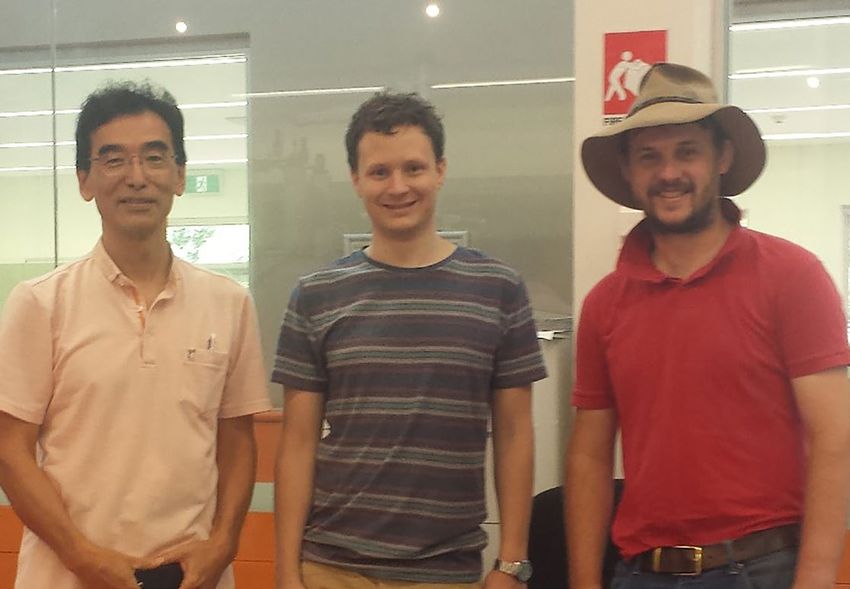
Dicom2Cloud - Developing a
                                            Graphical User Interface for
                                             Anonymizing and Uploading
                                              Clinical Brain Scans to an
                                               Image Processing Cloud
 Project
Research

                                                       Instance

           M
                    odern image processing algorithms aim to have              features, and upload this data to cloud platforms such as
                    clinical impact and help diagnose a variety of             Amazon AWS and Google cloud, where a wide range of
                    diseases, such as Multiple sclerosis, Parkinson, and       processing algorithms can be applied.
           neurodegeneration. However, many state-of-the-art post-
           processing techniques are not applied in a clinical setting,        Dicom2Cloud was developed as a platform-independent
           because the software developed by scientists is difficult           toolchain with a Python-based graphical user interface (GUI)
           to use, often operating-system specific, and might require          (Fig. 1). The implementation is open source and available
           extensive hardware resources. This limits the translation           on GitHub (https://github.com/CAIsr/dicom2cloud). The
           of research into clinical applications. One potential solution      application (Fig. 2) has three major processing steps: (i) file
           would be to upload the medical data to a cloud instance             selection, (ii) anonymization, and (iii) upload, processing and
           where all tools are installed and sufficient computational          download via a cloud instance. In detail, the File Panel (Fig.
           resources are available. The problem, however, is that              1B) is used to select a directory containing DICOM files, from
           medical data contains sensitive information that cannot             which the DICOM header information is read and an image
           be easily removed, such as facial features in magnetic              series is selected for processing. The Process Panel (Fig.
           resonance imaging data of the brain. During the last Health-        1C) allows the user to select the required cloud service and
           Hack event held in Brisbane (https://www.healthhack.com.            processing pipeline. Each image series is first anonymized
           au/), a team of enthusiastic developers (Saskia Bollmann,           using the MINC toolkit packaged inside a local Docker
           Isaac Lenton, Aswin Narayanan, Elizabeth Cooper-Williams,           container build using Neurodocker. In particular, we use
           and Yixia Peng) led by National Imaging Facility (NIF) Fellow,      dcm2mnc to convert the DICOM files into the MNC format,
           Dr Steffen Bollmann, approached this problem and developed          and mincanon to remove header information, such as scan
           Dicom2Cloud - a graphical user interface for Windows,               date and time, name, date of birth and information not critical
           Linux and Mac OS that can read DICOM data, remove facial            for the image.

                                                                                       Figure 2 – Dicom2Cloud’s application design overview. The
                                                                                   application consists of 3 panels that are used to select files, control
                                                                                    the pre-processing and pipeline selection and deliver information
                                                                                            about the processing status in the cloud instance.

              Figure 1 - Components of Dicom2Cloud GUI: A) Application
             overview, provides overview of the application and information
           about updates, B) File Panel, allows selection of DICOM datasets,
             C) Process Panel, allows selection of cloud service and type
              of processing, D) Cloud Panel, monitors submitted jobs and
                       downloads processed files from the cloud.

           4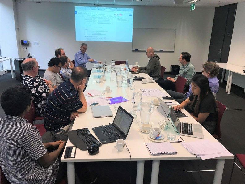
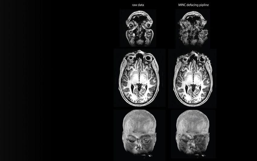
However, the facial features remain in the data and the
patient could potentially be re-identified. We therefore
apply a de-facing pipeline that robustly removes facial
features without introducing artificial edges (Fig. 3), which
otherwise would have negative effects on registration
algorithms. Next, the fully de-identified image is transferred
to cloud storage where an image processing pipeline is
automatically started on file arrival. Finally, the resulting
output files are downloaded back to the client and re-
associated with the patient ID. The Cloud Panel (Fig.
1D), allows the user to check the status of files sent for
processing in the cloud.

The design of Dicom2Cloud would also allow local
docker images to perform the full processing of data for
less intensive operations and therefore, could even be
used without a cloud backend. The team is currently
developing pipelines for running a brain segmentation
using FreeSurfer and the computation of quantitative
susceptibility maps for the first release of the software
and is looking for beta testers to test these pipelines.
The team is currently also seeking funding to pay for
cloud computing costs that would allow to offer the image
processing cloud backend free of charge for users.

       Figure 3 – This figure illustrates the result of the de-facing pipeline
      that was implemented. The goal is to remove facial features without
      introducing artificial edges that could cause problems in later steps.

For more details on the study, please contact Dr Steffen
Bollman (steffen.bollmann@cai.uq.edu.au).

                                          Dr Steffen Bollmann, NIF Fellow, is a post doctoral research fellow at the Centre for Advanced Imaging,
                                           UQ. He obtained a bachelor’s degree in science / biomedical engineering at the Ilmenau University of
                                       Technology, followed by a Masters degree in biomedical engineering & bioelectromagnetism. Following this,
                                       Steffen completed a PhD investigating multimodal imaging in ADHD children, adolescents and adults at the
                                        Neuroscience Centre Zurich and the Centre for MR-research, University Children’s Hospital Zurich. Steffen
                                      joined the Centre for Advanced Imaging, University of Queensland, in October 2014, where he is applying his
                                        expertise in multimodal imaging in the group of A/Prof. Markus Barth combining high resolution quantitative
                                        imaging (susceptibility, T1, T2*), functional MRI (fMRI), and electroencephalography (EEG) with the goal to
                                         understand the relationship between functional networks and to work towards identifying early biomarkers
                                         for neurodegenerative diseases. Exploiting the high signal levels of ultra-high field 7 Tesla MRI he aims to
                                                           investigate and quantify disease processes on a single subject level.

                                                                                                                                                        5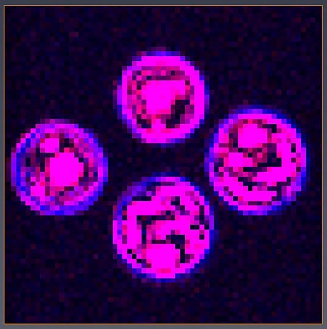
National Network of Trusted
                                              Data Repositories

                            D
                                    uring 2017 the National Imaging Facility (NIF)               data), the process by which data is moved from the
National Cross-Capability
                Project

                                    nodes at the University of Western Australia (UWA),          instrument to the digital repository service and the
                                    University of Queensland (UQ), University of New             format(s) of the data.
                            South Wales (UNSW) and Monash University collaborated
                            on a national project to enhance the quality, durability and    2.   The NIF requirements for a trusted data repository
                            reliability of data generated by NIF. The Project, Delivering        service - Provides a platform-agnostic checklist of
                            durable, reliable, high-quality image data, was jointly              requirements that a basic NIF trusted data repository
                            funded by the Australian National Data Service (ANDS) and            service should satisfy, including: identification of
                            Research Data Services (RDS). It was motivated both by               data by a unique Project identifier, ingestion of data
                            NIF’s desire to enhance the quality of the data associated           from NIF-compliant instruments, authentication via
                            with the use of its facilities, and the desire of ANDS/RDS           the Australian Access Federation (https://aaf.edu.
                            to facilitate the establishment of Trusted Data Repositories         au), interoperability and easy deployment across NIF
                            that enable access to data for at least 10 years and includes        nodes.
                            metadata that documents both the quality of the data and its
                            provenance.                                                     3.   Implementations of trusted data repository services for
                                                                                                 two exemplars:
                                                                                                     •  Preclinical MRI data (with mouse brain data
                      •         Qu a l i t y p e r ta i ns to a N IF use r ’s                           as an example) acquired across three NIF
                                                                                                        nodes—UNSW, UQ and UWA—using a Bruker
                                e x p e c t a t i o n th a t a n a n i ma l , pl ant                    BioSpec 9.4T MRI. The services have been
                                                                                                        implemented using the open source MyTardis/
                                o r m a t e r i a l can b e sca n n e d a n d                           ImageTrove (https://www.mytardis.org)
                                fr o m t h a t da ta rel i a b l e o u tco mes/                         platform.
                                                                                                     •  Clinical ataxia MRI data acquired using a
                                c h a r a c t e r i sa ti on s ca n b e o b ta i ned                    Siemens Skyra 3T MRI scanner in support of a
                                ( e . g . s i g n al , vol ume, morph o l ogy) over                     Monash-proposed International Ataxia Imaging
                                                                                                        Repository (IAIR). The service has been
                                ti m e a n d a cross N IF si te s.                                      Implemented using the open source XNAT
                                                                                                        (https://www.xnat.org) platform.
                      •         D u r a b i l i t y refe rs to g u a ran te e d long-
                                te r m a v a i l ab i l i ty of th e d a ta .                    Software developed to support the implementation
                                                                                                 of the repository services includes: Docker (https://
                      •         R e l i a b i l i t y me a n s th a t th e d a ta is             www.docker.com) Compose scripts to permit easy
                                                                                                 deployment at differents sites, client-side scripts for
                                u s e f u l f o r futu re rese a rche rs, i.e.                   uploading NIF-certified data to ImageTrove/MyTardis
                                s t o r e d i n on e o r mo re o p e n d a ta                    and an XNAT plugin for uploading non-DICOM files.

                                fo r m a t s a nd w i th su ffi ci e n t e vi dential       4.   Assessments of the resulting trusted data repository
                                                                                                 services against a relevant international metric, the
                                metadata.                                                        CoreTrustSeal (https://www.coretrustseal.org) Core
                                                                                                 Trustworthy Data Repositories Requirements.

                            The scope of the Project was limited to MRI data with the
                            understanding that the developed requirements and trusted
                            data repository services could be adapted to, or serve as a
                            basis for other instruments/modalities.

                            The key outcomes from the Project include:
                            1. The NIF agreed process for acquiring trusted data
                                (NAP) - Lists the requirements that must be satisfied
                                to obtain high-quality data, i.e. NIF-certified data,
                                suitable for ingestion in a NIF trusted data repository
                                service. They cover provisioning of a unique instrument
                                identifier, instrument registration with Research Data
                                Australia (https://researchdata.ands.org.au), Quality
                                Control (QC), quality assurance measures, requisite
                                metadata (including cross-reference to the QC                    Above: The team met throughout the year to tackle the issue of
                                                                                                      long term reliable imaging data storage and access.

                            6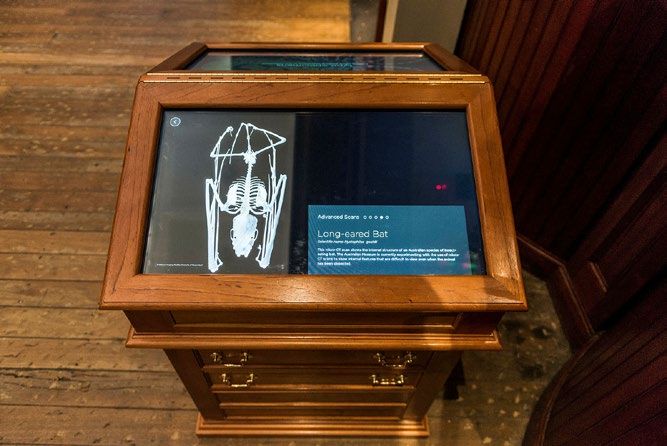
Dr. Andrew Mehnert, NIF Informatics Fellow is the Project manager and UWA lead

                 Andrew is Senior Lecturer in Data Management, Analysis and Visualisation at the Centre for Microscopy,
                 Characterisation and Analysis (CMCA) at the University of Western Australia (UWA). His position is jointly
                       funded by NIF and the Australian Microscopy & Microanalysis Research Facility (AMMRF).

For NIF users and the broader imaging research community the benefits and impact of this Project include:
        •   Reliable and durable access to data
        •   Improved reliability of research outputs and the provenance associated with it
        •   Making NIF data more FAIR (Findable, Accessible, Interoperable, Reusable - https://www.ands.org.au/working-
            with-data/the-fair-data-principles)
        •   Easier linkages between publications and data
        •   Stronger research partnerships

For research institutions they include:
        •   Enhanced reputation management
        •   A means by which to comply with the Australian Code for the Responsible Conduct of Research
        •   Enhanced ability to engage in multi-centre imaging research projects

For NIF they include:
        •    Improved data quality
        •    Improved international reputation
        •    The ability to run multi-centre trials

The transition plan post-funding includes: maintenance of existing services for 10 years; the integration of additional
instruments; creation of a project web portal; planned new national and international service deployments; refinements and
improvements; and CoreTrustSeal certification.

Project documents have been archived in the NIF Customer Relationship Management (CRM) system (accessible by NIF staff).
Project software is hosted on GitHub and is freely available for download here: https://github.com/NIF-au/TDR. For further
information please contact either the national Project Manager (andrew.mehnert@uwa.edu.au) or NIF (admin@anif.org.au).

                   Project Manager and UWA lead: Andrew Mehnert (NIF Informatics Fellow, Centre for
                   Microscopy, Characterisation and Analysis)
                   NIF lead - Graham Galloway (Chief Executive Officer, NIF)
                   UQ lead - Andrew Janke (NIF Informatics Fellow, Centre for Advanced Imaging)
                   UNSW lead - Marco Gruwel (Senior Research Associate, Mark Wainwright Analytical Centre)
                   Monash lead - Wojtek Goscinski (Associate Director, Monash eResearch Centre)

           A t rus t e d d a t a r e p o s i t o r y se r vice is e sse n tia l fo r sh a r in g
           dat a and e n s u r e s t h a t p ro je ct d a ta cr e a te d a n d u se d b y
           res earc h e r s i s “ m a n a g e d , cu r a te d , a n d a r ch ive d in su ch a
           way t o p r e s e r v e t h e i n i t i a l in ve stm e n t in co lle ctin g th e m ”
           and t hat t h e d a t a “ r e m a i n u se fu l a n d m e a n in g fu l in to th e
                        f u t u r e ” ( h t t p s : / / www.co r e tr u stse a l.o r g ) .

                                                                                                                              7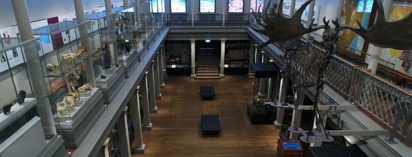
MRI Studies of Freezing in Cold
Hardy Plants
F
reezing is one of the extreme environmental factors affecting plants. Cold hardy plants have evolved a variety of complex
strategies to control water behaviour under freezing conditions. These include strategies to survive water deficit and
subfreezing temperatures that could cause lethal intracellular freezing of water. Many plants spontaneously reduce their
International
Collaboration
water content during formation of seeds in a controlled manner without losing the integrity of the cells or vice versa during
rehydration of the seeds.
Some mechanisms involve special compounds for controlling water behaviour (e.g., supercooling stabilizing compounds, SSC;
anti-nucleation compounds, ANC; and ice nucleation agents, INA) and regulate phase changes of water (water to gas, water to ice
or vice versa). SSCs include some flavonoids, polyphenols, and anthocyanins. ANCs include SSCs plus some compounds specific
to each ice nucleator. INAs include some proteins and small organic substances. These compounds may possibly regulate the
behaviour of aqueous solutes including the avoidance of unwanted precipitation/bubble formation and promotion of preferential
precipitation/aeration both in solution and on surrounding components such as membranes and cell walls (gel to sol or vice versa).
Figure 1 (left): Colour-coded axial cross sections
of Fagus (left) stems/(right) leaf buds constructed
from a series of 1H magnetic resonance 3D datasets
conducted at various temperatures of 4 Fagus
leaf buds. The field of view is 9 × 9 mm with pixel
dimensions of 134 × 134 μm. Red shows “high
temperature” freezing (> -14 °C) and blue shows
tissues that didn’t freeze, even at -19.5 °C. (left) The
blue interior of the stem (pith) remains unfrozen.
(right) The exterior (the surface of bud scales) in this
case remains unfrozen.
The distribution of water throughout plant tissues can be visualised using MRI. In an MRI scan, a radiofrequency pulse excites
the aggregate nuclear magnetisation giving it a transverse component which is then acquired as a signal and processed to give
an image. The transverse magnetisation disappears at a rate quantified by the T2 relaxation time. Typically, after excitation of the
magnetisation, there is a delay of a few milliseconds before acquisition of the signal. Ice water has a significantly lower transverse
relaxation time than liquid water and so the signal from ice vanishes giving an image that only shows the liquid water. The freezing
of plant tissues can be studied by conducting a series of experiments at different temperatures. At any particular temperature
only the unfrozen water in tissues will contribute to the image. The sequence of images can then be combined to show freezing
behaviour of the tissues.
Figure 2 (left): Another figure created from the same 3D dataset showing the unfrozen interior (pith) of the Fagus stem and
the unfrozen exterior (the surface of bud scales) of the leaf bud. The field of view is 9 × 37.5 mm with pixel dimensions of
134 × 250 μm.
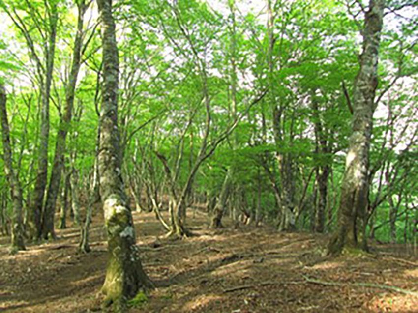
Figure 3 (left):Fagus leaf buds.
8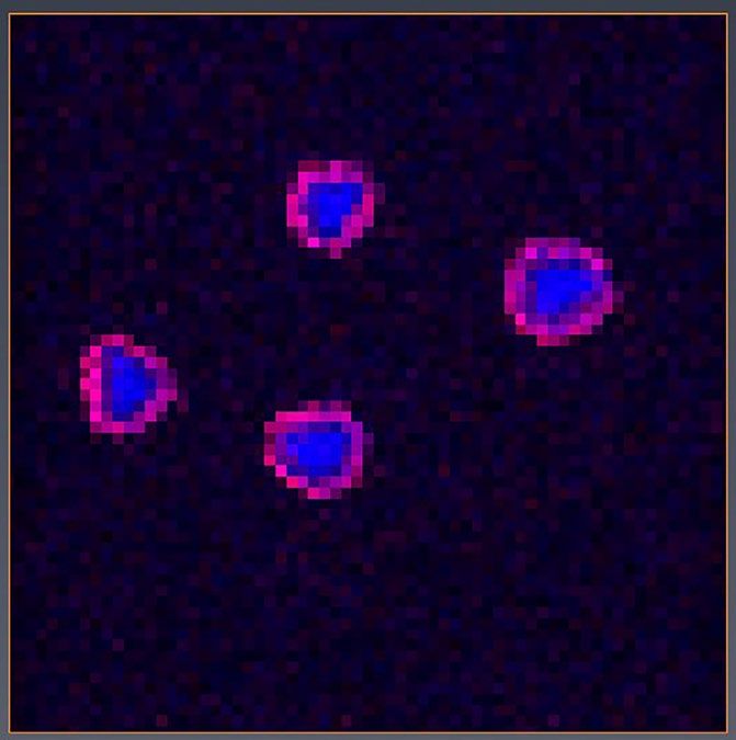
The colour-coded images (figures 1 and 2) of the Fagus leaf bud (a species of Japanese mountain vegetation, figures 3 and 4)
were constructed from fourteen experiments conducted at temperatures ranging from 1 °C to -19.5 °C (specifically 274, 270, 268,
266, 264, 261, 259, 258, 257, 256, 255.5, 255, 254.5, 254 K). The plant was given an hour to equilibrate at each temperature
prior to scanning using a gradient echo sequence to produce a 3D dataset with resolution 134 × 134 × 250 μm. The temperature
was then lowered and the procedure repeated. Subtracting an image acquired at a lower temperature from an image acquired
at a higher temperature gives an image showing the tissues that froze between those two temperatures – if a tissue doesn’t
freeze then subtracting mostly cancels the tissue from the image, whereas if the tissue does freeze it is absent from the lower
temperature image and is therefore not cancelled by the subtraction. In this way, images of the tissues that froze between each of
the successive temperatures in the list above can be created. The images in figures 1 and 2 were created by colour coding these
“difference images” red for freezing that occurs at higher temperatures and blue for freezing that occurs at lower temperatures.

                                                                            The interior of the stem and exterior of the bud did not
                                                                            freeze at all, even at -19.5 °C – these tissues appear
                                                                            blue in the images. From differential thermal analysis
                                                                            and MRI there are three different freezing behaviours
                                                                            seen in different tissues with freezing events occurring
                                                                            at warmer temperatures than -11 °C, between -11 and
                                                                            -17 °C and lower than -17 °C. Similar experiments have
                                                                            been conducted at the Western Sydney University
                                                                            Node of NIF on Azalea flower buds and Cornus species
                                                                            (C. officinalis, C. japonicam, C. florida). Another set of
                                                                            experiments examines where and how freezing starts at
                                                                            set temperatures; these experiments additionally include
                                                                            blueberry and forsythia stems.

                                                                            Interestingly, drought and freezing lead to similar
                                                                            behaviours of water at the plant cell level. Under freezing
                                                                            stress, most typically, the cells undergo extracellular
                                                                            freezing where the primary freezing is initiated in the
                                                                            intercellular spaces and the cell water (>70%) migrates
                                                                            to the extracellular spaces during cooling to -7 °C, which
                Figure 4 (above): Fagus in its natural habitat.
                                                                            results in extreme dehydration of the cells during further
                                                                            cooling.

                                                                            MRI is proving to be a powerful tool for studying freezing
                                                                            of cold hardy plants and identifying tissues containing
                                                                            powerful freeze regulating compounds which may be
                                                                            valuable resources for food industries, clinical use,
                                                                            cryopreservation of embryos and oocytes for IVF,
                                                                            endangered species, etc.

                                                                          Figure 5 (left): Dr Masaya Ishikawa; WSU NIF Fellow, Dr Tim Stait-
                                                                          Gardner; and WSU Biomedical Magnetic Resonance Facility Manager,
                                                                          Dr Scott Willis.

                                                           Collaborators
                  Biomedical Magnetic Resonance Facility, Western Sydney University node of National Imaging Facility
                                          Tokyo University of Science, Noda, Chiba, Japan
                                                       The University of Tokyo

                                                                                                                                           9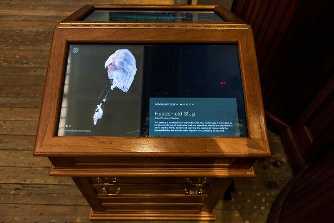
Imaging Uncovers Internal
                                           Structure of Australian
                                                  Treasures

       M
               icro-CT imaging data collected by NIF Fellow, Dr.          permanent installation, features 100 invaluable treasures
News

               Karine Mardon, using the Inveon PET-CT scanner at          from the Australian Museum collection, and the stories of
               the Centre for Advanced Imaging (CAI), the University      100 people who have had a profound influence on Australian
       of Queensland node of NIF, has been transformed into an            history.
       interactive display as part of the 200 treasures exhibition at
       the Australian Museum's newly restored Long Gallery (now           The gallery has a rich history, as the first gallery in Australia's
       Westpac Gallery).                                                  first museum. The 19th century theatre has been extensively
                                                                          restored over the past two years to preserve and adapt
       The data has been reconstructed to create a multimedia,            the space. While respecting the historical significance of
       interactive exhibit where visitors can see a 3D model of the       the gallery, it has embraced a modern spirit reflecting the
       internal structures of several specimens.                          museum's current and future collections.

       The Westpac Gallery 200 Treasures, which will be a

       The Museum has been experimenting with CT to view internal         Acknowledgements
       structures of a specimens without the need for dissection.         Micro-CT data collected at the Centre for Advanced Imaging, The
       The Headshield Slug micro-CT was able to show the internal         University of Queensland
                                                                          Multimedia display in the Westpac Long Gallery developed by the
       shell and entire digestive tract - including the creature's last
                                                                          interactive design company Holly.
       meal! AMRI scientists are using the scans to collect and           All images are copyright of the Australian Museum
       interpret data from a number of specimens.                         Article, written by Nina Moore, is available at https://cai.centre.
                                                                          uq.edu.au/article/2017/12/imaging-data-treasured-asset

                        Long Eared Bat multimedia display. Image ©           Headshield Slug multimedia display. Image ©
                                   Australian Museum                                     Australian Museum

       10
Impact
Australian research communities are well familiar with the word Impact and the recent emphasis and requirement
for identifying and demonstrating research impact. Until recently, the evaluation of research has focused largely on
publications, patents, and grants. Much less attention was paid to the relative impact beyond outputs and outcomes. The
move towards impact measurement encourages engagement beyond a particular academic discipline and awareness
of the interests and needs of the people that fund research. It also focuses effort on clearly articulating the many ways in
which investments in research deliver benefits for society.
In response to this requirement and to demonstrate the impact of research infrastructure, the National Collaborative
Research Infrastructure Strategy (NCRIS) capabilities have initiated a working group to develop and define metric that
indicate the Impact of NCRIS at program level. The group holds regular teleconference meetings and had a face-to-face
workshop in Canberra, February this year. The main objective of the workshop and meetings is to come to a common view
of the Impact Pathway, which can be used by individual capabilities. Following these efforts, NIF has reviewed its major
performance indicators to align with the Impact Framework and will continue to work closely with other NCRIS capabilities
to refine them.
nother initiative of the NCRIS capabilities was to establish 'The NCRIS Communications Group' early 2017 for the
A
purpose of collaborating and sharing ideas and resources between the NCRIS funded projects, as well as the essential
enabling infrastructure services of AARNet and AAF. The group, which comprises of Communication and Engagement
Managers of NCRIS capabilities, has developed NCRIS Network website. The website exposes the value of NCRIS
capabilities and provides a central hub for stories and successes of the projects. Visit www.ncris-network.org.au to learn
more about the Impact of NCRIS capabilities.
11You can also read