Studies of Guinea Pig Tumors I. Report of Fourteen Spontaneous Guinea Pig Tumors, with a Review of the Literature
←
→
Page content transcription
If your browser does not render page correctly, please read the page content below
Studies of Guinea Pig Tumors
       I. Report of Fourteen Spontaneous Guinea Pig Tumors,
                  with a Review of the Literature*

                            JAMESB. ROGERSANDHERMANT. BLUMENTHAL
             (Department of Anatomy, University of Louisville, School of Medicine, Louisville, Ky., and the Institute
                               of Experimental Pathology, The Jewish Hospital, St. JMUÕS,  Mo.)

                                                          SUMMARY
            Fourteen spontaneous tumors of an inbred strain of guinea pigs have been described.
         No tumors were found in guinea pigs under 3 years of age, thus supporting the ob
         servation of Papanicolaou and Olcott (43) that spontaneous tumors occur rarely in
         guinea pigs which do not survive to senility. While this constitutes an incidence of
         only 2.4 per cent of the total population of this colony, it represents an incidence of
         14.4 per cent of the animals surviving 3 years or longer.
            These fourteen tumors bring the total number of reported spontaneous neoplasms
         in guinea pigs to 138. The latter have been tabulated as to site of origin and micro
         scopic characteristics, and they are reported for the convenience of investigators
         encountering additional instances of spontaneous tumors or as a frame of reference
         for those studying the induction of tumors in guinea pigs.

   Despite the prevalent use of the guinea pig as an                 assemble a total number prior to the present report
experimental animal, surprisingly few spontaneous                    of only 124 spontaneous tumors. This figure in
tumors have been reported in this species. Re                        cludes ten cases described in detail by Papanicolaou
views dealing with this subject have been pub                        and Olcott (41-43) but does not include approxi
lished by Heim and Schwartz (21), Maury (36),                        mately 90 which these investigators collected but
Leader (28), Warren and Gates (53), and most re                      have not reported.1 It also does not include 23
cently by Tamaschke (51). The reviews preceding                      cases of an ovarian "tumor" described by Loeb
that of Warren and Gates in 1941 were incomplete                     (32) and several additional ones by others (8, 11,
in that they did not include a number of reports                     30) cited by Loeb. This "tumor" consists of em
dealing with spontaneous tumors of the respiratory                   bryonal structures, apparently of parthenogenetic
tract (Sternberg [49], Spronck [48], Goldberg [16],                  origin, which should not be considered as a true
and seven other spontaneous tumors observed by                       neoplasm, since, as Loeb has pointed out (32), they
Woglom [54]). The most recent review by Ta                           have limited duration of life, being destroyed at an
maschke (51) is incomplete in that it includes only                  early date and replaced by connective tissue.
seventeen spontaneous tumors, a number far                               The rarity of spontaneous tumors in guinea pigs
smaller than that reported by Warren and Gates                       can be appreciated also by other reports. Thus, in
14 years earlier, although it mentions a spon                        three inbred families of guinea pigs at the Belts-
taneous tumor reported by Gouyon, not men                            ville Agricultural Station of the U.S. Department
tioned in previous reviews. Since the review by                      of Agriculture, studied by Wright and Eaton (56),
Warren and Gates (53), a number of additional                        no neoplasms were found in 15,000 animals. Simi
cases have been reported, and we have been able to                   larly, Haagensen and Krehbiel (19) have stated
     * This work was supported by Research Grant #C-1590             that in their colony of several thousand guinea pigs
(Cl-6) from the National Cancer Institute of the National In         only two spontaneous tumors were found. Other
stitutes of Health, Department of Health, Education, and             reports indicate a somewhat higher incidence.
Welfare.                                                             Thus, Spronck (48) was able to collect 56 spon-
   Received for publication July 10, 1959.                              ; Personal communication from G. N. Papanicolaou.

                                                               191

          Downloaded from cancerres.aacrjournals.org on February 18, 2021. © 1960 American Association for Cancer
                                                        Research.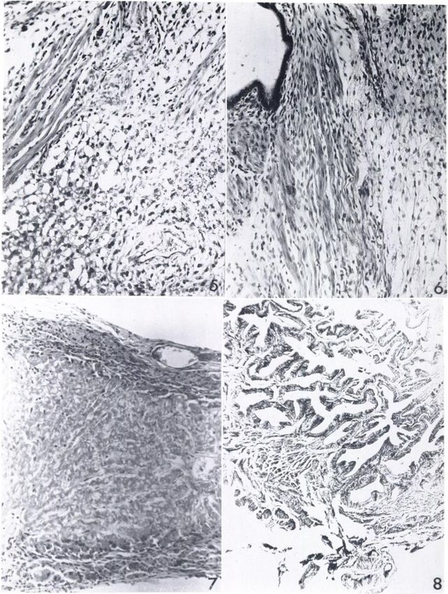
192                                                    Cancer Research                       Vol. 20, February 1960
       taneous papillary adenomas of the bronchus, and                    2,000 in the nonsusceptible strain. Each animal of
       Papanicolaou    and Olcott (41-43) have found                      the two strains, on death, was perfused with 20 per
       about 100 tumors in a group of 7,000 guinea pigs                   cent formaldehyde     and subsequently   autopsied.
       (1.4 per cent). Congdon and Lorenz (10) have re                    When tumors were encountered,        they were de
       ported an incidence of leukemia of 5 per cent in an                scribed, measured,    and blocks were taken for
       inbred strain, and Loeb (32) has reported an inci                  microscopic study. The latter were embedded in
       dence of parthenogenetic    "tumors" of at least 5
                                                                          paraffin, sectioned at 6 m/j. and stained with
       per cent in female guinea pigs. This latter lesion                 hematoxylin and eosin.
       probably occurs with even greater frequency, since
       their limited duration of life with replacement by                                      RESULTS
       scar tissue undoubtedly  results in a failure to de                    Incidence of spontaneous tumors.—Spontaneous
       tect many of them. Papanicolaou and Olcott (43)                    tumors were detected in fourteen of the 4,000 ani
       have attributed their relatively high incidence to                 mals (0.4 per cent) in the susceptible strain (Table
       the fact that many of the autopsied animals were                   1). However, since no tumors were found in ani
       senile, since the tumors rarely occurred in guinea                 mals under 3 years of age, data reflecting this fact
       pigs less than 4-5 years old.                                      are perhaps more pertinent. Slightly less than 2.5
          Between the years 1941 and 1951 one of us                       per cent of guinea pigs of the susceptible strain
       (JBR) maintained two separate inbred strains of                    survived over 3 years, and of this group the finding
       guinea pigs. During that period no animals were                    of fourteen tumors represents an incidence of 14.4
                                                                          per cent. The frequency in males surviving over 3
                           TABLE 1                                        years was 12 per cent and in females, 15.3 per cent.
               THE FREQUENCY
                           OFSPONTANEOUS
                                      TUMORS                                  The proportion of the nonsusceptible strain sur
                                                                          viving over 3 years was only about one-third that
                                                                          of the susceptible strain. The females of the non-
                             strain4,00097           strain2,00017
                                                                          susceptible colony are known to be susceptible to a
       Total no. live
       births:Total                                                       toxemia of pregnancy, and this would account, to
                                                                          some extent at least, for the smaller number of live
             no. surviving
         over 3 years:          (2. 4 per cent)         (0 .9 per cent)   births and for the smaller proportion of females
         Males               257214                   4                   surviving over 3 years; however, it would not ac
       FemalesNo.                                    130
                                                                          count for the smaller proportion of males. For rea
           with tumors:        (14.4 percent)*                            sons not yet known, the life span of the non-
         Males               8(12.0      " )*
                                                                          susceptible strain is shorter than that of the sus
         FemalesSusceptible 11(15.3      " )*Nonausceptible
                                                                          ceptible strain.
                                                                              Distribution and characteristics of the tumors.—
          * Per cent of total number of animals surviving over 3 years.
                                                                          Seven primary anatomic sites were represented
       withdrawn from these colonies for experimental                     among the fourteen spontaneous tumors. The or
       purposes, and they were permitted to live out their                gan most frequently involved in this strain was the
       life span. In one of these strains spontaneous tu                  uterus, where seven tumors were found. Two of the
       mors first appeared in 1948, and in the period                     latter were benign leiomyomas, and one was a
       1948-1951 fourteen spontaneous tumors were de                      benign adenomyoma.        Sarcomatous    degeneration
       tected. This has been designated the tumor-sus                     was found in the remaining four, one of which was
       ceptible strain. No tumors developed in the second                 a leiomyosarcoma,      one a fibrosarcoma,      one a
       strain during this period of observation, nor have                 myxosarcoma, and one a mesenchymal mixed tu
       any developed since 1951, although animals have                    mor. Two tumors were found in the liver, a hepat
       been withdrawn for experimental        purposes. The               ic-cell adenoma in one case, and a cavernous he-
       present report deals primarily with the character                  mangioma in another. There was one case each of
       istics of these fourteen spontaneous tumors, and a                 an embryonal carcinoma of the testis, a papilloma
       comparison is drawn as to frequency and type                       of the gall bladder, papillary pulmonary adeno-
       with guinea pig tumors heretofore reported. Fur                    matosis, a subcutaneous fibrolipoma, and a case of
       thermore, all the tumors reported to date have                     lymphoblastic    leukemia. In none of the tumors
       been classified as regards histologie type and organ               considered histologically malignant was evidence
       of origin for purposes of subsequent comparison                    of metastasis found.
       with induced tumors in the guinea pig.                                 The leiomyomas of the uterus were discrete, en
                                                                          capsulated tumors showing the typical pattern of
                 MATERIALS          AND METHODS                           intermingling bundles of elongated smooth muscle
          There were 4,000 live births in the tumor-sus                   cells. Differing from the latter only in that the tu
       ceptible colony during the period 1941-1951 and                    mor contained endometrial glands was the single

Downloaded from cancerres.aacrjournals.org on February 18, 2021. © 1960 American Association for Cancer
                                              Research.
ROGERSANDBLUMENTHAL—Spontaneous
Tumors in Guinea Pigs 193
case of adenomyoma (Fig. 1). The one tumor con ameter; microscopically, it showed a thin, intact
sidered to be a leiomyosarcoma was also encap fibrous capsule enclosing liver cells, with evidence
sulated grossly, and microscopically it retained a of sinusoids and the usual cordlike pattern, but
lamellated structure (Fig. 2), with the bundles devoid of bile duct structures (Fig. 7). Between the
compactly arranged and with little or no inter capsule and the mass of liver cells there was a thin
stitial structure. The cells were also closely packed, layer of large vacuolated phagocytes containing
and mitotic figures were fairly numerous. The tu brown pigment. This was considered to be a small
mor diagnosed as a fibrosarcoma was confined hepatic-cell adenoma. The gall bladder papilloma
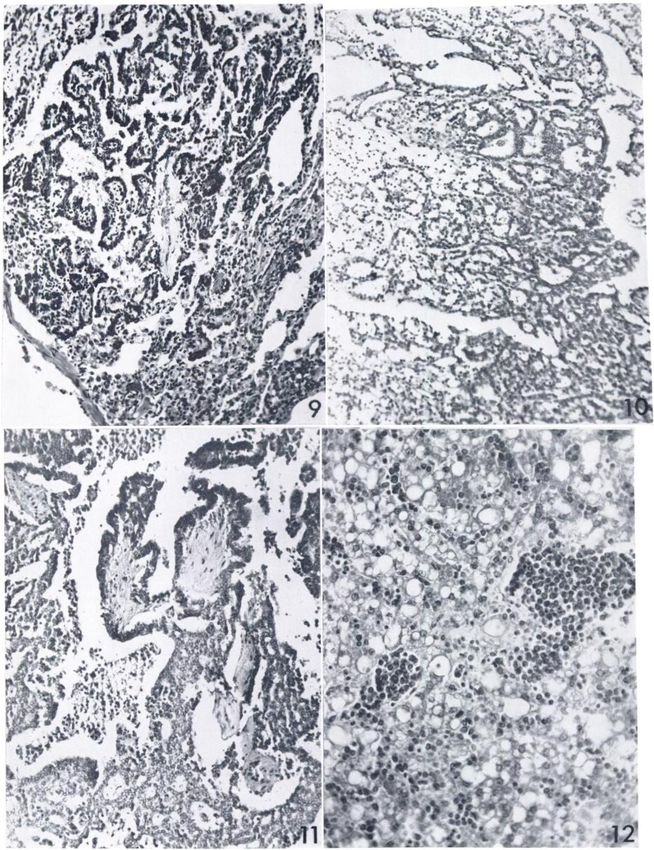
within the serosa (Fig. 3). In the latter tumor the presented as a small cauliflower-like growth on
cells were somewhat more loosely arranged and
were spindle-shaped rather than straplike, as in TABLE 3
the tumors of smooth muscle origin (Fig. 4). This REPORTEDSPONTANEOUS
TUMORSOFTHE
was taken to be an indication that this tumor arose RETICULOENDOTHELIAL
SYSTEM
from the interstitial connective tissue of the uterus
Tumor No. cases Author and date
TABLE2
Spleen:
REPORTEDSPONTANEOUS
TUMORSOF Splenoma (lymphosarco-
THE REPRODUCTIVE
TRACT ma?) Guérinand Guérin,
1925 (18)
TumorOvary:Embryoma cases11112111111113Author
yearMontroni, and Lymph glands:
Cervical, lymphosarcoma Dickson, 1915 (14)
Miguenz, 1918 (37)
teratomaU
or citedby 1930,
(28)Giordano,
Leader Leukemia Snijders, 1926 (47)
tÃ-u ti
(15)Haranghy
1931 Heston and Deringer,
uUterus:I^iomyoma u
1954(20)Papanicolaou
et al., 1952 (22)
Congdon and Lorenz,
1954 (10)
Lorenz et al., 1954 (33)
(gross Ol-cott, and
only)uFibromyomaAdenomyomar.eiomyosarcomaFibrosarcomaMyxosarcomaMesenchymal 1 Present report
1942(42)Present
reportLipschutz, Total 13
(31)Present 1941
reportu
uu
uU TABLE 4
tÃ-u
mixed tu REPORTEDSPONTANEOUSTUMORSOF
morTestis:Embryonal uu THE RESPIRATORY
TRACT
u
carcinomaTotalNo. TumorBronchus:Papillary cases2561131165Author
yearSternberg, and
Adenomaa (49)Spronck,1903
ttu
(48)Norris,1907
tt"
(40)Heston
1947
rather than from smooth muscle. The same inter "tÃ-
1952(22)Lorenz
and Deringer,
pretation was placed on the case of myxosarcoma, UtÃ-
(33)Present
el al., 1954
in which the tumor cells exhibited an even looser UAdenocarcinomaTotalNo.
ReportGoldberg,
arrangement (Fig. 5). The latter tumor was grossly 1920 (16)
more diffuse and less well demarcated than in the
foregoing cases, but had also not extended through
the serosa. The tumor designated as a mesenchy-
inal mixed tumor of the uterus differed from the gross inspection, and microscopically showed
myxosarcoma only in the fact that cystic endo- fronds and glands lined by tall hyperchromatic
metrial glands were incorporated in the neoplastic epithelial cells (Fig. 8). There was no evidence of
mass (Fig. 6). invasion into muscle.
The two tumors of the liver were both consid The single case of papillary adenomatosis of the
ered benign. One was a typical, grossly purple, lung presented grossly as two tiny white nodules in
cavernous hemangioma, and microscopically the left upper lobe. Microscopically these growths
showed numerous dilated vascular channels con consisted of numerous papillary structures, with a
sisting of a single layer of endothelium and filled core of loose connective tissue covered by a single
with blood. The second grossly consisted of a small layer of somewhat hyperchromatic cuboidal epi
white nodule measuring a few millimeters in di thelium (Fig. 9).
Downloaded from cancerres.aacrjournals.org on February 18, 2021. © 1960 American Association for Cancer
Research.194 Cancer Research Vol. 20, February 1960
The embryonal carcinoma of the left testis was when examination of all the autopsied guinea pigs
detected at autopsy as a yellow-gray, irregular in this group has been completed it is possible that
nodule occupying about two-thirds of that struc other microscopic tumors may be encountered. It
ture. Microscopically it consisted of areas of com is evident from the data in Table 1 and the char
pactly arranged small cells with scanty vacuolated acteristics of the tumors that females are more sus
cytoplasm, other areas with pseudogland forma ceptible than males, and that the most striking
tions, and in a few foci papillary-like structures feature in females is the susceptibility of the mus
(Figs. 10 and 11). cular and fibrous elements of the uterine wall to
Finally, the single case of leukemia was discov the development of neoplasms. The three tumors
ered at routine autopsy. Suspicion was first found in males were the papillary adenomatosis of
aroused by an enlarged liver and spleen and the the lung, the testicular tumor, and the hepatic-cell
diagnosis confirmed by sections. Infiltration with adenoma of the liver.
leukemic cells was particularly marked in the Since no grossly visible tumors were found in
liver, spleen, kidneys, and bone marrow. The leu guinea pigs under 3 years of age, the present report
kemic cells were characterized by large vesicular supports the observation of Papanicolaou and
nuclei and scant basophilic, agranular cytoplasm Olcott (41-43) that in this species tumors rarely
(Fig. 12). occur in animals which do not survive to senility.
Although in the present study no tumors were
DISCUSSION found in the nonsusceptible inbred strain, the sur
The frequency of 14.4 per cent found in the tu vival rate beyond 3 years in the latter colony was
mor-susceptible strain represents a minimum fig so low that the absence of tumors may have been a
ure, since these were all grossly visible tumors; a reflection of a shorter life span.
TABLE5
REPORTED MESENCHYMAL TUMORS NOTINSPECIFIC ORGANS
TumorFibrosarcoma, yearDickson,
casesiiiiii13Author and
subcutaneousu (14)Wood,1915
au (55)Lubarsch,
1916
itu
(34)Lubarsch,
1919
au (34)Haagensen
1919
aFibrolipomaSarcoma (19)Present and Krehbiel, 1936
reportRoffo,
?)Glioma (fibro (44)Bablet
1025
wallPsoas
(schwannoma)LipomyxofibromaXeurogenic (4)Kröning
and Block, 1934
muscleJawMesenteryChest (27)Warren
and Wepler, 1938
fibrosarcomaXeurilemmoma (53)Papanicolaou
and Gates, 1941
(multiple)Fibrosarcoma (42)Papanicolaou
and Olcott, 1942
withliposarcomaTotalSiteNeckForelegBackChestpAbdomen?Abdominal
coexistent
wallNo. and Olcott, 1942 (42)
TABLE 6 In Table 2 are listed the reported spontaneous
REPORTEDSPONTANEOUSMAMMARY tumors of the reproductive tract, including those
TCMOHSIN GUINEAPIGS of the present study, but exclusive of the ovarian
parthenogenetic "tumors" of Loeb (32). While we
cases2111ä yearKatase, and
TumorAdenocarcinoma""""•LipofibrosarcomaAdenomaPapillary have added no tumors of the ovary, the group of
(24)Sternberg,
1912
seven uterine tumors constitutes a sizable addition
(50)Jones, 1913 to those previously reported, since only two tu
(23)Blumensaat
1916 mors of the uterus have been reported prior to the
Cham-py, and
(7)Migunow,
1948 present study. The testicular tumor found in the
males)1
(in (38)Twort 1931 present study appears to be the only reported in
1932(52)Murray,
male)11212Author
(in and Twort,
stance of a spontaneous tumor of the male repro
(39)Anderson
1916 ductive system in the guinea pig.
Lumbro-sa,
and In Table 3 are listed the tumors of the reticulo-
(1)Apolant,
1933
cystadeno-maTotalNo. endothelial system, to which we have added only a
1908 (2) single case of leukemia. The number of cases of
spontaneous leukemia may actually be greater
than that shown, since in the study of Congdon
Downloaded from cancerres.aacrjournals.org on February 18, 2021. © 1960 American Association for Cancer
Research.ROGERSANDBLUMENTHAL—Spontaneous
Tumors in Guinea Pigs 195
and Lorenz (10), dealing in part with the induction greater number of spontaneous tumors of this or
of this disease by irradiation, there was evidence gan. The cavernous hemangioma and the hepatic-
suggesting that the latter agent may not have been cell adenoma of the liver also appear to be the first
responsible for the leukemia cases which appeared reported instances of a spontaneous origin of these
in the experimental group. tumors in this organ.
In Table 4 are tabulated the tumors of the re In Table 5 are shown the various tumors of
spiratory tract, to which we have again added only mesenchymal origin, not located in specific organs,
a single case of papillary adenomatosis of the which have been reported. Here we have added
bronchus. It is noteworthy that Grumbach (17) only a single benign fibrolipoma to the group of
claims to have induced such lesions in the lungs of subcutaneous tumors.
guinea pigs by inoculating a diphtheroid bacillus It is evident from Tables 2 to 7 that a large
TABLE7
MISCELLANEOUS
REPORTED
SPONTANEOUS
TUMORS
OrganSkinEyeBrainHeartaGastrointestinal cases1111 yearHaranghy and
cysticumDermoid
adenoides (20)Brunschwig,
et al., 1953
corneaTeratoma
of (9)Lutz, 1928
ponsRoundof (35)Bender,
1910
cell sarcoma (lymphosarcoma ?) 1925 (6)
FibrosarcomaFibromyoma 14 (3)Papanicolaou
Athias, 1937
tractu of stomach and Olcott, 1940 (41), 1942 (42)
tÃ-u Lipoma of stomach 111 " « « 1942(42)
uKidneyuEndocrine Liposarcoma
intestineOsteosarcoma
of 1942(42)Twort «
and Twort, 1932 (52)
?)Carcinoma
Round cell sarcoma (lymphosarcoma 11 Ball
(5)Roskin,
and Pagnon, 1935
systemfi of adrenal cortex 1930 (45)
tiU
Adenoma of thyroid (gross only) 1 Papanicolaou and Olcott, 1942 (42)
UBoneuNot
cortexOsteosarcoma
Adenoma of adrenal (41)Leader,
111111122Author " 1940
1937 (28)
sarcomaCarcinomaHepatic
Chondro Papanicolaou
(43)Gouyon, and Olcott, 1943
statedLiveruGall (51)Present1876, cited by Tamaschke
cell adenoma report
hemangiomaPapillomaNo.
Cavernous reportPresent
Present
bladder report
TotalTumorEpithelioma
isolated from the lymph nodes of patients with variety of spontaneous tumors have been observed
Hodgkin's disease; he is particularly impressed by in the guinea pig. Of the 138 such tumors reported
their similarity to jagziekte of sheep, an opinion (including the fourteen in the present study), 88
shared by Cowdry and Marsh (12). The infre- (62.4 per cent) were epithelial in origin. An addi
quency of this tumor in our colony, however, fails tional fifteen (10.9 per cent), including the leuke-
to support transmission by an infectious agent. mias, were reticulo-endothelial in origin, and in
The single instance of a papilloma of the gall this figure we have also included the two instances
bladder is also worthy of comment. This appears of round-cell sarcoma of the heart and of the kid
to be the first reported instance of a spontaneous ney, which were, in all likelihood, lymphosar-
tumor of this organ, despite the fact that the comas. The remaining 34 (24.8 per cent) were tu
biliary tract of the guinea pig appears to be unu mors of mesenchymal origin, and this includes the
sually susceptible to the development of induced cavernous hemangioma of the liver. While this
malignancy (Kazama [25, 26], Leitch [29], Delbet compilation thus shows a predominance of tumors
and Godard [13], Schmid [46]). Perhaps more care of epithelial origin, Papanicolaou found a large
ful examination of the gall bladder would yield a percentage of fibrous tumors in his colony.1
Downloaded from cancerres.aacrjournals.org on February 18, 2021. © 1960 American Association for Cancer
Research.196 Cancer Research Vol. 20, February 1960
REFERENCES 19. HAAGENSEN, C. D., and KHEHBIEL,L. F. Liposarcoma pro
1. ANDERSON,C., and LUMBROSO, U. Note sur une "néo- duced by 1:2 benzpyrene. Am. J. Cancer, 27:474-84, 1936.
production" intramammaire constatéechez un cobaye 20. HARANGHT, L.; GYORGYAY, F.; ANTALFFY, A.; and MERIE,
d'expériences.Arch. Inst. Pasteur de Tunis, 21:504-9, G. Meerschweinchentumoren. Acta Morphol., 4:301-7,
1983. 1954.
2. APOLANT,H. Referat Ãœberdie Genese des Carcinoms. 21. HEIM, F., and SCHWARTZ,P. Die Spontantumoren der
Verhandl. d. deutsch, path. Gesellsch., 12:3-12, 1008. Meerschweinchen. In: R. JAFFE,Anatomie und Pathologie
3. ATHIAS,M. Sarcome du cœurchez un cobaye aprèsinjec der Spontanerkrankungen der kleinen Laboratoriumstiere,
tion, dans le cerveau, de méthylcholantrène. Compt. rend. pp. 734-39. Berlin: Springer, 1931.
Soc. de hiol., 126:585-87, 1937. 22. HEBTON,W. B., and DERINGER,M. K. Induction of Pul
4. BABLET,J., and BLOCK,F. Sur un cas de tumeur maligne monary Tumors in Guinea Pigs by Intravenous Injection
spontanéedu cobaye. Bull. Assoc. franc, l'étudecancer, of Methylcholanthrene and Dibenzanthracene. J. Nat.
23:686-96, 1934. Cancer Inst., 13:705-18, 1952.
5. BALL,V„and PAGNON,F.: Sarcome à cellules rondes du 23. JONES,F. S. A Transplantable Carcinoma in the Guinea
rein chez un cobaye. Bull. Soc. se. vêt.Lyon, 38:49, 1935 Pig. J. Exper. Med., 23:211-18, 1916.
(cited by Leader). 24. KATASE,T. Demonstration verschiedener Geschwulste bei
6. BENDER,L. Sarcoma of the Heart in a Guinea Pig. J. Can Tieren. Verhandl. d. jap. path. Gesellsch., 2:89, 1912
cer Research, 9:384-87, 1925. (cited by Leader).
7. BLUMENSAAT, C., and CHAMPY,C. Un cas de tumeur mam 25. KAZAMA,Y. The Studies on the Artificial Production of
Tumors in Viscera. Jap. M. World, 2:309-12, 1922.
maire chez le cobaye, coincident avec le présencede
nématodes.Bull. Assoc. franc, l'étudecancer, 17:716-23, 26. . Experimentelle Untersuchung Über Geschwulstbil
dung an den Eingeweiden. II. Mitteilung. Gann, 17:51-58,
1928.
8. BRANCA,A. L'ovocyte atrésiqueet son involution. Arch. 1924.
Biol., 36:325-440, 1925-26. 27. KHÖNING, F., and WEPLER, W. Ein histologisch beach
9. BRUNSCHWIG, A. Dermoid of Cornea in Guinea Pig. Am. J. tenswerter Tumor des Meerschweinchens. Ztschr. f.
Path., 4:371-74, 1928. Krebsforsch., 48:246-51, 1938.
10. CONQDON,C. C., and LORENZ,E. Leukemia in Guinea 28. LEADER,S. A. Osteogenic Sarcoma of Femur in Guinea
Pigs. Am. J. Path., 30:337-51, 1954. Pig. Am. J. Cancer, 29:546-50, 1937.
11. COURMER,R., and OBERLING,C. Parthénogenèse 29. LEITCH,A. Gall Stones and Cancer of the Gallbladder: An
sponta
née dans l'ovaire du cobaye. Bull. Soc. Anatomique de Experimental Study. Brit. M.J., 2:451-54, 1924.
Paris, 93:724-30, 1923. 30. LsLiEvRE, P. A., and CORSY,F. La parthénogenèse dans
l'ovaire des mammifèreset le problèmede l'origine des
12. COWDRY,E. V., and MARSH,H. The Comparative Pathol embryomes. Bull, l'étudecancer, 16:711-43, 1927.
ogy of South African Jagziekte and Montana Progressive
Pneumonia in Sheep. J. Exper. Med., 46:571-85, 1927. 31. LIPSCHUTZ,A. Spontaneous Fibromyoma in a Female
Guinea Pig. Arch. Path., 31:702-5, 1941.
13. DELBET,P., and GODARD,H. Inclusion de calcule biliares
humains dans la vésicule 32. LOEB,L. The Parthenogenetic Development of Eggs in the
chez le cobaye. Bull. Assoc. franc,
l'étudecancer, 17:347-61, 1928. Ovary of the Guinea Pig. Anat. Ree., 61:373-408, 1932.
14. DICKSON,E. C. Sarcoma Occurring in a Guinea Pig. Proc. 33. LORENZ,E.; JACOBSON,L. O.; HESTON,W. E.; SHIMKIN,
Soc. Exper. Biol. & Med., 13:26-27, 1915. M.; ESCHENBRENNER, A. B.; DERINGER,M. K.; DONIGER,
15. GIORDANO, A. Un caso di teratoma spontaneo dell'ovaia in J.; and SCIIWEISTAL, R. In: R. E. Zirkle (ed.). Biological
una cavia gravida. Sper. Arch, biol., 93:407-21, 1939. Effects of External X and Gamma Irradiation, pp. 24-148.
16. GOLDBERG, S. A. The Occurrence of Epithelial Tumors in New York: McGraw-Hill Book Co., 1954.
the Domesticated Animals. J. Am. Vet. Med. Assoc., 34. LUBARSCH,0. Ueber spontane Impfsarkome bei Meersch
58:47-63, 1920. weinchen. Ztschr. f. Krebsforsch., 16:315-17, 1919.
17. GRUMBACH, A. Tumeurs epithélialesdu pneumon chez le 35. LUTZ,B. Ein Teratom am Kleinhirnbrückenwinkelbeim
cobaye à la suite d'injection d'un corynebacille diph- Meerschweinchen. Arb. a.d. neurol. Inst. a.d. Wien. Univ.,
théroide.Bull. Assoc. franc, l'étudecancer, 16:213-37, 18:111-17, 1910.
1926. 36. MAURY,A. I/es tumeurs chez le cobaye. Revue critique.
18. GUÉRIN, M., and GUÉRIN, P. Contribution à l'étudede Versailles: J. Aubert et Cie, 1931.
l'hérédité
du cancer, basée
sur l'observation d'un splénome 37. MIGUENZ,C. Sarcoma espontáneo transplantable en el
malin chez le cobaye. Néoplasmes, 4:276-86, 1925. cobaye. Rev. d. Inst. bact., 1:147-54, 1918.
FIG. 1.—Adenomyoma of the uterus. Hematoxylin-eosin FIG. 3.—Grossspecimen of fibrosarcoma of the uterus. The
stain, Mag. approx. X100. The top represents the serosal sur nodule has been bisected and arranged so that one-half shows
face of the uterus. In the center there is a dense, demarcated the intact serosal surface (A) and the other half the cut surface
nodule of interweaving bundles of muscle containing a few of yellow-gray tumor with small, dark foci of hemorrhage (B).
cystic endometrial glands and groups of smaller glandular FIG. 4.—Fibrosarcoma of the uterus. Hematoxylin-eosin
structures. stain, Mag. approx. X250. This is a microscopic section of
FIG. 2.—Leiomyosarcoma of the uterus. Hematoxylin- gross tumor (Fig. 3) and shows compactly arranged spindle
eosin stain, Mag. approx. X100. There are bundles of smooth cells. Arrows indicate mitotic figures.
muscle coursing in several directions. Muscle fibers are com
pactly arranged, and there is a marked paucity of stromal con
nective tissue.
Downloaded from cancerres.aacrjournals.org on February 18, 2021. © 1960 American Association for Cancer
Research.;.
y -
f^. *?vT
>f" ' **../" ¿A
' •¿ ' \ *i -y^jTl^^i
.•'..
' i -
•¿'
•¿
r.^»>' **
WERS3 * N»-«
Downloaded from cancerres.aacrjournals.org on February 18, 2021. © 1960 American Association for Cancer
Research.FIR. 5.—Myxosarconiü of the uterus. Hematoxylin-eosin
stain, Mag. iipprox. X100. The tumor is composed of loosely
arranged stellate cells which can be seen causing a separation
of muscle bundles.
FIG. 6.—Mesenchymal mixed tumor of the uterus, llcina-
toxylin-eosin stain, Mag. ¡ipprox.X100. The connective tissue
element of this tumor is similar to that seen in Figure .). At
the upper left margin there is a cystic endornetrial gland.
Flo. 7.—Hepatic cell adenoma of the liver. Ilematoxylin-
eosin stain, Mag. approx. X100. At top and bottom edges can
be seen the capsule of the nodule. Hepatic cells are arranged in
cords, but there is a total absence of bile duct structures.
FIG. 8.—Papilloma of the gall bladder. Heinatoxylin-eosin
stain, .Mag. approx. XlOt). The upper part of the photomicro
graph shows a papillary-glandular arrangement of epithelial
structures. Serosa is at the lower edge.
Downloaded from cancerres.aacrjournals.org on February 18, 2021. © 1960 American Association for Cancer
Research.êW-yl/M'
£&¿fe*r:
•¿;<
^ -
sfe--fiâi5^A^ *
v:-Ã-,¿*X
8
Downloaded from cancerres.aacrjournals.org on February 18, 2021. © 1960 American Association for Cancer
Research.FIG. 9.—Papillary adenoma of the bronchus. Hematoxylin-
eosin stain, Mag. »p]>ro\.XK'II. In the «niterthere are nu
merous papillary fronds covered by cuboidal epithelium. Along
the right edge are pulmonary alveoli.
FIG. 10.—Kinbryonalcarcinoma of the testis. Ilcmaloxylin-
eosiu stain. Mag. approx. X100. The photomicrograph shows
glands and compactly arranged small cells with scanty cyto
plasm.
FIG. 11.—Same tumor as Figure 10. Ilcmatoxylin-eosin
stain, Mag. approx. X100. Papillary structures are seen cov
ered by hyperchromatic cuboidal epithelium.
FlCi. H.—Liver with lenkcinic infiltration. Ileinatoxylin-
eosin stain, Mag. approx. Xl"0. Hepatic cords are evident,
with marked vaeuolization of hepatic cells. There are several
nodular leiikeinic infiltrations, as well as thin chains of leiike-
mic cells.
Downloaded from cancerres.aacrjournals.org on February 18, 2021. © 1960 American Association for Cancer
Research.3PM& &W& Wf ?j&*'?i :>?ÄäBtaS
iß^FM *wïvtf&Ë^^tâ
...«¿H
»iJ.vx •¿.
.TA..JA -.»s1*/..
'.Sitfr*^aa-*L _^..v. \ .,-
'
'*> v: •¿
12
Downloaded from cancerres.aacrjournals.org on February 18, 2021. © 1960 American Association for Cancer
Research.ROGERSANDBLUMENTHAL—Spontaneous
Tumors in Guinea Pigs 197
88. MIGUNOW,B. I. Primäre spontane Neubildungen bei verwandter Stoffe auf Epithelgewebe. Ztsclir. f. Krebs
Versuchstieren. Centralbl. f. allg. Patii, u. path. Anat., forsch., 34:335-46, 1937.
61:417-20, 1931. 47. SNIDJEHS,E. P. Over een overentbare leukaemie bij
cavia's. Nederl. tijdschr. v. geneesk., 70:1256-62, 1926.
39. MtFRRAY,J. A. A Transplantable Sarcoma of the Guinea
Pig. J. Path. & Bact., 20:260-68, 1916. 48. SPRONCK,C. H. H. Over adenoma destruens bij cavia co
baya; eei bijdrage tot de erfelijkheid van kanker. Nederl.
40. NoRRis, R. F. Pulmonary Adenomatosis Resembling tijdschr. v. geneesk., 1:1033-40, 1907.
Jagziekte in the Guinea Pig. Arch. Path., 43:553-58, 1947.
49. STERNBERG, C. Adenomähnliche Bildungen in der
41. PAPANICOLAOU, G. N., and OLCOTT,C. T. Studies of Spon Meerschweinchenlunge. Verhandl. d. deutsch, path. Ge-
taneous Tumors in Guinea Pigs. I. A Fibromyoma of the sellsch., 6:134-36, 1903.
Stomach with Adenoma (Focal Hyperplasia) of the Right 50. . Ein Adenokarzinom der Mamma bei einem
Adrenal. Am. J. Cancer, 40:310-20, 1940. Meerschweinchen. Ibid., 16:362-65, 1913.
42. . Studies of Spontaneous Tumors in Guinea Pigs. 51. TAMASCHKE, C. Die Spontantumoren der kleinen Labora
II. Tumors of the Stomach and Intestine. Arch. Path., toriumssäugerin ihre Bedeutung fürdie experimentelle
34:218-28, 1942. Onkologie. Strahlentherapie, 96:150-68, 1955.
43. -. Studies of Spontaneous Tumors in Guinea Pigs. 52. TWORT,C. C., and TWORT,J. M. Sarcoma and Carcinoma
HI. A Chondrosarcoma of Iliac Bone with Metastasis to in a Guinea Pig. J. Path. & Bact., 35:976, 1932.
Mammary Region. Cancer Research, 3:321-25, 1948. 53. WARREN,S., and GATES, O. Spontaneous and Induced
Tumors of the Guinea Pig. Cancer Research, 1:65-68,
44. ROFFO, A. H. Sobre un sarcoma transplantable en un 1941.
cobaye. Biol. Inst. de med. exp. para el estud. y. trat. d. 54. WoGLOM,W. H. Review of Thesis by Maury. Am. J. Can
cancer, 1:546-61, 1925.
cer, 23:844, 1935.
45. ROSKIN, G. Eine bösartige Geschwulst beim Meersch 55. WOOD,F. C. A. A Sarcoma in a Guinea Pig. Proc. N.Y.
weinchen zur vergleichenden Histologie und Cytologie der Path. Soc., 16:1-6, 1916.
normalen und pathologischen Nebenniere. Virch. Arch. f. 56. WRIGHT,S., and BATON,O. N. Persistence of Differentia
path. Anat., 277:466-88, 1930. tion among Inbred Families of Guinea Pigs. Technical
46. SCHMID,H. O. Die Wirkung bestrahlten Ergosterine und Bull., Vol. 103: U.S. Dept. of Agriculture, 1929.
Downloaded from cancerres.aacrjournals.org on February 18, 2021. © 1960 American Association for Cancer
Research.Studies of Guinea Pig Tumors: I. Report of Fourteen
Spontaneous Guinea Pig Tumors, with a Review of the
Literature
James B. Rogers and Herman T. Blumenthal
Cancer Res 1960;20:191-197.
Updated version Access the most recent version of this article at:
http://cancerres.aacrjournals.org/content/20/2/191
E-mail alerts Sign up to receive free email-alerts related to this article or journal.
Reprints and To order reprints of this article or to subscribe to the journal, contact the AACR Publications
Subscriptions Department at pubs@aacr.org.
Permissions To request permission to re-use all or part of this article, use this link
http://cancerres.aacrjournals.org/content/20/2/191.
Click on "Request Permissions" which will take you to the Copyright Clearance Center's (CCC)
Rightslink site.
Downloaded from cancerres.aacrjournals.org on February 18, 2021. © 1960 American Association for Cancer
Research.You can also read