Three-step management of a newborn with a giant, highly vascularized, cervical teratoma: a case report
←
→
Page content transcription
If your browser does not render page correctly, please read the page content below

Hochwald et al. Journal of Medical Case Reports            (2019) 13:73
https://doi.org/10.1186/s13256-019-1976-0

 CASE REPORT                                                                                                                                    Open Access

Three-step management of a newborn with
a giant, highly vascularized, cervical
teratoma: a case report
Ori Hochwald1* , Ziv Gil2, Arie Gordin2,3, Zeev Winer4, Ron Avrahami4, Eitan Abargel5, Asaad Khoury6, Amit Lehavi7,
Philippe Abecassis7, Liron Eldor8, Ofer Ben-Izhak9, Liron Borenstein-Levin1, Ran Stienberg10 and Amir Kugelman1

  Abstract
  Background: A giant congenital cervical teratoma is often highly vascularized; thus, in addition to a life-threatening
  airway occlusion at birth it comprises a high risk for significant and lethal blood loss during resection. In the case
  presented, an endovascular embolization of the carotid artery that supplied a giant congenital cervical teratoma
  was done as part of a three-stage treatment soon after birth and contributed to an overall good outcome.
  Embolization in cases of cervical teratomas was not described previously.
  Case presentation: We present a case of a preterm newborn from a Sephardic jewish origin with a giant, highly
  vascularized, congenital cervical teratoma that was managed successfully in three stages: (1) delivery by an ex utero
  intrapartum treatment procedure after extensive preoperative planning and followed by tracheostomy, (2)
  endovascular embolization of the carotid artery that supplied the tumor in order to decrease blood loss during
  resection, and (3) complete surgical resection. The parents were involved in all the ethical and medical decisions,
  starting just after the cervical mass was diagnosed prenatally.
  Conclusion: The management of giant congenital cervical teratoma is often challenging from both a medical and
  ethical prospective. Meticulous perinatal planning and parents’ involvement is crucial. Endovascular embolization of
  the tumor feeding vessels can significantly improve the resection outcome and overall prognosis.
  Keywords: Newborn, Congenital, Cervical, Teratoma, Endovascular embolization, Ex utero intrapartum treatment

Introduction                                                                           The pathophysiology of teratoma remains unknown. Nei-
Congenital teratomas (from the Greek “téras oma” mean-                               ther maternal age nor specific ethnicities were found to be
ing “monstrous tumor”) are rare tumors (1 per 40,000                                 associated with this tumor. In some cases, genetic changes
births [1]) containing tissues derived from all three primor-                        in the tumor cells were demonstrated and include 1p21.1
dial embryonic layers [2]. The most common site is the                               amplification, 9p22 deletion, and 17q21.33 1-copy gain [5].
sacrococcygeal region. Less than 10% of all congenital tera-                         Associated malformations are rare and include gastrointes-
tomas are located in the neck [2, 3]. Although these lesions                         tinal (for example, imperforate anus), cardiac (for example,
are usually histologically benign, they are usually large, and                       hypoplastic left heart syndrome), and neurological (for
when they are located in the neck, perinatal mortality can                           example, absence of corpus callosum and arachnoid cysts)
be high as a result of upper airway obstruction [3]. Once                            defects [1].
the airway is secured, the resection of a highly vascularized,                         Prenatal diagnosis, anticipation of neonatal upper
congenital giant teratoma can still be a fatal procedure due                         airway obstruction, delivery by ex utero intrapartum
to massive bleeding of the tumor [4].                                                treatment (EXIT) procedure, and multidisciplinary team
                                                                                     care can result in improved perinatal outcome [6]. We
                                                                                     present a case of a highly vascularized, giant congenital
* Correspondence: Ori.inbal@gmail.com
1
 Neonatal Intensive Care Unit, Ruth Rappaport Children’s Hospital, Rambam
                                                                                     cervical teratoma that was managed successfully in three
Health Campus, Haifa, Israel                                                         stages: (1) delivery by an EXIT procedure after extensive
Full list of author information is available at the end of the article

                                       © The Author(s). 2019 Open Access This article is distributed under the terms of the Creative Commons Attribution 4.0
                                       International License (http://creativecommons.org/licenses/by/4.0/), which permits unrestricted use, distribution, and
                                       reproduction in any medium, provided you give appropriate credit to the original author(s) and the source, provide a link to
                                       the Creative Commons license, and indicate if changes were made. The Creative Commons Public Domain Dedication waiver
                                       (http://creativecommons.org/publicdomain/zero/1.0/) applies to the data made available in this article, unless otherwise stated.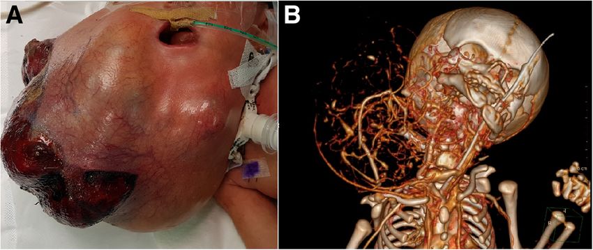
Hochwald et al. Journal of Medical Case Reports   (2019) 13:73                                                      Page 2 of 5

preoperative planning and followed by tracheostomy, (2)          possible event of maternal hypotension in order to main-
endovascular embolization of the carotid artery that             tain good fetal perfusion. After epidural catheter place-
supplied the tumor in order to decrease blood loss               ment while lying on her left side, the parturient returned to
during resection, and (3) complete surgical resection.           lie on her back. Immediately, a severe hypotension (65/30
The parents were involved in all the ethical and medical         mmHg) with tachycardia (150 beats/minute) appeared. We
decisions, starting before the delivery, just after the diag-    related this complication to the polyhydramnios causing
nosis of the cervical mass.                                      a significant decrease in the vena cava flow. After left
                                                                 uterine displacement and bolus of phenylephrine, her
Case presentation                                                blood pressure and heart rate returned to normal.
An otherwise healthy 33-year-old woman, gravida 3, para 2,       General anesthesia with rapid sequence induction was
from a Sephardic Jewish origin, was initially referred to our    induced with succinylcholine (100 mg) and propofol
institution at 30.6 weeks of gestation due to a large neck       (150 mg). During the EXIT procedure an appropriate
mass found on prenatal ultrasonography (US). Her previous        uterine relaxation was maintained to prevent placenta
two pregnancies were uncomplicated. The fetal sonogram           expulsion. We used a high dose of inhaled anesthetics
showed a 10 by 8 cm mass on the right side of the neck,          and minimal nitroglycerine intravenous drip. No bleeding
which was not present in detailed scans taken at 14 and 22       occurred during the procedure.
weeks. The mass was composed of a cystic portion and a              A classical uterine incision was made and only the
solid portion containing blood vessels and was growing rap-      fetal head and upper chest with the cervical mass were
idly in subsequent ultrasound studies. A significant polyhy-     delivered through the uterine incision. The rest of the
dramnios with amniotic fluid index (AFI) of 50 suggested         body and the cord were left in situ to avoid placental
an upper gastrointestinal obstruction and a highly possible      separation. The amniotic fluid was slowly drained to
airway obstruction as well. Findings were confirmed by fetal     avoid an abrupt drainage of the fluid and an early separ-
magnetic resonance imaging (MRI). In anticipation of the         ation of the placenta.
difficulty in establishing a secured airway at birth and the        The multidisciplinary team had planned and rehearsed
potential complicated resection of the giant tumor after         the following escalating step-by-step scenarios for establish-
birth, the mother was referred to our hospital for               ing a secured airway: (1) direct laryngoscopy and intubation
consultation.                                                    attempt by a pediatric anesthesiologist with the aid of a
   The parents were in consultation with the maternal fetal      neonatologist, (2) rigid bronchoscopy by an otolaryngolo-
team, neonatologist, anesthesiologist, pediatric surgeon,        gist and possible aid of flexible bronchoscopy by a pediatric
and otolaryngologist. The parents were presented with a          pulmonologist, (3) if laryngoscopy and bronchoscopy failed,
guarded prognosis but insisted that the pregnancy continue       a tracheostomy was planned by an otolaryngology team. As
with maximal efforts during delivery and during the neo-         the tumor was highly vascularized, any debulking proced-
natal period.                                                    ure was impractical and would have imminently put both
   Therefore, a planned EXIT procedure, which provides           the mother and newborn at the risk of death. Analgesia for
the best chance to establish a patent airway, was offered        the newborn using intramuscular fentanyl was prepared in
to our patient, presenting the risks [7]. Specifically, we       advance.
informed the parents about the risks for the mother,                After the head of the newborn emerged, a direct laryn-
including significant hemorrhage from the uterus due to          goscopy was attempted; however, the larynx was not visible
the uterine relaxation necessary to avoid placental separ-       as the tumor obstructed the pharynx. Attempts at direct in-
ation, with a possible uterine resection in the case of a        tubation were abolished after 1 minute. Next, a rigid bron-
life-threatening hemorrhage.                                     choscopy was performed and only the tip of the epiglottis
   Knowing the risk of an unplanned preterm delivery             was visualized. At 11 minutes from delivery, endotracheal
due to polyhydramnios and uterine contractions, we               intubation was successfully performed. From the time of
scheduled our patient for a planned cesarean delivery at         delivery to the time of intubation, the presence of a good
34 weeks organizing and preparing a multidisciplinary            heart rate of the fetus was monitored by echocardiography.
team ready to perform the EXIT procedure.                        After the airway was secured, the female newborn was de-
                                                                 livered, the cord was clamped, and the placenta extracted.
The EXIT procedure                                               The arterial cord pH was 7.01 with CO2 of 71 mmHg and
A multidisciplinary team including obstetricians, anes-          lactate of 9.5 mmol/L. Immediately after delivery, a com-
thesiologists, neonatologists, otolaryngologists, pediatric      puted tomography (CT) scan was performed under general
surgeons, pulmonologists, cardiologists, and nursing             anesthesia that demonstrated that a tracheostomy could be
staff participated. A combined epidural and general              performed without interfering with the tumor. During the
anesthesia was planned. Our patient’s blood pressure             tracheostomy, a biopsy was taken from the tumor that
was monitored continuously to detect and treat a                 demonstrated both mature and immature teratoma (Fig. 1).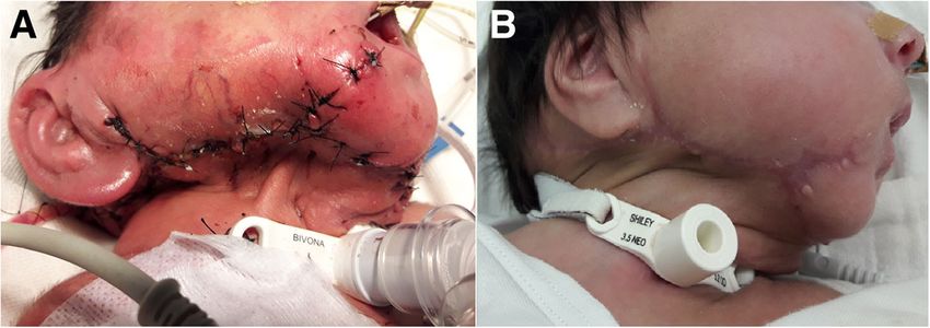
Hochwald et al. Journal of Medical Case Reports      (2019) 13:73                                                          Page 3 of 5

                                                                       demonstrated the highly vascularized tumor supplied by
                                                                       the external right carotid artery (Fig. 3a). A carotid oc-
                                                                       clusion test was performed and showed good blood supply
                                                                       from the contralateral internal carotid artery (ICA). Next,
                                                                       the origin of the external carotid was occluded using de-
                                                                       tachable platinum coils. Since resection of the tumor im-
                                                                       plied the sacrifice of all cervical carotid branches it was
                                                                       mandatory to also occlude the cervical ICA and common
                                                                       carotid artery to achieve minimal blood loss during surgery
                                                                       (Fig. 3b). The procedure was done under general
                                                                       anesthesia.
                                                                         Upon withdrawal of the angiography catheter a sudden
                                                                       deterioration of our patient was noted. Fluoroscopy and
                                                                       echocardiography demonstrated cardiac tamponade, prob-
                                                                       ably from a small laceration in the aorta. An urgent peri-
                                                                       cardiocentesis retrieving 3 ml of blood from the pericardial
                                                                       space allowed fast and full hemodynamic recovery (Fig. 3c,
                                                                       d). There was no re-accumulation of pericardial blood.

                                                                       The tumor resection
                                                                       Less than 24 hours after the embolization, the surgical
                                                                       team, which included a head and neck surgeon, pediatric
                                                                       otolaryngologist, and a plastic surgeon, performed the sur-
                                                                       gery with a pediatric anesthesiologist. Using LigaSure™
                                                                       Sealer/Divider (Medtronic), the tumor was dissected in-
                                                                       cluding a section of the lacerated skin. Because the main
                                                                       blood supply of the tumor was embolized, the surgery was
                                                                       performed from its distal part along our patient’s mandible
 Fig. 1 The tumor biopsy histology. a Teratoma containing mature       towards the proximal part at the junction of her neck and
 epithelial elements, cartilage, and mesenchyme. b Immature teratoma   thorax. The tumor impinged into her pharynx, and her
 containing neuroepithelium and primitive small round blue cells
                                                                       larynx and epiglottis were identified and preserved. Her
                                                                       mandible and neck muscles were preserved; however, her
  The birth weight of the newborn girl was 3 kg including              vagal nerve was surrounded by the tumor and was sacri-
the tumor. A giant submental mass that protruded from                  ficed with the carotid artery. The tumor weighted 800
the right side of her neck was noted. The tumor was                    grams and the overall blood loss was 300 ml. After tumor
covered with a thin skin layer with a large cystic and solid           resection and closure of the skin (Fig. 4), the tracheostomy
appearance. There were occasional bleedings from several               tube was changed. This was followed by a pneumothorax
lacerated areas on the tumor surface (Fig. 2a). A CT study             that required a chest tube insertion.
demonstrated the abundant blood supply of the tumor
including the fact that the right carotid artery was supply-           After the procedure
ing this tumor (Fig. 2b). During the first 2 days of life,             Gradually, after the tumor resection, our patient was
before the next procedure, the tumor continued to grow                 weaned from mechanical ventilation. She was fed initially
significantly, probably partially due to internal bleeding.            using an orogastric tube and gradually learned to feed or-
Her heart function was normal but prerenal azotemia                    ally. After the resection, a right vocal cord paralysis was ob-
evolved due to loss of large amounts of serotic fluid as               served using a flexible fiberoptic laryngoscopy; therefore,
well as blood from the lacerated mass.                                 we decided to leave the tracheal tube in place. We attrib-
                                                                       uted this finding to an injury of the recurrent laryngeal
Endovascular embolization                                              nerve during the resection. Another neurological sequela
Because resection of the giant, highly vascularized, cer-              was an abduction weakness of her right shoulder which
vical teratoma could be a fatal procedure due to massive               slowly recovered. Neck teratomas can arise from and com-
bleeding, we used endovascular embolization a day prior                pletely replace the thyroid tissue [1]. Thyroid function tests
to the tumor resection. The embolization was done by a                 demonstrated hypothyroidism and she started receiving
pediatric cardiologist and an interventional neuroradiol-              thyroid replacement therapy. A follow-up ultrasound of her
ogist. Cervical angiography via a femoral line catheter                head was normal. A brain MRI done a month after the final
Hochwald et al. Journal of Medical Case Reports (2019) 13:73 Page 4 of 5
Fig. 2 a The giant congenital teratoma on the third day of life. The tumor was covered with a thin skin layer with a large cystic and solid
appearance. There were occasional bleedings from several lacerated areas on the tumor surface. b A computed tomography study demonstrated
the abundant blood supply of the tumor including the fact that the right carotid artery was supplying this tumor
surgery demonstrated normal brain appearance. She was immature lungs and respiratory drive. Planning secure
discharged to her home at 3 months of age (Fig. 4). resuscitation at birth needs to take into account the avail-
ability of multidisciplinary teams and the issue of prema-
Discussion turity; thus finding the best timing for intervention.
Cervical teratomas can acquire gigantic dimensions in EXIT is a surgical delivery procedure used to deliver
late gestation. The compression over the esophagus fetuses who have airway compression. Failure to achieve
causes polyhydramnios and the associated compression airway under controlled and safe conditions and in a
over the trachea can cause airway obstruction at birth. reasonable time might result in asphyxia.
Severe polyhydramnion may lead to preterm delivery, Giant congenital cervical teratomas are challenging
thus complicating the upper airway obstruction with lesions. After making the in utero diagnosis, and following
Fig. 3 a Pretreatment lateral view of the right common carotid, notice the large and rich vascularization of the tumor from the external carotid
branches (white arrow). b Post embolization lateral view, platinum coils in common carotid (white arrow), internal carotid (arrow head), and
external carotid (black arrow); notice massive reduction in tumor vascularization. c Contrast medium in the pericardial space. d Pericardiocentesis
wire in the pericardial spaceHochwald et al. Journal of Medical Case Reports (2019) 13:73 Page 5 of 5
Fig. 4 a One day after the resection. b Before discharge, at 3 months of age
thorough assessment and consultations, our team felt that Publisher’s Note
the risk for the mother during the EXIT procedure and Springer Nature remains neutral with regard to jurisdictional claims in
published maps and institutional affiliations.
the risks and possible sequelae for the infant during and
after the different procedures after birth were significant Author details
1
enough that offering no treatment was ethically reason- Neonatal Intensive Care Unit, Ruth Rappaport Children’s Hospital, Rambam
Health Campus, Haifa, Israel. 2Department of Otolaryngology Head and Neck
able. The parents, fully aware of all options and risks, de- Surgery, Rambam Health Campus, Haifa, Israel. 3The Pediatric ENT service,
cided on maximal care to their daughter. Rambam Health Campus, Haifa, Israel. 4The Obstetrics & Gynecology Division,
Rambam Health Campus, Haifa, Israel. 5Invasive Neuroradiology Unit,
Rambam Health Campus, Haifa, Israel. 6Department of Pediatric Cardiology &
Conclusions Congenital Heart Disease in Adults, Ruth Rappaport Children’s Hospital,
In the case of our patient, once stabilized with tracheos- Rambam Health Campus, Haifa, Israel. 7The Department of Anesthesiology,
tomy after EXIT, we found the tumor to have a highly Rambam Health Campus, Haifa, Israel. 8The Department of Plastic Surgery,
Rambam Health Campus, Haifa, Israel. 9The Department of Pathology,
abundant blood supply. Although pre-excision embolizat Rambam Health Campus, Haifa, Israel. 10The Department of Pediatric Surgery,
ion was described sporadically in cases of sacral teratoma Rambam Health Campus, Haifa, Israel.
[4, 8], we found no literature on embolization in cases of
Received: 25 June 2018 Accepted: 10 January 2019
cervical teratomas. We assumed that embolization prior to
definite surgical resection of the tumor could decrease sig-
nificantly the blood loss and risk for complications or death References
1. Mohanty MK, Sahu P, Jaiswal AA, Singal R, Gupta S, Kohli G, et al. A huge
during the procedure. immature cervical teratoma; antenatal diagnosis, and its management - an
The clinical challenges reported in this case highlight unusual entity. J Clin Neonatol. 2013;2:42–5.
the importance of multidisciplinary teams in the man- 2. Elmasalme F, Giacomantonio M, Clarke KD, Othman E, Matbouli S.
Congenital cervical teratoma in neonates. Case report and review. Eur J
agement of congenital cervical tumors. This case also Pediatr Surg. 2000;10:252–7.
emphasizes the ethical issues and the need for an open 3. Johnson N, Shah PS, Shannon P, Campisi P, Windrim R. A challenging
discussion with the parents in a similar life-threatening delivery by EXIT procedure of a fetus with a giant cervical teratoma. J
Obstet Gynaecol Can. 2009;31:267–71.
prenatal diagnosis. 4. Rossi UG, Cariati M, Toma P. Giant sacrococcygeal teratoma embolization.
Indian J Radiol Imaging. 2013;23:145–7.
Abbreviations 5. Miliaras D, Grimbizis G, Conroy J, Psarra N, Miliaras S, Nowak N, et al. Novel
CT: Computed tomography; EXIT: Ex utero intrapartum treatment; karyotypic changes detected by comparative genomic hybridization in a
ICA: Internal carotid artery; MRI: Magnetic resonance imaging; case of congenital cervical immature teratoma. Birth Defects Res A Clin Mol
US: Ultrasonography Teratol. 2005;73:572–6.
6. Sichel JY, Eliashar R, Yatsiv I, Moshe Gomori J, Nadjari M, Springer C, et al. A
Funding multidisciplinary team approach for management of a giant congenital
No honorarium, grant, or other form of payment was given to anyone to cervical teratoma. Int J Pediatr Otorhinolaryngol. 2002;65:241–7.
produce this manuscript. 7. Lazar DA, Olutoye OO, Moise KJ Jr, Ivey RT, Johnson A, Ayres N, et al. Ex-
utero intrapartum treatment procedure for giant neck masses—fetal and
Authors’ contributions maternal outcomes. J Pediatr Surg. 2011;46:817–22.
All authors listed have been involved in the management of this case. All 8. Lahdes-Vasama TT, Korhonen PH, Seppanen JM, Tammela OK, Iber T.
authors contributed to drafting of the article and approved the final version Preoperative embolization of giant sacrococcygeal teratoma in a premature
and the submission of the manuscript. newborn. J Pediatr Surg. 2011;46:e5–8.
Consent for publication
The parents gave consent to the publication and approved the images
attached. Written informed consent was obtained from the patient’s parents
for publication of this case report and any accompanying images. A copy of
the written consent is available for review by the Editor-in-Chief of this
journal.
Competing interests
The authors declare that they have no competing interests.You can also read