Detection of scorpion venom by optical circular dichroism method
←
→
Page content transcription
If your browser does not render page correctly, please read the page content below
www.nature.com/scientificreports
OPEN Detection of scorpion venom
by optical circular dichroism
method
Y. Mazhdi & S. M. Hamidi*
Various efforts have been made to detect minimum amounts of any toxic materials in water or the
neurotoxic effect of venom (Odontobuthus Doriae Scorpion) in the human’s blood serum nerve by high-
sensitivity, accurate, and low-cost sensors in order to enhance life style. Therefore, the present study
was done to investigate reliability of two-dimensional plasmonic structure and circular dichroism (CD)
in toxic samples in order to measure and determine venom concentrations and its neurotoxic effect
on humans҆ blood serum Neurotransmitter analytes. Our results confirmed dependency of CD signal to
neurotoxic effect of venom concentrations and good sensitivity of this sensor with the help of achiral
plasmonic structure.
Chirality as a useful and key aspect in molecular biology can be used for biosensing a pplications1. Given impor-
tance of high-resolution and low-cost biosensors, many studies have been conducted to enhance chirality through
the use of new materials and chiral and achiral nanostructures2. A wide range of these nanostructures consist of
symmetry breaking ones like chiral o ligomers3, chiral n
anoparticles4, nanohelix a rrays5, twisted n
anorods6, three-
7 8
dimensional helices ; or symmetric ones like square array of nanoparticle p latform that are used to enhance
circular dichroism(CD) signal. It is well known that the absence of mirror symmetry or quasi two- or three-
dimensional thin films lacking plane mirror symmetry is the main key factor in achieving the above-mentioned
chirality9. Currently, experimental and theoretical evidence shows that surface plasmon polariton (SPP) waves
in achiral structures can show chirality due to asymmetry in field d istribution10,11.
Scorpion venom causes various complications including local pain, inflammation, necrosis, and blood-related
effects and envenomation affects nervous system quickly and within a few h ours12. It causes temporal paralysis,
involving a wide range of injuries, from mild to moderate and severe, affecting one muscle group or the whole
body13. Considering these warning and dangerous effects of this venom, recognizing its cholinergic and neuro-
logical effects on humans online and in the short time, is of great importance in very low concentrations. So far,
many chemical methods have been used to detect and distinguish this kind of venom in different concentrations;
but determination of the minimum amounts by invasive methods like optical ones is so important now. Among
optical methods, there is an optical sensor based on surface plasmon resonance (SPR) that is very famous and is
used to determine the minimum concentrations of any venoms with higher sensitivity based on online changes
in metal refractive index and dielectric adjacent environment.
Odontobuthus doriae scorpion is one of the most dangerous species of scorpion in Iran, venom of which was
detected its neurotoxic effect on humans҆ blood serum in this study using the SPR sensor designed and fabricated
based on the above-mentioned chirality in metallic substrate.
Experimental setup
Fabrication of 2D grating using the soft imprint lithography method can offer some advantages, such as rea-
sonable cost, high efficiency, and reproducible manufacturing at large scale, so here, this method was used for
fabrication of 2D g rating14. For this purpose, unique codes and corresponding resolution were extracted using a
2D charge-coupled device (CCD) of a camera. Then, this CCD was attached to a glass substrate by a two-sided
adhesive tape. Square unit cell of the CCD had the periodicity of 3.10 μm. After mixing polydimethylsiloxane
(PDMS) and curing agent (1:10) for 5 min, the mixture was poured on the template with an approximate thick-
ness of 1.4 mm. For fabricating our 2D plasmonic structures, the SYLGARD 184 elastomer kit provided by
Sigma-Aldrich Company was used. For eliminating the probable bubbles from the mixture, the sample was
inserted in a rotary vacuum pump for 15 min and then, the samples were put on a heater for 30 min at 50 °C,
15 min at 75 °C, and 15 min at 100 °C, respectively. After one day, the PDMS samples were peeled off from their
Magneto‑Plasmonic Lab, Laser and Plasma Research Institute, Shahid Beheshti University, Tehran, Iran. *email:
m_hamidi@sbu.ac.ir
Scientific Reports | (2021) 11:15854 | https://doi.org/10.1038/s41598-021-95493-7 1
Vol.:(0123456789)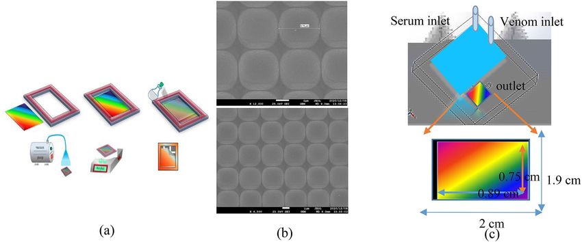
www.nature.com/scientificreports/
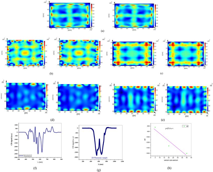
Figure 1. (a) schematic diagram of soft nanolithography, (b) SEM graph of samples and (c) main channel
without (top) and with (bottom) plasmonic sensor.
molds. At the next step, surfaces of the samples were approximately coated with about 35 nm of gold by a physi-
cal vapor deposition machine.
Figure 1a schematically shows fabrication process of a 2D plasmonic grating. Accordingly, the patterned
PDMS as a grating was observed with periodicity of 3.11 μm in scanning electron microscopy (SEM) image taken
from the control sample as presented in Fig. 1b. In addition, schematic of the 2D regular structure covered with
gold is also shown in Fig. 1b. Actually, this structure was selected since 2D grating acts like a Bragg reflector due
to its bandgap in reflection spectrum in visible regim 15,16 as well as providing a good surface lattice resonance
because of gold nanorods at the interface of each unit cell. For investigating the effect of scorpion venom on
stimulation of nervous system in humans҆ neurons in transmission measurement setup, fluidic channel was
required according to Fig. 1c. For this purpose, a transparent flow cell with two inlet channels of the same size
(top image) was designed for simultaneous and equal entry of humans҆ serum and different concentrations of
scorpion venom using laser incisions on a transparent plexiglas sheet with a thickness of 2 mm. A circular cav-
ity of 2 mm thickness was embedded inside the flow cell and in passage of blood serum and scorpion venom
solution to combine the two inputs in the flow cell. Blood serum was collected from laboratory with External
Quality Assurance Services (EQAS) with identification number 2740. The blood sample was taken as a blood
clot in a gel tube for 20 min at ambient temperature and then centrifuged at 2500 rpm for 10 min, the serum was
removed and poured into a falcon.
In addition, the generated sensor chip was attached to the cube embedded inside the flow cell so that, its
gold-coated surface was adjacent to the material passing through the flow cell (bottom image).
For testing sensor’s capability and sensitivity to venom, three concentrations of 5, 20, and 40 ppm were
needed, which were prepared as follows: first, 2 mg of scorpion venom was dissolved, as a white powder in 1 ml
of phosphate-buffered saline (PBS) biological solution. After 10 min of centrifugation, 0.05, 0.2, and 0.4 mg of the
solution were dissolved in three equal amounts of 20 ml of PBS to prepare concentrations of 5, 20, and 40 ppm
as S1, S2 and S3 samples, respectively.
Moreover, for preparing 50 ml of healthy humans҆ blood serum, 150 ml of blood was collected and after
clotting, it was centrifuged and then, the serum was separated. These prepared samples were studied in CD
experimental setup, in which sample’s transmission was documented after excitation by right and left circularly
polarized (RCP and LCP) light and difference between them was recorded in the visible region.
Sample guideline. All methods were carried out in accordance with relevant guidelines and regulations in
Noor Pathobiology and Genetics Laboratory with registration number 30859/4/26/P by the Ministry of Health
and Medical Education of Iran has a certificate of participation in External Quality Assurance Services (EQAS)
with identification number 2740.
Human sample guideline. This study was approved by the Ethics Committee of the "Ethical committee of
Vice president of research of Shahid Beheshti university/IR.SBU.REC.1400.
Results and discussions
CD spectra of the sample as difference in transmission spectra between LCP and RCP light are shown in Fig. 2.
Figure 2 shows CD spectra for two-dimensional (2D) bare sample, serum placed onto 2D plasmonic sample,
three different S 1–S3 samples and also, normalized CD signal as a difference between CD signals in the presence
of venom concentrations and serum sample in (a), (b), (c), and (d) sections, respectively.
Observed chirality due to achiral structure and tilt with respect to normal sample’s surface appeared by
asymmetric field distribution pattern as a result of asymmetric plasmon modes excited by RCP and LCP light.
These interesting features can result from nanowires in each motif of plasmonic square array. In fact, in each
Scientific Reports | (2021) 11:15854 | https://doi.org/10.1038/s41598-021-95493-7 2
Vol:.(1234567890)
www.nature.com/scientificreports/
Figure 2. CD signal of (a) bare 2D plasmonic sample, (b) 2D plasmonic sample as serium sensor, (c) 2D
plasmonic sample as Venom senor in three different concentrations and (d) normalized CD signal or three
different venom concentrations.
angle, a percentage of wires’ projection and thus, plasmonic field yield changes in transmission consequently,
leading to chiro-optical effects.
As shown in Fig. 2a, there is overlap between the localized surface plasmon resonances (LSPR) due to
nanowires and diffraction orders by lattice yielding to surface lattice resonance (SLR) in CD signal resonance as
explained in our previous r eports17. This can be attributed to the fact that transmission spectrum of the sample
shows two resonance dips at 550 and 673 nm because of SLR and extraordinary optical transmission (EOT) of
nano plasmonic structure.
Importantly, the signal appears near SLR and when unknown sample is placed on surface of sensor, which
can be sensed through any change in CD signal. Furthermore, sign of chirality is reversed after passing from SLR
wavelength that is contrary to the fact that exactly in resonance region, trend of enhanced chirality was stopped
due to the increase in venom concentration that the main and dominant effect of this increase in concentration
is directly on neurotoxicity, as a result of changing the concentration of the main neurotransmitter analytes in
the blood serum. According to research, this complication is the predominant cause of many diseases caused
by scorpion bites13.
As mentioned in the introduction, scorpion venom, due to being a neurotoxin, has many effects on the nerv-
ous system at the presynaptic level (β-neurotoxin) and postsynaptic level (α-neurotoxin)12, which β-neurotoxin
inhibit the release of acetylcholine or noradrenaline which are neurotransmitters and α-neurotoxin reversibly
blocks acetylcholine receptors at the postsynaptic level13. The main reason for the effect of scorpion venom
on the central and peripheral nervous system as well as irritable tissues in muscles is the ability of this toxin
to interact with N a+, K+, Ca2+, and C
l− channels, which causes metabolic impacts18. We used Iranian yellow
scorpion venom in this study, which is a species of doriae in the family Buthidae. The O.doriae venom is able to
interact with some of voltage-dependent channels of sodium ( VNa) and potassium ( VK). According to a study
conducted in 2003 (at Shahid Sadoughi University of Medical Sciences in Yazd C ity21). In this way, after inject-
ing scorpion venom into the living organism, its blood serum is separated and at different times, the amount of
total protein, total bilirubin, uric acid, cholesterol, amylase enzyme and electrolytes ( Cl−, K+, Na+) are analyzed.
Evaluation of statistical results of serum biochemical parameters shows that the electrolytes ( Cl−, K+, Na+) in the
tested samples compared to the control group, at a time between 5 to 15 min after injection, had a significant
decrease (P < 0.05)22. This decrease is due to cholinergic effects and vomiting. Also, no significant difference was
observed in other biochemical parameters. Therefore, it can be said that in poisoning with Odontobuthus doriae
scorpion venom, the first cholinergic effects of the venom occur and it is completely dominant over the other
effects. Therefore, scorpion venom has a direct effect on the amount of serum electrolytes. It reduces amount of
electrolytes and because of neuronal signal of the humans҆ central nervous system causes changes in concentration
Scientific Reports | (2021) 11:15854 | https://doi.org/10.1038/s41598-021-95493-7 3
Vol.:(0123456789)www.nature.com/scientificreports/
of these electrolytes. When these electrolytes in an analyte are placed near metal surface of the plasmon sensor,
they form a layer near metal surface with an action potential of V 0, which is known as Stern layer. This created
neuronal activity and action potential that causes physical changes is the basis of our sensory approach. This
action potential is unique to each specific neurotoxin and its different injected concentrations. In other words,
any neurotoxin that binds to tissues or human neurotransmitters creates its own unique action potential, which in
our sensing method determines the type and concentration of the toxin. Therefore, with changes in electrolytes,
the potential size of the Stern layer changes and according to the Drude–Stern relation, refractive index of the
metal also changes so that, resonant frequency of surface plasmons varies and reveals the neurological effects of
toxin on the humans҆ central nervous system19.
2
ǫ0 ωP∗ 3LSP
� LSP = − 2 2 1−L
V0 (1)
8π C Nedd TF (ǫ∞ + L )
where, ǫ0 is the electric permittivity of vacuum and d is the distance between the plates (the plasmonic template is
assumed to be at ground potential). dTF is the Thomas–Fermi screening length and N and e are electron number
density and elementary charge, respectively, ωp* is the gold plasma frequency, and static dielectric constant ε∞
accounts for background polarization because of the presence of core electrons. λLSP is the resonance wavelength
of LSP and c is the speed of light in vacuum. L is the geometrical factor in polarization direction of incident
electromagnetic wave, and V 0 is an applied voltage.
Our simulation results approved that CD takes place in the same resonance wavelength as SLR of the sample
for normal incidence and 5-degree tilt angle, confirming the main role of SPR in this finding.
Symmetry breaking in tilt incidence angle is very stronger due to SPR in corners of each motif. Besides, it
approves enhancement factor of signal in incidence angle, which is a bit more than normal ones, for example
by 5 degrees. This fact is the result of non-symmetric SPR excitation in each nanowire in corners of motifs as
shown in Fig. 3.
In our 2D plasmonic sample with perfect mirror symmetry in tilt angle, while vector of incident radiation
wave was in the sample plane by any of the unit cell lattice and formed a triangle with two structure lattice con-
stants, SPR appeared as a result of plasmonic dipole excitation. In direction of the radiation wave vector in the
sample plane, known as direction of dipole screw, this total wave vector is the result of constructive interference
of the individual SPPs of wires with each other, showing dependence of this final wave vector on phase of each
single SPP wave vector.
Consequently, asymmetry near field created by this SPP wave causes plane mirror symmetry of the array
structure to be broken and makes this structure to have an optical chirality effect.
Since, the SPP vector of array in metal includes both real and imaginary parts; this structure is expected
to produce a significant CD signal due to the presence of imaginary part of the wave vector. Obviously, the
interfering effect of SPP of each hole at metal-dielectric boundary is directly dependent on properties of both
environments, so change in dielectric environment alters phase matching conditions of the SPP interference and
produces a different CD signal as explained in the literature20.
As can be seen in the diagram, the amplitude of the circular dichroism signal is amplified by increasing dif-
ferent concentrations of scorpion venom in the blood serum, which is a sign of the sensitivity of the proposed
sensor to nerve stimulation by scorpion venom in the blood serum. The values of the CD signal at the 520 nm
wavelength, which is approximately the same as the SLR wavelength and near the initial wavelength of the EOT
wavelength rang, are plotted for three different concentrations of venom in the blood serum to get the sensitivity
of S = 27.4 as shown in Fig. 3h.
As discussed, the results clearly show that any change that affects the amount of neurotoxicity of venom,
whether changes in the type of toxin or changes in its concentration, will affect the sensor response because any
change in the toxin will cause a change in the action potential, which depends on the type and concentration
of the toxin. Therefore, this approach, which is done completely online, is sensitive to any changes in the toxin
and only delay in the measurement is related to the time of effect of the toxin on serum analytes, which lasts
between 5 and 15 min.
Conclusion
In conclusion, achiral plasmonic structure was used in this study as highly sensitive and low-cost sensor to detect
neuronal activity of venom of the Iranian Odontobuthus doriae scorpion in humans҆ blood serum. Our results
showed that changes in venom concentrations caused alterations in amount of electrolytes and thus, action
potential of the Stern layer and according to the Drude–Stern relation, refractive index of the metal changed
in our 2D plasmonic substrate. Resonant frequency of SLR in the main sample was altered by the neurologi-
cal effect of the toxin on the humans҆ central nervous system and thus, it can be said that any change in venom
concentration can be sensed. Finally, in this achiral sensors, resonance wavelength and thus CD signal has been
affected by any change in the lattice plasmon polaritons by creating action potential at the sample surface. This
action potential is unique to each specific neurotoxin and its different injected concentrations. In other words,
any neurotoxin that binds to tissues or human neurotransmitters creates its own unique action potential, which
in our sensing method determines the type and concentration of the toxin.
Scientific Reports | (2021) 11:15854 | https://doi.org/10.1038/s41598-021-95493-7 4
Vol:.(1234567890)www.nature.com/scientificreports/
Figure 3. Magnetic and Electric field distributions of sample for right and left circular polarizations for (a)
LCP-T-E and RCP-T-E and (b) LCP-T-H and RCP-T-H and (c) LCP-R-H and RCP-R-H and (d) LCP-T-E and
RCP-T-E for normal incidence and (e) LCP-T-H and RCP-T-H for 5-degree incidence angle. (f) Simulation of
CD signal, (g) measured CD signal in normal incidence and (h) sensitivity diagram of the sensor.
Received: 4 May 2021; Accepted: 19 July 2021
References
1. Hendry, E. et al. Ultrasensitive detection and characterization of biomolecules using superchiral fluids. Nat. Nanotechnol. 5, 783
(2010).
2. Gordon, R. et al. Strong polarization in the optical transmission through elliptical nanohole arrays. Phys. Rev. Lett. 92, 037401
(2004).
3. Le, K. Q. Enhanced circular dichroism via symmetry breaking in a chiral plasmonic nanoparticle oligomer. J. Electron. Mater. 47,
2836–2840 (2018).
4. Petronijevic, E. et al. Chiral effects in low-cost plasmonic arrays of elliptic nanoholes. Opt. Quant. Electron. 52, 176 (2020).
5. Gibbs, J. G., Mark, A. G., Eslami, S. & Fischer, P. Plasmonic nanohelix metamaterials with tailorable giant circular dichroism. Appl.
Phys. Lett. 103(21), 213101 (2013).
6. Zhao, S.-X. & Zhang, W. Plasmonic chirality of one-dimensional arrays of twisted nanorod dimers: The cooperation of local
structure and collective effect. Opt. Express 27, 38614–38623 (2019).
7. Cao, T., Wei, C., Mao, L. & Li, Y. Extrinsic 2D chirality: Giant circular conversion dichroism from a metal-dielectric-metal square
array. Sci. Rep. 4, 7442 (2014).
8. Mohammadi, E. et al. Nanophotonic platforms for enhanced chiral sensing. ACS Photon. 5, 2669–2675 (2018).
9. Wang, X. & Tang, Z. Circular dichroism studies on plasmonic nanostructures. Small 13, 1601115 (2017).
10. Horrer, A. et al. Local optical chirality induced by near-field mode interference in achiral plasmonic metamolecules. Nano Lett.
20, 509–516 (2020).
11. Maoz, B. M., Moshe, A. B., Vestler, D., Bar-Elli, O. & Markovich, G. Chiroptical effects in planar achiral plasmonic oriented nano-
hole arrays. Nano Lett. 12, 2357–2361 (2012).
12. Jalali, A. et al. OD1, the first toxin isolated from the venom of the scorpion Odonthobuthus doriae active on voltage-gated Na+
channels. FEBS Lett. 579, 4181–4186 (2005).
Scientific Reports | (2021) 11:15854 | https://doi.org/10.1038/s41598-021-95493-7 5
Vol.:(0123456789)www.nature.com/scientificreports/
13. Vatanpour, H., Jalali, A., Rowan, E. G. & Rahim, F. Effects of odontobuthus doriae scorpion venom on mouse sciatic nerve. IJPR
12(supplement), 145–151 (2013).
14. Saeidifard, S. et al. Two-dimensional plasmonic biosensing platform: Cellular activity detection under laser stimulation. J. Appl.
Phys. 126(10), 104701 (2019).
15. Asgari, N. & Hamidi, S. M. Exciton-plasmon coupling in two-dimensional plexitonic nano grating. Opt. Mater. 81, 45 (2018).
16. Sohrabi, F. et al. Phase-sensitive optical neural recording of cerebellum tissue on a flexible interface. J. Appl. Phys. 127(11), 113101
(2020).
17. Mbarak, H., Ghahrizjani, R. T., Hamidi, S. M., Mohajerani, E. & Zaatar, Y. Reversible and tunable photochemical switch based on
plasmonic structure. Sci. Rep. 10(1), 1–7 (2020).
18. Maertens, C. et al. Potent modulation of the voltage-gated sodium channel Nav1.7 by OD1, a toxin from the scorpion Odonthobu-
thus doriae. Mol. Pharmacol. 70, 405–414 (2006).
19. Zhang, J., Atay, T. & Nurmikko, A. V. Optical detection of brain cell activity using plasmonic gold nanoparticles. Nano Lett. 9(2),
519–524 (2009).
20. Wang, X. et al. Theoretical investigation of subwavelength structure fabrication based on multi-exposure surface plasmon interfer-
ence lithography. Results Phys. 12, 732–737 (2019).
21. Jafari, S. S. & Khamesipour, V. R. The effects of different doses of scorpion (Odontobuthus) venom on some changes of biochemical
factors of blood serum in dogs. Daneshvar 8, 15–24 (2001).
22. Ismail, M. The treatment of the scorpion envenoming syndrom. Toxicon 34, 1014–1026 (1994).
Author contributions
Y.M. did the optical measurement, analyzed the results and wrote the main text of the manuscript. S.M.H.
supervised the measurement part and writing the results of the study. All authors read and approved the final
manuscript.
Competing interests
The authors declare no competing interests.
Additional information
Correspondence and requests for materials should be addressed to S.M.H.
Reprints and permissions information is available at www.nature.com/reprints.
Publisher’s note Springer Nature remains neutral with regard to jurisdictional claims in published maps and
institutional affiliations.
Open Access This article is licensed under a Creative Commons Attribution 4.0 International
License, which permits use, sharing, adaptation, distribution and reproduction in any medium or
format, as long as you give appropriate credit to the original author(s) and the source, provide a link to the
Creative Commons licence, and indicate if changes were made. The images or other third party material in this
article are included in the article’s Creative Commons licence, unless indicated otherwise in a credit line to the
material. If material is not included in the article’s Creative Commons licence and your intended use is not
permitted by statutory regulation or exceeds the permitted use, you will need to obtain permission directly from
the copyright holder. To view a copy of this licence, visit http://creativecommons.org/licenses/by/4.0/.
© The Author(s) 2021
Scientific Reports | (2021) 11:15854 | https://doi.org/10.1038/s41598-021-95493-7 6
Vol:.(1234567890)You can also read