Radiographic lumbosacral vertebral abnormalities and constipation in cats
←
→
Page content transcription
If your browser does not render page correctly, please read the page content below
Veterinary World, EISSN: 2231-0916                                                                     RESEARCH ARTICLE
Available at www.veterinaryworld.org/Vol.14/February-2021/22.pdf                                             Open Access

  Radiographic lumbosacral vertebral abnormalities and constipation in
                                 cats
 Chutimon Thanaboonnipat1        , Kamonchanok Kumjumroon2 , Kamonwan Boonkwang2             , Natthacha Tangsutthichai2    ,
                                      Wassapon Sukserm2 and Nan Choisunirachon1

1. Department of Surgery, Faculty of Veterinary Science, Chulalongkorn University, Bangkok, 10330, Thailand; 2. Faculty
                     of Veterinary Science, Chulalongkorn University, Bangkok, 10330, Thailand.
                      Corresponding author: Nan Choisunirachon, e-mail: nan.c@chula.ac.th
          Co-authors: CT: chutimon.th@chula.ac.th, KK: kmchn.k@gmail.com, KB: ammyvetcu@gmail.com,
                      NT: lunatic-xchild@hotmail.com, WS: the-legend_gazzkung@hotmail.com
                  Received: 06-10-2020, Accepted: 20-01-2021, Published online: 23-02-2021

doi: www.doi.org/10.14202/vetworld.2021.492-498 How to cite this article: Thanaboonnipat C, Kumjumroon K,
Boonkwang K, Tangsutthichai N, Sukserm W, Choisunirachon N (2021) Radiographic lumbosacral vertebral abnormalities
and constipation in cats, Veterinary World, 14(2): 492-498.

                                                            Abstract
Background and Aim: Lumbosacral intervertebral disk disease (IVDD) in cats usually develops concurrent with
constipation, spondylosis deformans, and sacralization. However, the prevalence of lumbar IVDD in cats was considered
low, and there was less information on the incidence of non-traumatic lumbosacral vertebral abnormalities that may affect
large bowel dysfunction. This study aimed to retrospectively investigate the relationship between non-traumatic lumbosacral
vertebral abnormalities, both congenital and acquired, and large bowel dysfunction in cats.
Materials and Methods: Of 3108 cats that were presented to the Diagnostic Imaging Unit, the Small Animal Teaching
Hospital, Faculty of Veterinary Science, Chulalongkorn University, between March 2016 and February 2018, 1365 cats
met the inclusion criteria. All abdominal radiographs were reviewed, and all subsequent data were recorded, including the
presence of congenital or acquired lumbosacral lesions, number of lumbar vertebrae, and length of the second, fifth, and last
lumbar vertebrae, including the type of lumbar abnormalities. Moreover, radiographic information relating to constipation
and megacolon was also collected.
Results: Non-traumatic lumbosacral vertebral abnormalities were observed in 29.74% of cats. The most common congenital
lumbosacral vertebral abnormalities were six lumbar vertebrae, sacralization, and lumbarization, whereas most common
acquired lumbosacral abnormalities were bone spur, narrowing disk space, spondylosis deformans, and lumbosacral
degeneration, respectively. Cats with abnormal lumbosacral vertebrae are prone to have more problems with the large
bowel (p=0.0057; odds ratio=1.731). Moreover, congenital and acquired lumbosacral abnormalities were also at risk of
large bowel abnormalities (p=0.0069; odds ratio=1.920 and p
Available at www.veterinaryworld.org/Vol.14/February-2021/22.pdf
peritoneal effusion, dysautonomia, and abdominal Animals and experimental design
foreign bodies, can increase the risk of large bowel The data of the cats were considered and included
dysfunctions [2,14-16]. Studies on the association if they revealed complete signalment and there were at
between these problems and any of the non-trau- least two orthogonal radiographic views of the whole
matic or congenital vertebral abnormalities, including abdomen that covered the region of interest (ROI)
the incidence of latter abnormalities, are rare in cats, from the caudal thoracic vertebrae to ischial tuberos-
whereas they are well described in other species. In ity. Cases of incomplete clinical information, uncov-
humans, either congenital or acquired spinal stenosis ering ROI of abdominal radiograph than the former
and bone spur may increase the risk of cauda equina information, evidence of previous traumatic injuries
syndrome (CES), subsequently causing atonic bowel, from abdominal radiographs, pregnancy, skeletal
which develops to severe constipation [17]. In dogs, immaturity, and other conditions that might cause col-
lumbosacral transitional vertebrae are believed to be orectal obstruction or constipation, such as intra-ab-
one of the risk factors of fecal incontinence because of dominal mass, were excluded from the study. Then, all
its association with CES [18]. Degenerative lumbosa- clinical information were collected.
cral stenosis (DLSS), which causes fecal incontinence, Radiographic analysis
is also commonly found in dogs. DLSS includes inter-
vertebral disk disease (lumbosacral intervertebral disk Characterization of lumbosacral vertebrae
disease [IVDD]), spondylolisthesis, and discospon- Abdominal radiographs of all included cats were
dylitis [19]. Despite several lumbosacral vertebral collected as digital information and communication
abnormalities in cats, such as hemivertebrae, tran- system (DICOM) files and were reviewed using the
sitional vertebrae, bone spurs, and spondylosis, the DICOM viewer software (OsiriX, Switzerland). First,
association of these abnormalities to CES in cats has all abdominal radiographs were reviewed and cat-
not been previously reported [20]. The previous stud- egorized into the normal group (Group 1; Gr.1) or
ies showed that lumbosacral IVDD was concurrently the non-traumatic lumbosacral vertebral abnormality
found with constipation [21] and spondylosis defor- group (Group 2; Gr. 2). Briefly, abdominal radio-
mans and sacralization [22]. graphs of the cats were considered abnormal if they
Since the prevalence of lumbar IVDD in cats presented either congenital lesions (Group 2A; Gr.
was considered uncommon [23] and there was less 2A), such as less or excessive lumbar vertebral num-
information on the incidence of non-traumatic, lum- ber, hemivertebrae, lumbarization, and sacralization,
bosacral vertebral abnormalities that may affect large or non-traumatic acquired lesions (Group 2B; Gr. 2B)
bowel dysfunction. This study aimed to retrospec- of spondylosis deformans, ventral bone spur, narrow-
tively investigate the incidence of non-traumatic lum- ing disk space, and lumbosacral degeneration. In the
bosacral vertebral abnormalities in cats and the rela- case of a single presentation of lumbosacral abnormal-
tionships of lumbosacral vertebral abnormalities and ities, the lesion was categorized to be the cranial area
constipation and/or megacolon in cats on abdominal if the lesion was located at the area from the first to the
radiographs. third lumbar vertebrae or caudal area if the lesion was
Materials and methods located at the area from the fourth lumbar vertebrae to
the first sacrum.
Ethical approval and informed consent
This study used clinical and radiographic infor- Characterization of large bowel dysfunction
mation from the Small Animal Teaching Hospital, The lateral abdominal radiograph of each cat was
Faculty of Veterinary Science, Chulalongkorn observed for gastrointestinal (GI) tract appearance,
University database only (including owned or including characteristics of the large bowel contents.
unowned animals and data from retrospective studies). The colon diameter was recorded at the most dilated
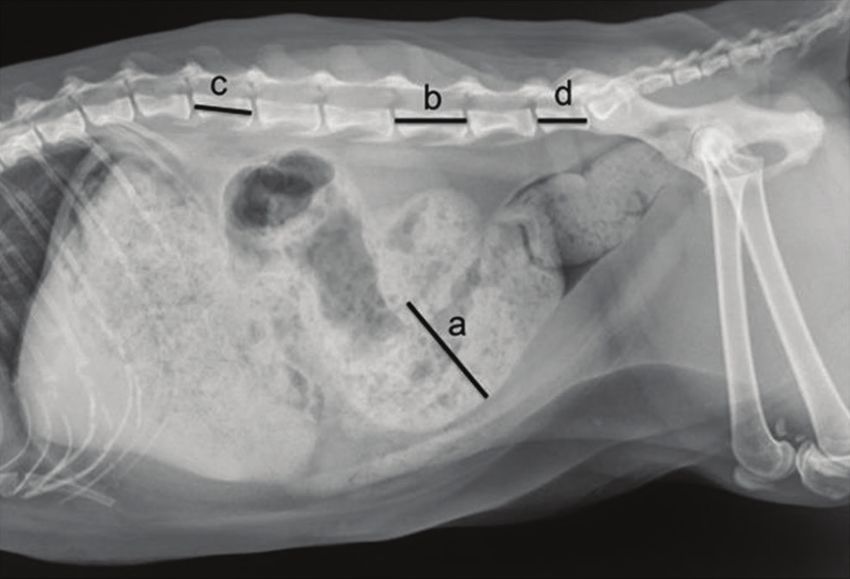
Established internationally recognized high standards portion (MD). Then, colon diameters were compared
of individual veterinary clinical patient care were to the length of the fifth lumbar (L5) vertebrae. In
followed. Ethical approval from a committee was, addition to the length of L5, the length of the second
therefore, not necessarily required. Informed consent (L2) and last (LL) lumbar vertebrae was measured
(either verbal or written) was obtained from the owner and compared (Figure-1). All measurements were
or legal custodian of all animals described in this work performed using digital calipers, and all parame-
for the procedures undertaken. ters were recorded. The cutoff values were applied
Study location and period to differentiate the types of large bowel dysfunction,
This study was designed as a retrospective followed previous information [9]. Briefly, the colon
study using patient information (both clinical and that contained large bowel contents and had an MD
radiographic data) of cats that were presented to diameter 1.28 times larger than the L5 length was
the Diagnostic Imaging Unit, the Small Animal considered to have constipation, whereas the colon
Teaching Hospital, Faculty of Veterinary Science, that contained fecal contents and had an MD diameter
Chulalongkorn University, in March 2016 and 1.48 times larger than the L5 length was classified as
February 2018. megacolon [9].
Veterinary World, EISSN: 2231-0916 493Available at www.veterinaryworld.org/Vol.14/February-2021/22.pdf
Radiographic lumbosacral vertebrae
Of 1365 cats, there were 959 and 406 cats in Gr.
1 and Gr. 2, respectively. In Gr. 1, the average lengths
of L2, L5, and LL were 17.78±0.04 mm, 21.51±0.05
mm, and 16.38±0.06 mm, respectively. The average
lengths of L2, L5, and LL in each group by sex and
gonadal status are shown in Table-2. Interestingly, the
lengths of L2, L5, and LL in male cats were signifi-
cantly greater than those of female cats (pAvailable at www.veterinaryworld.org/Vol.14/February-2021/22.pdf
Table-1: Clinical information such as number, sex, age (mean±SEM; months), and body weight (mean±SEM; kg) of the
cats, all of the overall, normal lumbar‑sacral vertebrae, and non‑traumatic lumbar‑sacral lumbar abnormalities.
Clinical Gonadal status
information
Overall Male Intact Castrated Female Intact Spayed
male male female female
Overall cats
Number 1365 781 424 357 584 269 315
Age (months) 53.71±1.40 45.93±1.53 33.14±1.60 61.11±2.53 64.11±2.48 45.26±3.17 80.20±3.48
Body weight (kg) 4.02±0.03 4.40±0.04 4.07±0.05 4.78±0.06 3.50±0.04 3.27±0.06 3.70±0.06
Normal vertebrae
Number 959 557 306 251 402 201 201
Age (months) 47.13±1.43 42.32±1.64 31.32±1.75 55.73±2.71 53.80±2.51 40.05±3.10 67.55±3.70
Body weight (kg) 4.01±0.04 4.39±0.05 4.02±0.06 4.85±0.08 3.48±0.05 3.25±0.07 3.70±0.07
Abnormal vertebrae
Number 406 224 118 106 182 68 114
Age (months) 69.23±3.13 54.90±3.36 37.86±3.49 73.87±5.39 86.86±5.35 60.63±8.3 102.51±6.56
Body weight (kg) 4.03±0.06 4.41±0.08 4.21±0.11 4.64±0.11 3.57±0.08 3.33±0.12 3.71±0.10
Congenital
Number 226 140 81 59 86 35 51
Age (months) 50.77±3.20 41.44±2.83 28.15±2.74 59.68±4.62 65.98±6.74 51.74±10.60 75.75±8.56
Body weight (kg) 4.06±0.08 4.35±0.10 4.10±0.13 4.69±0.15 3.58±0.11 3.33±0.16 3.76±0.14
Acquired
Number 180 84 37 47 96 33 63
Age (months) 92.40±5.33 77.35±7.00 59.14±8.45 91.68±10.18 105.57±7.69 68.71±12.55 124.17±8.79
Body weight (kg) 4.00±0.09 4.51±0.12 4.44±0.19 4.57±0.16 3.55±0.11 3.30±0.17 3.68±0.13
Table-2: Lumbar vertebral lengths (mean±SEM; mm) of the second lumbar vertebra (L2), the fifth lumbar
vertebra (L5), and the last lumbar vertebra (LL) of cats, all of the overall, normal lumbar‑sacral vertebrae, and
non‑traumatic lumbar‑sacral lumbar abnormalities.
Lumbar vertebral Gonadal status
lengths
Overall Male Intact Castrated Female Intact Spayed
male male female female
Overall cats
L2 17.78±0.04 18.26±0.05 17.95±0.07 18.63±0.06 17.15±0.05 16.90±0.08 17.36±0.07
L5 21.51±0.05 22.08±0.07 21.67±0.09 22.57±0.07 20.74±0.06 20.45±0.09 20.99±0.07
LL 16.38±0.06 16.93±0.07 16.75±0.01 17.14±0.10 15.66±0.08 15.51±0.12 15.78±0.11
Normal vertebrae
L2 17.75±0.05 18.25±0.06 17.94±0.08 18.62±0.08 17.07±0.06 16.81±0.09 17.32±0.08
L5 21.55±0.05 22.17±0.07 21.80±0.09 22.36±0.09 20.69±0.07 20.40±0.11 20.97±0.09
LL 16.23±0.06 16.78±0.08 16.60±0.12 16.99±0.11 15.47±0.09 15.32±0.13 15.61±0.13
Abnormal vertebrae
L2 17.86±0.07 18.30±0.10 17.97±0.16 18.67±0.11 17.32±0.10 17.17±0.15 17.41±0.13
L5 21.40±0.09 21.84±0.13 21.33±0.20 22.44±0.13 20.86±0.11 20.59±0.19 21.02±0.14
LL 16.76±0.11 17.31±0.14 17.15±0.21 17.48±0.19 16.08±0.16 16.06±0.27 16.09±0.19
Congenital
L2 17.95±0.11 18.25±0.14 17.90±0.20 18.74±0.16 17.46±0.15 17.20±0.22 17.64±0.20
L5 21.37±0.12 21.66±0.17 21.19±0.25 22.32±0.18 20.90±0.16 20.53±0.27 21.15±0.18
LL 17.40±0.14 17.72±0.18 17.42±0.26 18.13±0.24 16.87±0.22 16.60±0.35 17.05±0.27
Acquired
L2 17.75±0.10 18.38±0.15 18.11±0.26 18.59±0.16 17.20±0.12 17.00±0.25 17.23±0.16
L5 21.44±0.13 22.15±0.18 21.65±0.33 22.54±0.19 20.83±0.15 20.45±0.31 20.92±0.19
LL 15.95±0.15 16.61±0.21 16.56±0.33 16.65±0.27 15.36±0.20 15.37±0.40 15.31±0.23
increased risk of constipation and megalocon (38/275 body, intramural or extramural GI lesions, constipa-
cats, p=0.0044; odds ratio=1.865), while those with tion, obstipation, and/or megacolon. The latter GI
cranial location (2/21) did not show statistical associ- abnormalities could be induced by several underlying
ation (p=0.6804). causes, such as narrowing pelvic canal and idiopathic
conditions. Idiopathic constipation and/or megaco-
Discussion
lon may involve generalized dysfunction of colonic
Abdominal radiography is an efficient diag- smooth muscle. The etiology is still unclear [3].
nostic tool for detecting any abnormalities of the Although constipation was sometimes reported in
abdominal internal organs. Among these, GI diseases, cats, the incidence of non-obstructive constipation
especially of the distal GI tract, were periodically and its causes, such as non-traumatic lumbosacral ver-
detected clinically, for example, diarrhea, GI foreign tebral abnormalities, is low.
Veterinary World, EISSN: 2231-0916 495Available at www.veterinaryworld.org/Vol.14/February-2021/22.pdf
Figure-2: A diagram indicating the distribution number of the cats in each group, categorized by the presentation of
lumbar-sacral lumbar vertebral characteristic and the presentation of large bowel dysfunction.
This study aimed to demonstrate the incidence might affect bone maturation. However, further inves-
of non-traumatic lumbosacral vertebral abnormalities tigations should be undertaken to clarify this.
in cats and relationships of the abnormalities to large The age range of the congenital subgroup was
bowel dysfunction in cats observed on a retrospective significantly lower than that of the acquired subgroup.
model using abdominal radiographs. The statistical This could be due to the embryologic development of
analysis was performed only in the normal verte- the vertebrae that are closely related to spinal cord or
bral group since nontraumatic lumbosacral vertebral other organ abnormalities [30]. Owners could detect
abnormalities could affect the measurement of lum- abnormal clinical signs of various organs; then, cats
bar vertebral length and lead to unreliable results. The were presented earlier to the veterinarian. In contrast,
results revealed the statistical differences of lumbar cats with acquired bone abnormalities might gradu-
vertebral length between male and female cats, which ally reveal clinical signs over time, in accordance with
are consistent with the previous study in humans, indi- degenerative diseases of organs. As a result, the age
cating that gonadal hormones of both sexes play an at which the acquired lumbosacral vertebral abnormal
important role as initiators of bone growth spurt and cats were presented to the veterinarian might be later.
mineralization through complex mechanisms, such as Furthermore, 70.26% of cats in this study had
bone resorption inhibition and stimulation of active normal bone condition, while the other 29.74% were
Vitamin D3 metabolite production [24]. Androgens considered otherwise. In the abnormal group, 16.55%
can also enhance the skeletal sensitivity to calcitonin and 13.19% of cats had congenital and acquired lum-
in rats [25]. In addition to sex, gonadal status also has bar vertebral abnormalities, respectively. The most
an effect on lumbar vertebral length in both male and common congenital lumbar vertebral abnormality in
female cats. This effect might be caused by prepuber- this study was an abnormal number of lumbar verte-
tal gonadectomy. It has been reported that prepubertal brae, which accounted for 68.12%. There were also
gonadectomy has an effect on bone growth by delay- studies on abnormal vertebral numbers found in sev-
ing the closure time of physis [26]. Despite the retro- eral other species, such as ferrets and rabbits [31,32].
spective model indicating that the history of the gona- This abnormality may have clinical significance
dectomy period for each cat could not be obtained, from the possible radiographic misinterpretation and
authors assumed that most gonadectomized cats in inaccurate choice of surgical site. The most common
this study might have undergone gonadectomy before acquired bone abnormality found in this study was
the prepubertal period. Prepubertal gonadectomy ventral bone spur (50.56%). As bone spurs develop,
can assist in population control in cats, which are a they can create pressure on exiting spinal nerve roots
seasonal polyestrus species [27,28], and additionally and possibly result in neurologic deficits. Although
reduce the risk of feline mammary gland tumors, espe- the osteophytes typically do not project into the spinal
cially in female cats [29]. This phenomenon is more canal, the possibility of spinal cord pressure must also
clearly observed in male cats compared to female cats. be considered [33].
The discrepancy of the effect of gonadal status on phy- Overall, 52 (12.80%) cats with lumbar verte-
seal closure between sexes of the cat might be caused bral abnormalities had radiographic evidence of an
by sex hormonal imbalance in juvenile male cats that abnormal large bowel, in which colon diameter was
Veterinary World, EISSN: 2231-0916 496Available at www.veterinaryworld.org/Vol.14/February-2021/22.pdf
significantly larger than that of cats with normal lum- condition and provide proper management to improve
bar vertebrae of 75 (7.82%) (p=0.0057). The asso- the quality of their lives.
ciation of vertebral abnormalities and GI disorders Authors’ Contributions
in cats has never been clearly described. The feline
idiopathic megacolon has been attributed to primary CT and NC: Study conception and design. KK,
neurologic and degenerative neuromuscular disorders, KB, NT, and WS: Acquisition of data. CT, KK, KB,
and the most frequent cause is that these vertebral NT, WS, and NC: Analysis and interpretation of data.
abnormalities damage the lumbosacral plexus, which CT, KK, KB, NT, WS, and NC: Drafting of manu-
originates from the level of L4 through the sacrum, script. CT, KK, KB, NT, WS, and NC: Critical revi-
which may affect the autonomic function of the pel- sion. All authors have read and approved the final
vic viscera [2,34]. Sacralization or the abnormality of manuscript.
last lumbar vertebrae can also result in clinical signs Acknowledgments
of lumbosacral disease, including terminal colon
atony [34]. This study was conducted and supported
This retrospective study has some limitations. by Faculty of Veterinary Science, Chulalongkorn
The radiographic evaluation of lumbar vertebra should University, Bangkok, Thailand, for the senior project:
be obtained from spot radiographs to avoid distortions, 31040603 in the academic year of 2018, number 9.
which could cause misinterpretation. In addition, a Competing Interests
calibration ball should be applied for increased accu-
racy and to avoid variation in magnification among The authors declare that they have no competing
the measurements due to different distances between interests.
objects and films. Although this study revealed that Publisher’s Note
the lumbar vertebral abnormalities may be related to
Veterinary World remains neutral with regard
constipation or megacolon in cats, the association of
to jurisdictional claims in published institutional
the abnormal bone location and the affected nerves
affiliation.
supplying the colon should be further investigated to
clarify the pathogenesis of the large bowel abnormali- References
ties. In 2005, Chang et al. [35] reported that the inner- 1. Yam, P. (1997) Decision making in the management of con-
vations of rectal detrusor and external anal sphincter stipation in the cat. Practice, 19(8): 434-440.
in dogs are provided by the ventral roots of L7, S1, S2, 2. Rossi, G., Jergens, A., Cerquetella, M., Berardi, S., di
and S3, which originate from the sacral spinal cord Cicco, E., Bassotti, G., Pengo, G. and Suchodolski, J.S.
(2018) Effects of a probiotic (SLAB51™) on clinical and
parasympathetic and somatic centers. In addition, histologic variables and microbiota of cats with chronic
since large bowel dysfunction, both constipation and constipation/megacolon: A pilot study. Benef. Microbes.,
megacolon, could be induced by other causes, such as 9(1): 101-110.
concurrent diseases, dehydration, diet, and other med- 3. Foley, P. (2017) Constipation, tenesmus, dyschezia, and
ications, the complete clinical information through faecal incontinence. In: Textbook of Veterinary Internal
Medicine, Diseases of Dog and Cat. 8th ed. Elsevier,
physical examination, including multifactorial obser- Saunders, St. Louis. p633-638.
vation, could provide evidence. 4. Jergens, A.E. and Ahrens, F.A. (2008) Drugs acting on
the gastrointestinal tract. In: Hsu, W.H., editor. Handbook
Conclusion of Veterinary Pharmacology. Wiley-Blackwell, Iowa.
This study unveiled radiographic information p235-252.
5. Demetriou, J.L. and Welsh, E.M. (2000) Colonic obstruc-
concerning lumbosacral vertebral abnormalities and tion in an adult cat following open castration. Vet. Rec.,
large bowel dysfunction in cats. This information 147(6): 165-166.
could assist veterinarians in evaluating, monitoring, 6. Washabau, R.J. and Hasler, A.H. (1997) Constipation,
and obtaining awareness of constipation or megacolon obstipation, and megacolon. In: August, J.R., editor.
problems in cats with incidental findings of vertebral Consultations in Feline Internal Medicine. 3rd ed. Saunders,
Philadelphia, PA. p104-111.
abnormalities on abdominal radiographs. Although 7. Colopy-Poulsen, S.A., Danova, N.A., Hardie, R.J. and
the clinical signs are not immediately apparent, they Muir, P. (2005) Managing feline obstipation secondary to
continuously progress. Veterinary practitioners should pelvic fracture. In: Yardley, P.A., editor. Compendium on
perform an early, thorough diagnostic workup to Continuing Education for the Practising Veterinarian, North
define the potential of disease progression and pro- America. p662-669.
8. LeRoy, B.E. and Lech, M.E. (2004) Prostatic carcinoma
vide proper advice to cat owners for prevention and causing urethral obstruction and obstipation in a cat.
control of large bowel functions. Moreover, a neuro- J. Feline Med. Surg., 6(6): 397-400.
logical examination should also be performed to con- 9. Trevail, T.I.M., Gunn-Moore, D., Carrera, I., Courcier, E.
firm problems in cases diagnosed with both intestinal and Sullivan, M. (2011) Radiographic diameter of the colon
and lumbosacral vertebral abnormalities. Therefore, in normal and constipated cats and in cats with megacolon.
Vet. Radiol. Ultrasound, 52(5): 516-520.
the information found on abdominal radiographs of 10. Bertoy, R.W. (2003) Megacolon in the cat. Vet. Clin. North
cats, both distal GI and vertebral lesion, could assist Am. Small Anim. Pract., 32(4): 901-915.
the owners and veterinarians to undertstand the cat’s 11. Washabau, R.J. and Holt, D. (1999) Pathogenesis, diagnosis,
Veterinary World, EISSN: 2231-0916 497Available at www.veterinaryworld.org/Vol.14/February-2021/22.pdf
and therapy of feline idiopathic megacolon. Vet. Clin. North mineralisation: With observations in male delayed puberty.
Am. Small Anim Pract., 29(2): 589-603. Arch. Dis. Child., 54(12): 950-953.
12. Johnson, D.A. (2006) Treating chronic constipation: How 25. Ogata, E., Shimazawa, E., Suzuki, H., Yoshitoshi, Y.,
should we interpret the recommendations? Clin. Drug Asano, H. and Ando, H. (1970) Androgens and enhance-
Investig., 26(10): 547-557. ment of hypocalcemic response to thyrocalcitonin in rats.
13. Tack, J., Müller-Lissner, S., Stanghellini, V., Endocrinology, 87(2): 421-426.
Boeckxstaens, G., Kamm, M.A., Simren, M., Galmiche, G.P. 26. Root, M.V., Johnston, S.D. and Olson, P.N. (1997) The
and Fried, M. (2011) Diagnosis and treatment of chronic effect of prepuberal and postpuberal gonadectomy on radial
constipation-a European perspective. Neurogastroenterol. physeal closure in male and female domestic cats. Vet.
Motil., 23(8): 697-710. Radiol. Ultrasound, 38(1): 42-47.
14. Palmer, N.C. (1968) Osteodystrophia fibrosa in cats. Aust. 27. Stornelli, M.A., Reyna, J.C., Stornelli, M.C., Favre, R.N.,
Vet. J., 44(4): 151-155. Savignone, C.A., Tittarelli, C.M. and Sotaet, R.L. (2009)
15. Rosin, E. (1993) Megacolon in cats. Vet. Clin. North Am. Seasonal changes in testicular cell morphology in domes-
Small Anim. Pract., 23(3): 587-594. tic male cats (Felis catus). Reprod. Domest. Anim., 44(2):
16. Rosin, E., Walshaw, R., Mehlhaff, C., Matthiesen, D., 287-290.
Orsher, R. and Kusba, J. (1988) Subtotal colectomy for 28. Jennett, A.L., Jennett, N.M., Hopping, J. and Yateset, D.
treatment of chronic constipation associated with idiopathic (2016) Evidence for seasonal reproduction in UK domestic
megacolon in cats: 38 cases (1979-1985). J. Am. Vet. Med. cats. J. Feline Med. Surg., 18(10): 804-808.
Assoc., 193(7): 850-853. 29. Reichler, M. (2009) Gonadectomy in cats and dogs: A
17. Winge, K., Rasmussen, D. and Werdelin, L.M. (2003) review of risks and benefits. Reprod. Domest. Anim., 44(2):
Constipation in neurological diseases. J. Neurol. Neurosurg. 29-35.
Psychiatry, 74(1): 13-19. 30. Bailey, C.S. and Morgan, J.P. (1992) Congenital spinal mal-
18. Fluckiger, M.A., Damur-Djuric, N., Hassig, M., Morgan, J.P. formations. Vet. Clin. North Am. Small Anim. Pract., 22(4):
and Steffenet, F. (2006) A lumbosacral transitional verte- 985-1015.
brae in the dog predisposes to cauda equina syndrome. Vet. 31. Proks, P., Stehlik, L., Paninarova, M., Irova, K.,
Radiol. Ultrasound, 47(1): 39-44. Hauptman, K. and Jeklet, V. (2015) Congenital abnor-
19. Danielski, A., Bertran, J. and Fitzpatrick, N. (2013) malities of the vertebral column in ferrets. Vet. Radiol.
Management of degenerative lumbosacral disease in cats Ultrasound, 56(2): 117-123.
by dorsal laminectomy and lumbosacral stabilization. Vet. 32. Proks, P., Stehlik, L., Nyvltova, I., Necas, A., Vignoli, M.
Comp. Orthop. Traumatol., 26(1): 69-75. and Jekl, V. (2018) Vertebral formula and congenital abnor-
20. Newitt, A., German, A.J. and Barr, F.J. (2008) Congenital malities of the vertebral column in rabbits. Vet. J., 236(6) :
abnormalities of the feline vertebral column. Vet. Radiol. 80-88.
Ultrasound, 49(1): 35-41. 33. Morgan, J.P. and Biery, D.N. (1985) Spondylosis defor-
21. Harris, J.E. and Dhupa, S. (2008) Lumbosacral interver- mans. In: Newton, C.D. and Nunamaker, A.M., editors.
tebral disk disease in six cats. J. Am. Anim. Hosp. Assoc., Textbook of Small Animal Orthopaedics. Pennsylvania,
44(3): 109-115. Philadelphia, PA. p733-738.
22. Magi, G., Cherubini, G.B. and Taeymans, O. (2018) 34. Dyck, P.J. and Windebank, A.J. (2002) Diabetic and non-
Sacrocaudal (sacrococcygeal) intervertebral disc protrusion diabetic lumbosacral radiculoplexus neuropathies: New
in 2 cats. Can. Vet. J., 59(4): 388-392. insights into pathophysiology and treatment. Muscle Nerve,
23. Johanson, J.F., Sonnenberg, A., Koch, T.R. and 25(4): 477-491.
McCarty, D.J. (1992) Association of constipation with neu- 35. Chang, S.M., Yu, G.R., Diao, Y.M., Zhang, M.J., Wang, S.B.
rologic diseases. Dig. Dis. Sci., 37(2): 179-186. and Hou, C.L. (2005) Sacral anterior root stimulated def-
24. Krabbe, S., Christiansen, C., Rødbro, P. and Transbølet, ecation in spinal cord injuries: An experimental study in
I. (1979) Effect of puberty on rates of bone growth and canine model. World J. Gastroenterol., 11(11): 1715-1718.
********
Veterinary World, EISSN: 2231-0916 498You can also read