APPLICATION OF PANORAMIC T2 SEQUENCE FOR MAGNETIC RESONANCE IMAGING OF LUMBOSACRAL SPINE - SciELO
←
→
Page content transcription
If your browser does not render page correctly, please read the page content below
ISSN Versión Online: 2308-0531
                 Rev. Fac. Med. Hum. April 2021;21(2):283-291.
                                                                                                                                   Facultad de Medicina Humana URP
                 DOI 10.25176/RFMH.v21i2.3452
                 ORIGINAL PAPER

                APPLICATION OF PANORAMIC T2 SEQUENCE FOR
             MAGNETIC RESONANCE IMAGING OF LUMBOSACRAL SPINE
                                                    APLICACIÓN DE SECUENCIA T2 PANORÁMICA PARA RESONANCIA
                                                             MAGNÉTICA DE COLUMNA LUMBOSACRA

                                                                  Alexander Román-Meza1,2,3,a,b,c, Raúl Ruiz-Arias4,5,6,d,e

                   ABSTRACT
                   Introduction: The evaluation of the total spine by magnetic resonance imaging in a T2-weighted single sequence
                   using Software Composing previous planning, called panoramic, would be beneficial in the study of lumbosacral
                   spine for the additional information that could be obtained. Objectives: To analyze the application of T2 panoramic
                   sequence for magnetic resonance imaging of the lumbosacral segment. Method: Retrospective and cross-sectional
                   study executed with 186 cases selected by stratified probability sampling, between 18 and 60 years old, using a
                   form consisted of a general sheet and an analytical sheet obtained from the request for radiological study and the
                   examination itself. Results: Of the total, 80.1% were in the adult stage of life, and 53.8% were female, and 52.7%
ORIGINAL PAPER

                   had the suspicion of herniated disc as a specific diagnostic presumption. Degenerative pathologies were mainly
                   presented, with 93%, having herniated discs as the most frequent with 57%, which included extruded, protruded
                   and migrated hernias with 40.6%, 37.9% and 21.5 % respectively. Likewise, it was verified with hypothesis test that
                   this acquisition allows to localize 50% more findings in contrast to the usual examination, defining that this sequence
                   should be applied because it localizes findings in a greater number in comparison to the lumbosacral selective
                   examination. Conclusions: In general, it was defined that the application of this panoramic T2 sequence is more
                   usefulness for a better evaluation, because it achieves greater findings, resulting as main pathologies in lumbosacral,
                   cervical and dorsal segments those of a degenerative type, standing out the herniated discs.
                   Key words: Magnetic resonance imaging; Radiologic technology; Spine (source: MeSH NLM).

                   RESUMEN
                   Introducción: La evaluación de columna total mediante resonancia magnética en una sola secuencia potenciada
                   en T2 utilizando Software Composing previa planeación, denominada panorámica, sería beneficioso en el estudio
                   de columna lumbosacra por la información adicional que podría obtenerse. Objetivos: Analizar la aplicación de
                   secuencia panorámica en T2 para resonancia magnética del segmento lumbosacro. Métodos: Estudio retrospectivo
                   y transversal realizado con 186 casos seleccionados mediante muestreo probabilístico estratificado, entre 18 a 60
                   años, utilizándose una ficha conformada por una hoja general y una hoja analítica obtenidas de la solicitud de
                   estudio radiológico y el propio examen realizado. Resultados: Del total, un 80,1% era de etapa de vida adulta, y
                   53,8% era femenino, además que un 52,7% tuvieron como presunción diagnóstica específica la sospecha de hernia
                   discal. Se presentó principalmente patologías degenerativas, en un 93%, teniendo a las hernias como las más
                   frecuentes en un 57%, que a su vez comprendió hernias extruidas, protruidas y migradas con 40,6%, 37,9% y 21,5%
                   respectivamente. Así mismo, se comprobó con prueba de hipótesis que esta adquisición permite localizar un 50%
                   más de hallazgos a diferencia del examen habitual, definiéndose que esta secuencia debe aplicarse porque localiza
                   hallazgos en un mayor número en comparación al examen selectivo lumbosacro. Conclusión: De forma general,
                   se definió que la aplicación de esta secuencia T2 panorámica es de mayor utilidad para una mejor evaluación, por
                   lograrse mayores hallazgos, resultando como patologías principales tanto en segmento lumbosacro como cervical
                   y dorsal las de tipo degenerativas, resaltando las hernias discales.
                   Palabras clave: Imagen por resonancia magnética; Tecnología radiológica; Columna vertebral (fuente: DeCS
                   BIREME).

                  1 Servicio de Tomografía y Resonancia, Hospital Nacional Edgardo Rebagliati Martins, EsSalud, Lima-Perú.
                  2 Centro de Diagnóstico por Imágenes de Clínica Internacional - Sede San Borja, Lima-Perú.
                  3 Miembro de la Asociación Peruana de Tecnólogos Médicos en Calidad y Seguridad Radiológica (APTEMCSER), Lima-Perú.
                  4 Jefe de la Oficina de Estadística e Informática del Hospital de Emergencias José Casimiro Ulloa - Ministerio de Salud, Lima-Perú.
                  5 Docente de Posgrado de la Facultad de Tecnología Médica de la Universidad Nacional Federico Villarreal, Lima-Perú.
                  6 Docente de Estudios Generales de la Universidad San Ignacio de Loyola, Lima-Perú.
                  a
                    Graduate in Radiologic Technology; b Specialist in computed tomography (CT) and magnetic resonance imaging (MRI); c Master of Health
                  Services Management; d Graduate in Statistics; e Master in Statistics.

                  Cite as: Alexander Román-Meza, Raúl Ruiz-Arias. Application of Panoramic T2 Sequence for Magnetic Resonance Imaging of Lumbosacral
                  spine. Rev. Fac. Med. Hum. April 2021; 21(2):283-291. DOI 10.25176/RFMH.v21i2.3452

                 Journal home page: http://revistas.urp.edu.pe/index.php/RFMH

                   Article published by the Magazine of the Faculty of Human Medicine of the Ricardo Palma University. It is an open access article, distributed under the terms of the
                   Creative Commons License: Creative Commons Attribution 4.0 International, CC BY 4.0 (https://creativecommons.org/licenses/by/4.0/), that allows non-commercial
                   use, distribution and reproduction in any medium, provided that the original work is duly cited. For commercial use, please contact revista.medicina@urp.pe

                                                                                                                                                                              Pág. 283
Rev. Fac. Med. Hum. 2021;21(2):283-291.                                                                 Román A et al

                 INTRODUCTION                                               vary according to each sequence(7,8).
                 The Peruvian population has undergone a                    The technique is based on a direct application of
                 sociodemographic transition with a decrease in             the Composing Software, which allows to show an
                 the mortality rate, a larger economically active           image resulting from other sequences that were
                 population, and population aging, with a greater           acquired in advance. This was traditionally only
                 presentation of chronic-degenerative diseases to the       done in a later post-processing of images, but now
                 detriment of infectious diseases(1). Among this group      it can be obtained automatically by pre-planning
                 of now frequent diseases are dorsopathies, located in      the sequences of interest in alignment with what
                 a sixth place of morbidity for outpatient consultation,    your acquisitions are achieved with the composite
                 with 3.2%, and which occurs in all stages of adult         image minutes later. This possibility means a notable
                 life: 2.4% in young adults, 5.6% in adults and 6.9%        optimization in terms of the use of extra time and
                 in older adults, and even in teenagers, with 1.3%, a       additional platform for image post-processing(8,9).
                 situational context that had not occurred in recent
                                                                            Considering that the T2 sequence is a pattern and
                 decades(2).
                                                                            a basic part of any study of the spinal cord, the
                 In this way, an adequate evaluation of the spine is        fact of using a panoramic sequence enhanced in
ORIGINAL PAPER

                 of utmost importance, especially at the level of the       T2 would be of great contribution and utility to
                 lumbosacral spine, since in this region there is greater   the usual study of the lumbosacral spine; this is
                 symptomatology, being the objective of clinical and        because it would allow additional information to
                 especially radiological review because it is the most      be obtained. Therefore, its analysis would serve as a
                 useful diagnostic. Currently, magnetic resonance           recommendation to opt for its performance due to
                 images stand out for allowing the visualization of         its contribution and benefit in the diagnosis. In view
                 anatomical information at the spinal and vertebral         of this, the objective was to analyze the application
                 level with high contrast resolution, having as a           of this sequence for the study of the lumbosacral
                 fundamental part the T2-weighted image sequence            spine by magnetic resonance imaging (MRI).
                 that demonstrates the differentiation between
                 cerebrospinal fluid with the other structures of           METHODS
                 the vertebromedullary region. Therefore, it has a
                 sensitivity and specificity of 85.5%, close to 100% in     Study design
                 oncological and inflammatory pathology(3,4).               A retrospective and cross-sectional study was carried
                 However, traditionally the clinical and especially         out, from January to June 2016, in a group of adult
                 radiological evaluation of the spine in general has        patients whose ages were between 18 and 60
                 been limited to the region that is presumed to be          years of age. An MRI study of the lumbosacral spine
                 affected by various limitations, despite the fact that     from an outpatient consultation at a private health
                 not all pathological types will affect a single region,    services institution in Lima was performed.
                 such as herniated discs with a greater lumbosacral         Population and sample
                 settlement, spondyloarthropathies in the lower
                 back with the likelihood of involvement in adjacent        The population was made up of 357 patients who
                 segments, and others that are clinically silent such as    had MRI of the lumbosacral spine, whose sample
                 vertebral hemangiomas, located mainly at the dorsal        was obtained with the proper formula for a finite
                 level but also at other levels(5,6).                       population:

                 Based on this, some method is required to visualize
                 the total spine, such as the panoramic sequence or
                 also called panoramic image, which makes it possible
                 to see the spine in a single sequence with modern          In the formula, a prevalence equivalent to 0.5 was
                 magnetic resonance imaging systems. This allows            considered, with a precision of 0.05 and a significance
                 the acquisition of images in the sagittal plane of the     level of 0.05, resulting in a sample of n = 186 cases.
                 entire axis of the spine, taking only a few additional     The applied sampling was stratified probabilistic,
                 minutes compared to the traditional examination,           whose criterion was to consider the life stages
                 without the need for repositioning the patient, and        included in the cases, both young adults aged 18-30
                 only planning it from the acquisition station in the       years and adults properly speaking aged 31-60 years.
                 MRI Scanning Room, where the acquisition time will         The population comprised 71 young adults and 286

                 Pág. 284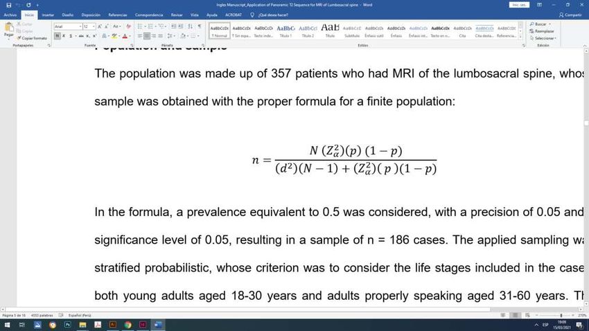
Rev. Fac. Med. Hum. 2021;21(2):283-291.                          Application of Panoramic T2 Sequence for Magnetic Resonance Imaging

adults, with which 37 cases of young adults and 149       Borja headquarters the radiological examinations
cases of adults were selected under the same degree       were obtained after coordination with the
of proportionality, all randomly assigned, thus           headquarters of the Center for Diagnostic Imaging
making up 186 cases of the total sample.                  and Supervision of the Magnetic Resonance Area,
Collection and evaluation instrument                      respecting the confidentiality of the information
                                                          collected for the exclusive use of this investigation.
A collection sheet was applied, consisting of a general
sheet and an analytical sheet, the data of which were     RESULTS
filled in considering the request for a radiological
study and the examination performed respectively,         The ages of the patients who had magnetic
using the systematic observation technique. The first     resonance imaging ranged from 18 to 60 years and
sheet allows collecting basic data such as gender and     the median achieved was 40 years. Reviewing the
stage of life of the patient, as well as the diagnostic   selected cases based on life stage, adults themselves
presumption to perform the examination, and the           (from 31 to 60 years) were 80.1%, while young adults
second sheet collects if findings were presented and      (from 18 to 30 years) represented 19.9%. Likewise,
in which pathological groups and subgroups they           the female gender constituted 53.8% and the male

                                                                                                                                       ORIGINAL PAPER
could be classified. The examinations were carried        gender 46.2%, and in which total of cases the most
out on a Siemens MAGNETOM Aera 1.5 Tesla MRI              frequent diagnostic presumption was the herniated
scanner, using the Spine 32 antennas (attached to         discs, with 52.7%.
the equipment table) and Body 18 (placed at the
abdominal level referring to the lumbosacral spine),      In general, of 186 examinations in total, there
in which the study was acquired specific in its usual     were 5 cases that presented a normal diagnosis in
form as well as panoramic T2 Sequence using just an       the traditional study and with the application of
additional 3:37 minutes.                                  panoramic T2 sequence. Therefore, the remaining 181
                                                          cases had some finding, regardless of the acquired
Statistical analysis
                                                          segment of the spine. Of these 181 mentioned, the
A data matrix was prepared using the Microsoft Excel      usual lumbosacral study had a total of 414 findings.
2016 software for the corresponding collection,
                                                          In the lumbosacral spine, degenerative pathologies
creating contingency tables with a subsequent
                                                          were seen as the most frequent findings, occurring in
verification of the data obtained. For processing,
the statistical software R for Windows, Version 3.1.2,    93% of cases. Of these, herniated discs were the most
was used, making the corresponding frequency              common with 57%, the majority of which comprised
distribution for the different findings and allowing      disc extrusion in 40.6%. Likewise, there were disc
the calculation of means and trends. Likewise, the        protrusion in 37.9% and in fewer disc migration
normal distribution was verified in the data obtained     with only 21.5%, continuing with lumbar disc
by means of the Kolmogorov-Smirnov test and               degeneration and modic changes with 19.5% and
with this apply the hypothesis test for a proportion,     9.9% respectively, having as a finding less frequent
considering:                                              to spondylolysis with only 0.5%. The other findings
                                                          were congenital anomalies (5.3%) and neoplastic
• Null hypothesis (H0): The acquisition of the
                                                          pathology (1%, represented by hemangiomas as
  panoramic T2 Sequence in the lumbosacral spine
  does not allow 50% more findings to be located in       the only type of tumor) and, with less than 1% of
  contrast to a routine examination.                      cases, there were inflammatory pathologies (0.5%)
                                                          and traumatic injuries (0.2%). Among the congenital
• Alternative hypothesis (H1): The acquisition of the     anomalies, the so-called transition anomalies stood
  panoramic T2 Sequence in the lumbosacral spine
                                                          out, such as lumbarizations in 50% (conversion of the
  does allow 50% more findings to be located in
                                                          S1 vertebra) and sacralizations in 36.4% (conversion
  contrast to a routine examination.
                                                          of the S5 vertebra). Herniated discs located in
Ethical aspects                                           the lumbosacral spine were 52.9%: disc extrusion
The study was carried out based on ethical                accounted for 21.5%, disc protrusion accounted for
considerations and good practices, with the               20%, while only 11.4% were disc migration, given in
authorization of the Academic and Research                167 patients (Table 1).
Directorate of Clínica Internacional, from whose San

                                                                                                                         Pág. 285
Rev. Fac. Med. Hum. 2021;21(2):283-291. Román A et al
Table 1. Findings obtained by routine study by MRI of Lumbosacral spine.
Type of Pathology in Lumbosacral Spine
Specific diagnostic Degenerative Congenital Neoplastic Total
findings n % n % n % n %
Disc protrusion 83 21.6% 83 20.0%
Disc extrusión 89 23.1% 89 21.5%
Disc migration 47 12.2% 47 11.4%
Disc degeneration 75 19.5% 75 18.1%
Modic type II 21 5.5% 21 5.1%
Modic type I 17 4.4% 17 4.1%
Schmorl's nodes 7 1.8% 7 1.7%
ORIGINAL PAPER
Spondylolysis 2 0.5% 2 0.5%
Other degenerative
44 11.4% 44 10.6%
types
Lumbarization 11 50.0% 11 2.7%
Sacralization 8 36.4% 8 1.9%
Other congenital
3 13.6% 3 0.7%
types
Hemangioma 4 100% 4 1.0%
Other types
0 0% 0 0%
Neoplastic
Other pathologies
3 0.7%
Vertebromedullary
Totals by types 385 100% 22 100% 4 100% 414 100%
Absolute total 385 93.0% 22 5.3% 4 1.0% 414 100%
Of the 181 previously mentioned cases, findings at On the other hand, in the dorsal spine there was also
the cervical and dorsal spine level were also noted a higher percentage of degenerative pathologies,
by application of panoramic T2 sequence, which in a frequency of 81.7%, where herniated discs
were 117 and 46, that is, 163 additional findings. predominated in more than half of the cases (51.7%),
At the cervical spine level, there were basically including protruding hernias in 70% (especially in
degenerative pathologies, in 99.1%, highlighting the D7-D8 disc) and 30% extruded hernias (Figure
herniated discs, with 59.5%, of which 58% included 1); apart from Schmörl's nodules with 24.1% and
protruding hernias (especially in the C5-C6 disc), disc degenerations with 20.7%. The remaining 18.3%
with extruded hernias in 39.1% and finally migrated in the dorsal segment corresponded to neoplasm
hernias in 2.9% (Figure 1), followed in 28.4% by pathologies, whose lineage was specifically the
disc degenerations. It should be mentioned that vertebral hemangioma. The dorsal hernias presented
congenital anomalies were also presented in 0.9%, were 42.3% of the total findings, comprising both
represented by hydrosyringomyelia. It should be protruding hernias, in 29.6%, and extruded hernias,
noted that localized hernias accounted for 59% of in 12.7%. Therefore, the main findings located
cervical lesions, which in turn included protruded outside the lumbosacral spine were hernias,
hernias in 34.2% while 23.1% consisted of extruded given in 77 patients, both at the cervical level, in
hernias and only 1.7% were migrated hernias. 59%, and at the dorsal level, in 42.3% (Table 2).
Pág. 286Rev. Fac. Med. Hum. 2021;21(2):283-291. Application of Panoramic T2 Sequence for Magnetic Resonance Imaging
Table 2. Findings obtained outside the Lumbosacral spine by Panoramic T2 sequence.
Type of Pathology outside the Lumbosacral spine
Degenerative Congenital Neoplastic Total
Specific diagnostic findings
n % n % n % n %
Disc protrusion 40 34.5% 40 34.2%
Disc extrusion 27 23.3% 27 23.1%
Disc migration 2 1.7% 2 1.7%
Disc degeneration 33 28.4% 33 28.2%
Findings in Other degenerative
ORIGINAL PAPER
the Cervical types 14 12% 14 12%
Spine
Hydrosyringomyelia 1 100% 1 0.9%
Other congenital types 0 0% 0 0%
Totals by type 116 100% 1 100% 117 100%
Absolute total at
116 99.1% 1 0,9% 117 100%
Cervical level
Disc protrusion 21 36.2% 21 29.6%
Disc extrusion 9 15.5% 9 12.7%
Schmorl's nodes 14 24.1% 14 19.7%
Disc degeneration 12 20.7% 12 16.9%
Findings in Other degenerative
2 3.4% 2 2.8%
the Dorsal types
Spine
Hemangiomas 13 100% 13 18.3%
Other neoplastic types 0 0% 0 0%
Totals by type 58 100% 13 100% 71 100%
Absolute total at
58 81.7% 13 18.3% 71 100%
Dorsal level
Regarding the location of herniated discs at the 39.7%, L3-L4 with 12.3%, L2-L3 with 3.2% and L1-
level of the lumbosacral spine, which has been the L2 with 2.3%. The L5-S1 disc was the main location
most frequent injury, it occurred mainly at the level of extruded and migrated hernias, with 44.9% and
of the L5-S1 disc in 42% of cases, although it also 55.1% respectively, unlike protruding hernias located
occurred in the other intervertebral discs: L4- L5 with mainly in the L4-L5 disc, with 44.6% (Graph 1).
Pág. 287Rev. Fac. Med. Hum. 2021;21(2):283-291. Román A et al
A B
C
ORIGINAL PAPER
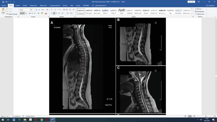
D E
F
Figure 1. MRI of 2 cases with application of panoramic T2 Sequence: 1. 29-years-old pt. (A), where disc
extrusion in L5-S1 is denoted as well as disc protrusion in C5-C6 (images B and C respectively). 2. 39-year-
old pt. (D), where disc extrusion in L4-L5 is denoted as well as disc protrusion in D7-D8 (images E and F
respectively).
Pág. 288Rev. Fac. Med. Hum. 2021;21(2):283-291. Application of Panoramic T2 Sequence for Magnetic Resonance Imaging
Disc migration
Disc extrusion
Disc protrusion
0.00% 20.00% 40.00% 60.00% 80.00% 100.00%
Disc protrusion Disc extrusion Disc migration
L1-L2 disc level 2.40% 3.40% 0%
L2-L3 disc level 7.20% 1.10% 0%
L3-L4 disc level 10.80% 19.10% 2.10%
L4-L5 disc level 44.60% 31.50% 46.80%
ORIGINAL PAPER
L5-S1 disc level 34.90% 44.90% 51.10%
Graphic 1. Anatomical location of Herniated lumbar disc.
The application of the panoramic T2 image for Analyzing, the hypothesis verification defined that
examination of the lumbosacral spine by magnetic 50% more findings are located with the panoramic
resonance imaging was verified using hypothesis sequence in question, unlike the usual study. This
testing for a proportion, taking into consideration is similar as reported by Méndez(16), which obtained
that, as H0, the technique does not allow 50% more 74.8% of additional findings using STIR sequence,
findings to be located than in a regular examination, applying post-process after examination and
and that, as H1, the technique does allow 50% more considering elderly patients. In addition, what is
findings to be located in contrast to a regular test. proposed by Schiappacasse(17) and Campos(18) is
In this way, the Sig value of the corresponding Zcal supported, where it was proposed to use a sagittal
was obtained from the findings at the level of the sequence for the entire column. It also confirms
lumbosacral segment in the conventional exam what was concluded by Burbano(19) and Campos(18),
and the amount of findings obtained by adding the where the T2-weighted image was superior for
panoramic T2 sequence, obtaining Sig = 0.031, which the various anomalies. In relation to this, what was
being less than 0.05, rejecting the null hypothesis and expressed by Tito(20), is corroborated, who indicated
therefore at 95% confidence level, it can be stated the importance of including this sequence in the
that the application of this. This sequence does allow examination of an image that reveals adjacent
50% more findings to be located, showing that it is lesions to those presented in the specific initial
useful for MR imaging of the lumbosacral spine. evaluation segment.
Reviewing the findings, there was the presence of a
DISCUSSION greater number of degenerative pathologies, with
The results demonstrate that the application of 93% of findings. The main ones were herniated discs,
the panoramic T2-weighted sequence for the with a frequency of 60%, which coincided with the
lumbosacral spine examination is valid. Therefore, studies by Ortega(21) at the Centro Médico Ecatepec, of
the importance of magnetic resonance imaging in the Instituto Mexicano de Seguridad Social, with the
the evaluation of the spine is first reaffirmed, as stated studies by González(22) at the Hospital Universitario
by Aroche(10) and Medina(11). Second, that requests for Nacional de Colombia, and with the findings of
examination with a specific presumption justify their Enríquez(23) in the Clínica Pichincha, whose results
performance for a due evaluation, since they allow to were also the most outstanding, with 40.2%, 33.7%
see early changes and/or incipient lesions and thus and 32.7% respectively. The highest percentage were
have a clinical relevance, as explained by Fretes(12), disc extrusion, in 40.6%, then protruding hernias,
Kovacs(13), Millán and Cols(14), and Rodríguez(15). with 37.9%. These findings were different and at
Pág. 289Rev. Fac. Med. Hum. 2021;21(2):283-291. Román A et al
the same time with a lower percentage compared with the greatest symptomatological settlement due
to the studies by Ortega(21) and González(22), as well to the presence of some typical risk factors of the
as the study of hernias carried out by Solano and activity, workload or rhythm of people's lives.
Ávila(24) at the Hospital Carrasco de Cuenca, of the
Instituto Ecuatoriano de Seguridad Social, in which CONCLUSION
disc protrusions were mainly presented, with figures
Through the present investigation, it was reaffirmed
equivalent to 73.3%, 87.6% and 96.7% in each case.
that the lumbosacral spine resonance is a valuable
Similarly, it was found that the main location of the exam because it allows the discard and/or evaluation
different types of hernias presented was mainly of multiple findings with precision through the
at the level of the L5-S1 disc in 42% of cases. This possibility of differentiation. Degenerative entities
result coincided with the theses of Gil(25) in a Sanitary are the most frequent, and it was shown that the
Production Company of Lima, with those of Rivero(26) acquisition of the panoramic image in T2 is useful
in the Hospital Nacional Dos de Mayo of the Ministry because it helps to locate a greater number of
of Health, with those of Quispe(27) in the Clínica La Luz conditions at the general level of the spine as in
de Lima, that of Medina(28) in the Hospital Nacional non-contiguous segments. Therefore, this sequence
ORIGINAL PAPER
“Ramiro Prialé Prialé”, of the Social Security - EsSalud, should be considered as a fundamental element
and with those of Román(29) in the Hospital Nacional to find additional information that contributes to a
“Luis N. Sáenz” of the Peruvian National Police, in better diagnosis, compared to the usual lumbosacral
whose results it was also the main location, with magnetic resonance.
66%, 62%, 39.1%, 34.4% and 34% respectively, fully
In this way, it was determined that the application
endorsing that it is the most affected intervertebral
of the panoramic T2 Sequence for lumbosacral
disc in people regardless of the activity that could
magnetic resonance is absolutely functional and
develop and the population group to which they
useful in the diagnostic evaluation because it allows
could belong.
locating 50% more additional findings compared
Likewise, findings were obtained in both the cervical to the usual study of the lumbosacral segment. The
and dorsal spine, acquiring the panoramic T2 main findings in the lumbosacral segment, as well as
sequence. In both segments, the highest frequency at the cervical and dorsal level, were degenerative
was also given by herniated discs presented in diseases, predominantly herniated discs in a greater
59.5% and 51.7% of cases, respectively. With this, it is proportion. The most frequent lumbosacral location
confirmed that herniated discs are the most frequent was the L5-S1 disc.
pathology of the entire spinal axis, and not only at
the lumbosacral level. Clinically, the latter is the area
Authorship contributions: ARM: Conception and contained in the manuscript has not been previously
design of the article, collection of information, review published or submitted to another biomedical journal.
of magnetic resonance examinations, analysis and Financing: Self-financed.
interpretation of results, and writing of the manuscript;
RRA: Planning and technical-administrative advice Interest conflict: The authors declare no conflict of
on the article, statistical advice, critical review of the interest.
manuscript, and final approval of the manuscript. Received: December 14, 2020
Declaration of Non-publication in another Approved: February 13, 2021
indexed journal: We declare that the material
Correspondence: Alexander H. Román Meza.
Address: Cl. Tarata 493. La Perla, Callao-Perú.
Telephone number: 949071089 - 4200066
E-mail: alexrom2490@gmail.com
Pág. 290Rev. Fac. Med. Hum. 2021;21(2):283-291. Application of Panoramic T2 Sequence for Magnetic Resonance Imaging
BIBLIOGRAPHIC REFERENCES
1. Ministerio de Salud. Análisis de la Situación de Salud del Perú, 2018. 18. Campos L y col. Valor de la resonancia magnética y del protocolo de
Lima: Ministerio de Salud; 2019. cuerpo completo en mieloma múltiple. Med Int Méx. 2014; 30(1): 745-
754.
2. Ministerio de Salud. Repositorio Único Nacional de Información en
Salud. Lima: Ministerio de Salud (Internet). 2020 (Citado 18 nov 2020). 19. Burbano H, Belalcázar E, Fernández S. Resonancia magnética de la
Disponible en: http://www.minsa.gob.pe/reunis/index.asp?op=5 columna lumbar: lo que el radiólogo debe conocer antes de elaborar
un reporte. An Rad Méx. 2014; 13(1): 292-305.
3. Fleckenstein P, Tranum J. Bases anatómicas del diagnóstico por
imagen. 3a ed. Barcelona: Elsevier; 2014. 20. Tito H. Importancia de la Secuencia T2 Panorámica en Columna
Vertebral – Instituto de Imágenes Médicas. Tesis de Especialidad. Lima,
4. Herring W. Radiología básica. Aspectos fundamentales. 4a ed. Madrid: Perú. Facultad de Tecnología Médica, Universidad Nacional Federico
Elsevier; 2020. Villarreal; 2019. 54 pp.
5. Burgos J, Izquierdo E, Sarramea H. Patología de la Columna Vertebral. 21. Ortega J. Hallazgos más frecuentes en Resonancia Magnética de
Madrid: Editorial Médica Panamericana; 2019. cambios osteodegenerativos en la Columna Lumbar en pacientes
6. Cura J del, Pedraza S, Gayete A, Rovira A. Radiología esencial. Tomo I. jóvenes con lumbago en el Centro Médico ISSEMYM Ecatepec. Tesis
2a ed. Madrid: Editorial Médica Panamericana; 2019. de Especialidad. Ciudad de México, México: Facultad de Medicina,
Universidad Autónoma de México; 2014. 61 pp.
7. Pastrana M, González C. Técnicas de imagen por resonancia magnética.
Madrid: Arán Ediciones S.L. 2015 22. González E. Hallazgos degenerativos de Columna lumbar en
Resonancia magnética de pacientes con dolor lumbar. Tesis de
8. Siemens. MAGNETOM Aera. Erlangen: Siemens; 2015. Especialidad. Bogotá, Colombia: Facultad de Medicina, Universidad
Nacional de Colombia; 2013. 49 pp.
9. Siemens. Imanes, espines y resonancias. Una introducción a los
ORIGINAL PAPER
fundamentos de la resonancia magnética. Erlangen: Siemens; 2015. 23. Enríquez D. Identificación de Patologías degenerativas del Disco
intervertebral de Columna lumbar en Pacientes mayores de 40 años
10. Aroche Y, Pons L, De La Cruz A, González I. Patogenia, cuadro clínico y por Resonancia magnética en la Clínica Pichincha, Junio–Diciembre
diagnóstico imagenológico por Resonancia magnética de las Hernias 2015. Tesis. Quito, Ecuador. Facultad de Ciencias Médicas, Universidad
discales. MEDISAN. 2015; 19(3): 391-492. Central del Ecuador; 2016. 171 pp.
11. Medina C. Correlación de los hallazgos tomográficos y en resonancia 24. Solano P, Ávila L. Prevalencia de Hernia de disco en Columna Lumbar
magnética en pacientes con discopatías de columna lumbar, atendidos diagnosticada por Resonancia Magnética en el Hospital José Carrasco
en el Centro de Alta Tecnología del Hospital Escuela Antonio Lenin Arteaga IESS Cantón Cuenca. Mayo 2014–Octubre 2014. Tesis. Cuenca,
Fonseca, enero 2014 a enero 2015. Tesis de Especialidad. Managua, Ecuador: Facultad de Ciencias Médicas, Universidad de Cuenca; 2014.
Nicaragua. Facultad de Ciencias Médicas, Universidad Nacional 43 pp.
Autónoma de Nicaragua; 2016. 61 pp.
25. Gil D. Alteraciones discales en Resonancias magnéticas de Columna
12. Fretes C. Papel de la RMN en diagnóstico, pronóstico y manejo de la lumbosacra en Postulantes asintomáticos a una Empresa de Sanitarios.
lumbalgia y radiculopatía. Rev Bol Dolor. 2015; 9(2): 33-39. Tesis de Maestría. Lima, Perú. Facultad de Medicina, Universidad
13. Kovacs F, Arana E. Patología degenerativa en la columna lumbar. Nacional Mayor de San Marcos; 2017. 45 pp.
Radiología. 2016; 58(1): 26-34. 26. Rivero R. Prevalencia de Hernia discal en Columna Lumbar según
14. Millán E, Cabrera A, Muñiz J, Sola C, Zubia J. Indicaciones de la Resonancia magnética, Enero–Abril 2015. Tesis. Lima, Perú. Facultad
resonancia magnética en la lumbalgia de adultos. Rev Calid Asist. de Tecnología Médica, Universidad Nacional Federico Villarreal; 2019.
2014; 29(1): 51-57. 49 pp.
15. Rodríguez C. Utilidad de la Resonancia magnética en pacientes con 27. Quispe J. Hernia de Núcleo Pulposo a través de Resonancia magnética
dolor lumbar inespecífico. Informes de Evaluación de Tecnologías en Columna lumbar Lima 2017. Tesis. Lima, Perú. Facultad de
Sanitarias. Madrid, España: Ministerio de Sanidad, Servicios Sociales Tecnología Médica, Universidad Nacional Federico Villarreal; 2018. 49
e Igualdad – Unidad de Evaluación de Tecnologías Sanitarias de la pp.
Comunidad de Madrid; 2013. 54 pp. 28. Medina M. Hallazgos radiológicos de la Hernia discal lumbar por
16. Méndez R. Beneficios de la inclusión de una secuencia panorámica de Resonancia magnética, en pacientes del Hospital Nacional “Ramiro
columna vertebral en STIR en los protocolos del raquis en pacientes Prialé Prialé”- EsSalud, Huancayo-2017. Tesis. Huancayo, Perú. Facultad
de 18 a 80 años en Clínica Tomonorte 2013-2014. Tesis. Lima, Perú. de Ciencias de la Salud, Universidad Peruana Los Andes; 2019 80 pp.
Facultad de Medicina, Universidad Nacional Mayor de San Marcos; 29. Román R. Hernia de Núcleo Pulposo de Columna lumbosacra de
2014. 49 pp. Policías en actividad según Resonancia. Tesis de Especialidad. Lima,
17. Schiappacasse G, Díaz J, Alvayay P. Protocolo abreviado de resonancia Perú. Facultad de Tecnología Médica, Universidad Nacional Federico
magnética en espondiloartritis: más allá de la sacroileítis. Rev Med Villarreal; 2018. 43 pp.
Chile. 2015; 143(1): 905-912.
Pág. 291You can also read