Morphology of the Esophageal Hiatus: Is It Different in 3 Types of Hiatus Hernias? - eScholarship.org
←
→
Page content transcription
If your browser does not render page correctly, please read the page content below
JNM
                             J Neurogastroenterol Motil, Vol. 26 No. 1 January, 2020
                             pISSN: 2093-0879 eISSN: 2093-0887
                             https://doi.org/10.5056/jnm18208
                            Journal of Neurogastroenterology and Motility                                                Original Article

Morphology of the Esophageal Hiatus: Is It
Different in 3 Types of Hiatus Hernias?

     Dushyant Kumar,1 Ali Zifan,1 Gary Ghahremani,2 David C Kunkel,1 Santiago Horgan,3 and Ravinder K Mittal1*
    Departments of 1Gastroenterology, 2Radiology, and 3Surgery, University of California San Diego, La Jolla, CA, USA

 Background/Aims
 The esophageal hiatus is formed by the right crus of the diaphragm in the majority of subjects. Contraction of the hiatus exerts a
 sphincter-like action on the lower esophageal sphincter (LES). The aim is to study the hiatal anatomy (using CT scan imaging) and
 function (using high-resolution manometry [HRM]), and esophageal motor function in patients with sliding and paraesophageal hiatal
 hernia.
 Methods
 We assessed normal subjects (n = 20), patients with sliding type 1 hernia (n = 18), paraesophageal type 2 hernia (n = 19), and mixed
 type 3 hernia (n = 19). Hernia diagnosis was confirmed on the upper gastrointestinal series. The hiatal morphology was constructed
 from the CT scan images. The LES pressure and relaxation, percent peristalsis, bolus pressure, and hiatal squeeze pressure were
 assessed by HRM.
 Results
 The CT images revealed that the esophageal hiatus is formed by the right crus of the diaphragm in all normal subjects and 86% of
 hernia patients. The hiatus is elliptical in shape with a surface area of 1037 mm2 in normal subjects. The hiatal dimensions were
 larger in patients compared to normal subjects. The HRM revealed impaired LES relaxation and higher bolus pressure in patients with
 paraesophageal compared to the sliding hernia. The hiatal pinch on HRM was recognized in significantly higher number of patients
 with sliding as compared to paraesophageal hernia.
 Conclusions
 Using a novel approach, we provide details of the esophageal hiatus in patients with various kinds of hiatal hernia. Impaired LES
 relaxation in paraesophageal hernia may play a role in its pathophysiology and genesis of symptoms.
 (J Neurogastroenterol Motil 2020;26:51-60)

 Key Words
 Esophageal peristalsis; Hiatal, hernia; Lower esophageal sphincter; Manometry; Tomography X-ray computed

Received: December 18, 2018 Revised: July 4, 2019 Accepted: July 23, 2019
  This is an Open Access article distributed under the terms of the Creative Commons Attribution Non-Commercial License (http://creativecommons.
   org/licenses/by-nc/4.0) which permits unrestricted non-commercial use, distribution, and reproduction in any medium, provided the original work
   is properly cited.
*Correspondence: Ravinder K Mittal, MD
                    University of California San Diego, Altman Clinical and Translational Research Institute Building, 9500 Gillman Drive, MC 0061, La
                    Jolla, CA 92093-0990, USA
                    Tel: +1-858-543-3328, E-mail: rmittal@ucsd.edu

                                                 ⓒ 2020 The Korean Society of Neurogastroenterology and Motility
                                             J Neurogastroenterol Motil, Vol. 26 No. 1 January, 2020                                                     51
                                                               www.jnmjournal.org
Dushyant Kumar, et al

                                                                              California San Diego (UCSD) between years 2013 to 2017. Only
                                                                              those patients who were operated for the first time for HH repair
Introduction                                                                  were included in this study. The demographics and symptoms of
     In normal subjects, the lower esophageal sphincter (LES)                 all patients were determined from the electronic medical records.
and stomach are located in the abdomen. When the stomach is                   Chief symptoms at the time of presentation, age, sex, weight (body
located in the chest (partially or totally), unrelated to swallows, the       mass index), smoking history, and any other pertinent information
condition is known as hiatus hernia (HH), an extremely common                 was noted. Twenty normal subjects were randomly chosen from the
medical condition. In 1952 Frank Nicholson classified HH into                 medical radiology database of UCSD. Their medical records were
sliding (type 1), which is the most common variety (85% cases)                reviewed to assure that they did not have a HH. These subjects had
and paraesophageal (type 2).1-3 The major difference between type             CT scan done for indication other than the pathology in the area of
1 and type 2 HH is that in the former the LES and a part of the               interest. All CT scans were performed using a GE HD 750 (In-
stomach migrate into the chest. On the other hand, in type 2, the             tegrity Medical, Fort Myers, FL, USA), 64 slice CT scanner and
stomach migrates into the thorax through the diaphragmatic hiatus,            images were acquired at 100-120 kilovolts and 200-300 milliamps.
alongside the LES and distal esophagus, and hence the term para-              The approval was obtained from the UCSD institutional review
esophageal. The latter is further categorized into types 2 and 3; the         board for the retrospective chart review for this study (HRPP#
difference between the two is that LES is located in a normal loca-           171538).
tion, ie, intra-abdominal in type 2 but similar to type 1, the LES is
intrathoracic in type 3.3                                                     Upper Gastrointestinal Series
     The esophageal hiatus is formed by the right and left crus of                 An upper gastrointestinal series, performed prior to surgery
the diaphragm, However, there are many variations of how 2 crus               was reviewed by a senior radiologist (G.G.) to determine the type of
come together to form the esophageal hiatus. A study by Collis et             HH present. The HH were classified into 3 types; type 1 (sliding
al4 in 64 cadavers found 15 different types of arrangements of right          HH), type 2 (paraesophageal HH), and type 3 (mixed: sliding and
and left crus in the formation of the esophageal hiatus. Listerud             paraesophageal HH). The definition of type 1 HH was when the
and Harkins5 dissected 204 fresh cadavers and described 11 differ-            esophagogastric junction (EGJ) and stomach were located above
ent types of arrangements of right and left crus in the formation of          the diaphragmatic hiatus and the EGJ was located above the gas-
esophageal hiatus. The most common type is the one in which the               tric fundus. For type 2 HH, the EGJ was located at or below the
right crus divides into 2 bundles to encircle the esophagus and the           level of diaphragmatic hiatus and part of the stomach alongside the
left crus joins the left branch of the right crus to form the left hiatal     esophagus (> 2 cm), above the diaphragm. For type 3 HH, the
margin.6                                                                      EGJ and stomach were located above the diaphragm and 2 cm or
     The right and left crus of the diaphragm and esophageal hiatus           more of the fundus was located cephalad to the LES and esophagus
can be visualized on CT scan imaging.7 We recently studied the                (Fig. 1).
anatomy of esophageal hiatus using CT scan imaging in normal
subjects.8 The goal of our current study is to determine the hiatal           High-resolution Manometry
anatomy in patients with different types of HH using our novel                     An HRM catheter with 36 pressure sensors (EAZ 4810;
approach of constructing the 3-dimensional (3D) anatomy of the                Medtronic Inc, Minneapolis, MN, USA) was used for the esopha-
esophageal hiatus. We also describe patterns of hiatal squeeze in dif-        geal manometry. The results of HRM study, performed as a part of
ferent types of esophageal HH using high-resolution manometry                 the preoperative work up in all patients was analyzed. The standard
(HRM).                                                                        protocol for the HRM at our institution is as follows: patient in the
                                                                              left lateral position, landmark ID for the LES pressure measure-
                                                                              ment, 10 wet swallows, and finally 3 deep breaths to determine
Materials and Methods                                                         the diaphragmatic squeeze location and pressure. The Manoview
                                                                              software (3.0) (Medtronic Inc, Minneapolis, MN, USA) was
Subjects                                                                      used for analysis and following parameters were extracted from the
    It is a retrospective study of patients having undergone surgery          generated report: basal LES pressure, percent LES relaxation with
for HH repair with a primary diagnosis of HH at the University of             swallows, incidence of peristalsis, simultaneous pressure waves,

52                                                   Journal of Neurogastroenterology and Motility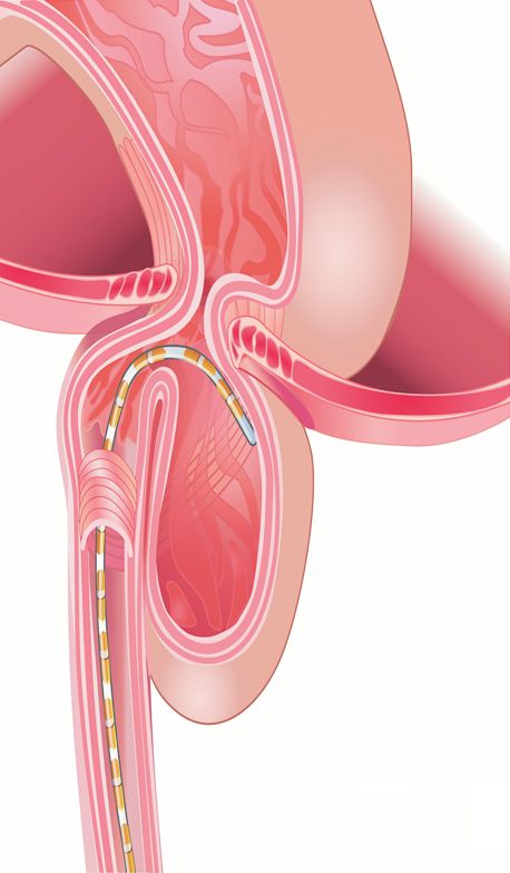
Esophageal Hiatus in Paraesophageal HH

distal contractile integral (DCI), patterns of stomach pressure, and          sensors below the LES showing thoracic (negative with inspiration)
diaphragmatic squeeze pressure with deep breaths. The LES pres-               or abdominal waveform (positive with inspiration), the 3 HRM
sure was referenced to stomach pressure when no hernia was pres-              EGJ patterns were identified: pattern 1, diaphragmatic pinch pres-
ent and to hernia pressure when the latter was present.                       ent at the same location as the LES (expected in normal subjects
     Based on the presence or absence of diaphragmatic pinch, loca-           and patients with type 2 HH); pattern 2, diaphragmatic pinch
tion of diaphragmatic pinch in relationship to LES and pressure               present distal to the LES, with at least 2 sensors below the LES

                    Esophagus
                                                                                           Esophagus
                    EGJ
                                                                                            Stomach
                                       Esophagus
                                                       Stomach

                                                                             EGJ
  Stomach              Diaphragm
                                                                                       Diaphragm

                                               EGJ

                                                                                                          Figure 1. Radiographic patterns in 3
                                                       Stomach
                                                                                                          types of hiatal hernia (HH) patients.
          Sliding/type 1                  Paraesophageal/type 2                 Mixed/type 3              EGJ, esophagogastric junction.

                           Pattern 1                                         Pattern 2                                  Pattern 3
          Normal                                Type 2 HH                                  Type 1 HH                                  Type 3 HH

              Diaphragmatic pinch present                         Diaphragmatic pinch present                Diaphragmatic pinch absent
              and stomach below the pinch                          and hernia above the pinch              and hernia above the diaphragm
                          7                                                    23                                        26

     Upper GI series

                     Type 1      Type 2                             Type 1    Type 2     Type 3               Type 1     Type 2     Type 3
                       3           4                                  12        3          8                    4          9          13

Figure 2. Esophagogastric junction (EGJ) patterns of hiatal hernia (HH) on manometry and relationship with the radiological diagnosis in 3 types
of hernia (see text for explanation). EGJ pattern 1 expected in patients with normal subjects and patients with paraesophageal hernia type 2. High-
resolution manometry (HRM) EGJ pattern 2 expected in patient with sliding HH (type 1) and pattern 3 expected in patient with type 3 HH.
GI, gastrointestinal.

                                                       Vol. 26, No. 1 January, 2020 (51-60)                                                      53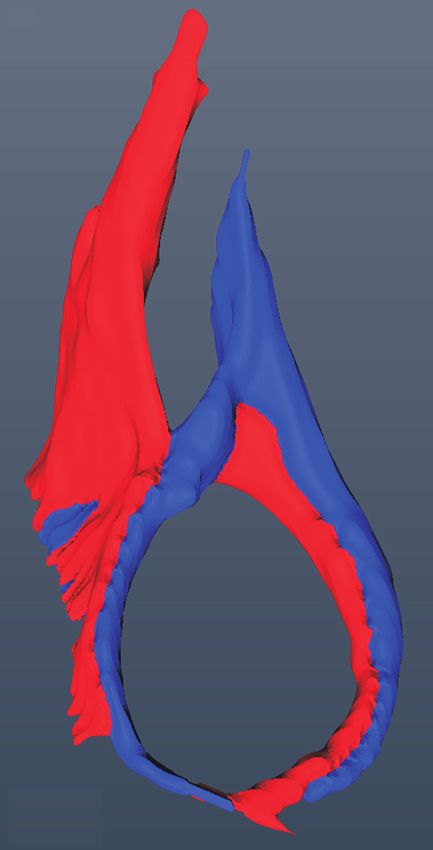
Dushyant Kumar, et al

showing intrathoracic and another 2 sensors below the pinch show-            and the 3D anatomy of the region was constructed. Once built, sev-
ing intra-abdominal waveform (expected in patients with type 1               eral dimensions of the hiatus were measured (long and short diam-
and type 3 HH);2,9 and pattern 3, diaphragmatic pinch absent and             eter, surface area, and angle of hiatus with vertebral column) (Fig. 3).
all sensors below the LES showing intrathoracic pressure pattern                  We prospectively studied one patient diagnosed with type 3
(catheter did not enter the abdomen) (Fig. 2).                               HH on the upper gastrointestinal (UGI) series and an HRM
     CT image analysis: the CT scan of chest and upper abdomen               pattern 2 seen on clinical HRM study. A manometry catheter was
performed prior to surgery (when available) was analyzed to deter-           placed trans-nasally and a CT scan was performed with the catheter
mine the anatomy of the diaphragmatic hiatus. The CT scan find-              in place.
ings of HH patients were compared to 20 subjects without HH on
the CT scan images. Coronal sections (30-40 slices), built from the          Statistical Methods
axial slices of 2.0 mm to 2.5 mm thickness were uploaded into the                Quantitative data are reported as mean (± standard devia-
computer software (Amira, Carlsbad, CA, USA). Following struc-               tion) or median (interquartile range 25-75) when appropriate. The
tures were identified and their margins marked in each of the CT             normality of distributions was checked using the Shapiro-Wilk test.
scan image: right and left crus of diaphragm, esophagus, stomach,            One-way analysis of variance (ANOVA) followed by Bonferroni’s
and vertebral bodies. Different colors were assigned to each structure       or Tukey’s post hoc test was performed for multiple comparisons, or

 A                                                                                         B
        Hiatal angle                                                                                  75                                 Angle
                                                                                                      70
                                                                                                      65
                                                                                                      60
                                                                                           Degrees

                                                                                                      55
                                                                                                      50
                                                                                                      45
                                                                                                      40

  [a]                   [b]           [c]           [d]             [e]                               35

        Normal           Type 1 HH      Type 2 HH     Type 3 HH            LCFH                       30
                                                                                                            Type 1   Type 2   Type 3     Normal

 C                                                                                       D
      70
                                                                              Small                                              Surface area
                                                                                             10 000
                                                                              Large
      60                                                                                             9000
                                                                                                     8000
      50
                                                                                                     7000
      40                                                                                             6000
 mm

                                                                                         mm

                                                                                                     5000
      30
                                                                                                     4000
      20                                                                                             3000
                                                                                                     2000
      10
                                                                                                     1000
                                                                                                       0
                   Type 1            Type 2          Type 3               Normal                            Type 1   Type 2   Type 3     Normal

Figure 3. Hiatal anatomy and hiatal dimension. (A) Anatomy of esophageal diaphragmatic hiatus in normal subjects and patients with various
types of hiatal hernia (HH). The hiatus is larger in dimension in 3 types of HH compared to normal subjects. (B-D) Hiatal dimensions (angle,
large diameter [Larged] and small diameter [Smalld], and surface area) in normal subjects, patients with type 1, type 2, and type 3 HH. (B) Angle
of the hiatus in relationship to spine, (C) long and short dimensions of hiatus, and (D) cross sectional area of the hiatus. Normal subjects have
significantly smaller surface area, large and small diameters compared to 3 types of hernia but there is no difference among 3 type of HH. Data
showed in median and interquartile range. LCFH, left crus forming right hiatal margin. +Outlier value in the group.

54                                                  Journal of Neurogastroenterology and Motility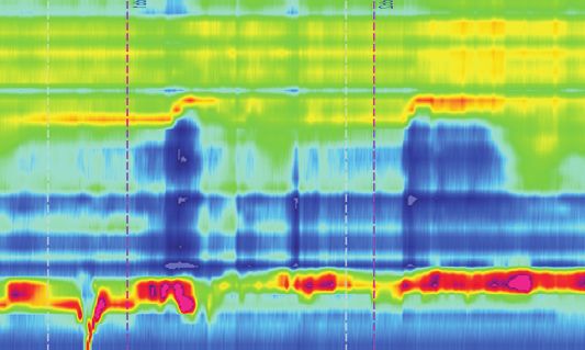
Esophageal Hiatus in Paraesophageal HH
the non-parametric one-way ANOVA with Kruskal-Wallis test with left, and posterior edges. The long and short diameters of the hiatus
Dunn’s multiple comparison tests was used. P < 0.05 was consid- in subjects with no HH were 18.0 ± 4.0 mm and 10.0 ± 3.0 mm
ered significant. respectively, with a cross sectional area of 810 mm2 (interquartile
range, 671).
The CT scan was available in 20 patients with HH, 6 patients
Results with type 1, 7 patients with type 2, and 7 patients with type 3. The
HH size by CT scan was 4.4 ± 2.0 cm, 9.9 ± 4.0 cm, and 8.7
Demographics and Symptoms ± 1.9 cm, for types 1, 2, and 3, respectively (Table 2). The type
Patient in all 3 HH types were predominantly females. Major of HH identified was the same between CT scan and UGI series
symptoms of type 1 HH patients were heartburn, epigastric dis- in 6/6 patients with type 1, 5/7 for type 2 and 7/7 for type 3. Two
comfort, chest pain, and sore throat. In addition to the above symp- patients diagnosed with type 2 HH on the UGI had type 3 HH
toms, patients with type 2 and type 3 HH also reported solid food on the CT scan. Similar to normal subjects, the hiatus was formed
dysphagia (sensation of food sticking in throat), bloating, nausea, by the right crus of the diaphragm in the majority (17/20 subjects)
vomiting, and regurgitation (Table 1). of HH patients (Fig. 3A[b-d]). In 3 subjects, one of each, type 1,
type 2, and type 3, both the left and right crus contributed to the
Upper Gastrointestinal Series formation of hiatus (Fig. 3A[e]). The shape of hiatus was elliptical
A total of 56 patients with the diagnosis of HH and 20 normal in all types of HH, with hiatal dimensions significantly larger (3
subjects without HH were part of this study. Based on the UGI to 4 times) in patients as compared to subjects without HH. The
evaluation, 18 patients were classified as sliding HH (type 1), 19 as hiatal margin appeared thinner, especially at the anterior end in all
paraesophageal HH (type 2), and another 19 as mixed HH (type 3). types of hernias. The angle of the hiatus with the spine, long and
The size of hernia as seen in the UGI series in 3 subtypes is shown short diameter, and cross-sectional area in 3 types of hernia and
in Table 2. normal subjects are shown in Figures 3B-D. The dimensions of the
hiatus was significantly smaller in type 1 HH compared to type 2
Esophageal Hiatus in Patients With and Without and type 3 HH (P < 0.05).
Hiatal Hernia
The 3D surface anatomy of the region of interest in 20 subjects
Table 2. Hernia Size
without HH revealed that the hiatus was formed by the right crus
Moda Type 1 Type 2 Type 3
of the diaphragm in all subjects. The right crus, after originating P -valuea P -valueb P -valuec
lities HH HH HH
from the right side of the lumbar vertebra (L1-L3), divided into
CT scan 4±2 10 ± 4 9±2 0.009 0.723 0.041
2 bundles and encircled the esophagus. The left crus reinforced
UGI 4±2 5±1 5±1 0.088 0.232 0.999
the left bundle of the right crus to form the left margin of the a
P -values are different between type 1 and 2 HH.
esophageal hiatus (Fig. 3A[a]). The oval-shaped hiatus was placed b
P -values are different between type 1 and 3 HH.
obliquely across the spine with the anterior end located at a more c
P -values are different between type 2 and 3 HH.
HH, hiatal hernia; UGI, upper gastrointestinal.
cranial location as compared to the posterior end. The hiatus wall Data are presented as mean ± SD.
is thinner at the cranial and anterior end as compared to the right,
Table 1. Demography and Symptoms
BMI Epigastric Chest Sore Dyspha- Regur
Hiatus hernia type Age (yr) M/F Heartburn Bloating Nausea
(kg/m2) discomfort pain throat gia gitation
Type 1 HH (n = 18) 56 ± 12 33/67 28 ± 5 27 4 5 2 11 0 2 5
Type 2 HH (n = 19) 66 ± 11 26/74 28 ± 4 16 5 4 0 11 2 5 4
Type 3 HH (n = 19) 68 ± 9 0/100 29 ± 5 27 9 7 2 8 2 9 9
M, male; F, female; BMI, body mass index; HH, hiatal hernia.
Data are presented as mean ± SD or %.
Vol. 26, No. 1 January, 2020 (51-60) 55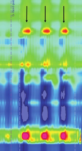
Dushyant Kumar, et al

                                                                                                     pared to type 2 and type 3 HH, with residual LES pressure higher
High-resolution Manometry                                                                            and percent relaxation lower in type 2 as compared to type 1 (Fig.
                                                                                                     4A). There was no difference in the above parameters between
Esophageal length                                                                                    types 2 and 3 patients. The intrabolus pressure was also higher in
    The esophageal lengths (distance between the lower edge of                                       patients with types 2 and 3 as compared to type 1 HH (Fig. 4A).
UES and upper edge of LES) of type 1, type 2, and type 3 HH
were 19 ± 3, 18 ± 3, and 17 ± 3 cm, respectively. These values                                       Diaphragmatic pinch pressure
were not statistically significant from each other.                                                       The mean hiatal squeeze pressure was 91 ± 31 mmHg (n =
                                                                                                     16) in type 1 HH, with an inverse (though not statistically signifi-
Lower esophageal sphincter pressure and swallow-                                                     cant) relationship between the size of hernia and diaphragmatic
induced relaxation                                                                                   pinch pressure (r = –0.5, P = 0.051). The mean hiatal squeeze
     The baseline LES pressure was not different in type 1, type 2,                                  pressure was 129 ± 85 mmHg in type 2 HH (n = 7), and 109 ±
and type 3 HH (11 ± 9, 19 ± 16, and 16 ± 14 mmHg, respec-                                            33 mmHg in type 3 HH (n = 7), with no difference as compared
tively). The swallow induced LES relaxation (median integrated                                       to type 1 HH (Fig. 4B).
relaxation pressure) was significantly different between type 1 com-

 A                 80        Bolus
                                                                    B                          Pinch
                                                                                                                             C                             DCI
                             LES                                                                                                                 5000
                   70        LES                                                      250

                                                                                                                             DCI (mmHg/cm/sec)
                   60                                                                                                                            4000
 Pressure (mmHg)

                                                                    Pressure (mmHg)

                   50                                                                 200
                   40                                                                                                                            3000
                                                                                      150
                   30
                                                                                                                                                 2000
                   20
                                                                                      100
                   10                                                                                                                            1000
                    0                     *               *                            50
                   10
                            Type 1   Type 2          Type 3                                 Type 1      Type 2    Type 3                                Type 1   Type 2   Type 3

Figure 4. Manometry data (median and interquartile range) in 3 types of hiatal hernia (HH) patients: (A) lower esophageal sphincter basal pres-
sure (LESB), residual pressure (LESR) with swallow and bolus pressure, (B) diaphragmatic pinch pressure, and (C) distal contractile integral (DCI).
+
  Sign corresponds to outliers, *P < 0.05 compared to type 1 HH.

                        UGI type 2       Pattern 1                              UGI type 2                  Pattern 2                             UGI type 3          Pattern 3

Figure 5. Manometry patterns in 3 different patients with type 2 paraesophageal hiatal hernia (HH) identified on the upper gastrointestinal (UGI)
series. EGJ, esophagogastric junction.

56                                                            Journal of Neurogastroenterology and Motility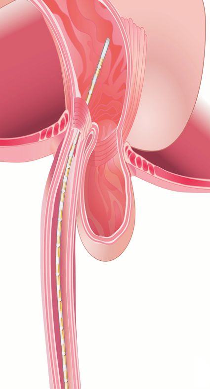
Esophageal Hiatus in Paraesophageal HH

                                                                          3 HH (7/19) patients. The prevalence of 3 EGJ manometry pat-
Peristalsis and contraction integral                                      terns and types of HH observed on UGI series is shown in the
    The esophageal peristalsis was normal and there was no differ-        flow diagram (Fig. 2). The majority of patients with type 1 HH (12)
ence in the peak contraction amplitude and DCI in the 3 types of          revealed HRM EGJ pattern 2. On the other hand, 50% of patients
HH (Fig. 4C).                                                             with types 2 and 3 HH revealed HRM EGJ pattern 3, ie, the
                                                                          catheter did not enter the abdomen; it was coiled in the herniated
Relationship Between Types of Hiatal Hernia on                            portion of the stomach.
Manometry, Upper Gastrointestinal Series, and                                 In 7 patients with type 2 HH on UGI series with an identifi-
Computed Tomography Scan Images                                           able diaphragmatic pinch on the HRM, 4 patients revealed HRM
     The diaphragmatic pinch was identified on HRM more often             EGJ pattern 1 (expected for 2 HH) but 3 patients revealed HRM
in type 1 HH (16/18) as compared to type 2 HH (7/19) and type             EGJ pattern 2, which would be expected in either type 1 HH or

mmHg
150.0

      140
      130
      120
      110
      100
      90
      80
      70
      60
      50
      45
      40
      35
      30
      25
      20
      15                                                                                              Figure 6. Changing manometry pat-
                                                                                                      terns of hiatal hernia (HH) in a patient
      10
      5
      0
        5
                                                                                                      with type 3 HH on the upper gastro-
  10.0                                                                                                intestinal series. EGJ, esophagogastric
 Set range        Diaphragmatic pinch        Diaphragmatic pinch              Barium swallow          junction.

  A                                              B

                                                                                                      Figure 7. CT scan finding with ma-
                                                                                                      nometry catheter in place. (A) Upper
                                                                                                      gastrointestinal (UGI) series show a pa-
                                                                                                      tient with type 3 hiatal hernia (HH). (B)
                                                                                                      High-resolution manometry (HRM)
  C                                              D                                                    esophagogastric junction (EGJ). Pat-
                                                                                                      tern 2 is consistent with the presence of
                                                                                                      stomach above the diaphragm and the
                                                                                                      tip of catheter in the abdomen. (C) On
                                                                                                      a separate day, the manometry catheter
                                                                                                      could not be advanced into the abdo-
                                                                                                      men as shown in the coronal image. (D)
                                                                                                      HRM pattern is consistent with HRM
                                                                                                      EGJ pattern 3.

                                                     Vol. 26, No. 1 January, 2020 (51-60)                                                    57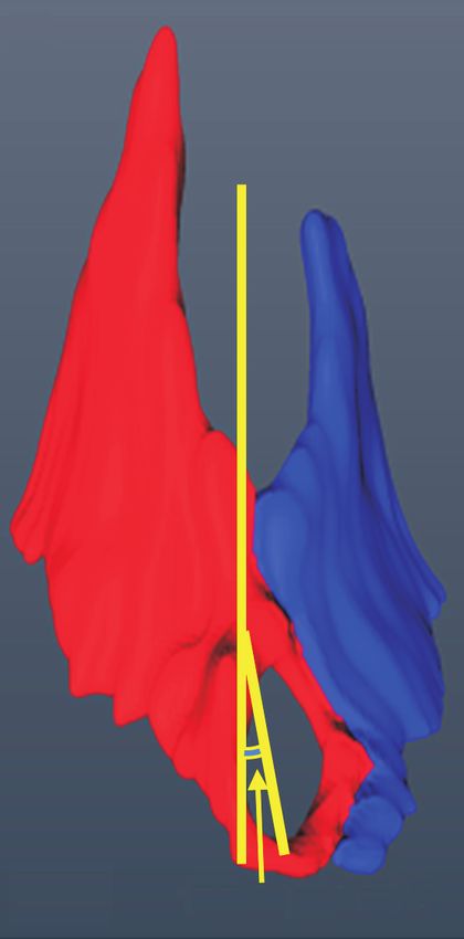
Dushyant Kumar, et al

type 3 HH on the UGI series (Fig. 5). In one patient with a di-               types 2 and type 3, as compared to sliding or type 1 HH. When the
agnosis of type 3 HH on UGI series, the HRM pattern changed                   HRM catheter tip did enter into the abdomen, the diaphragmatic
from HRM EGJ pattern 2 to HRM EGJ pattern 1, only a few                       squeeze pressure with deep inspiration was not different among
minutes later (Fig. 6), suggesting that similar to type 1 HH, the             3 types of HH. Difficulty with the catheter crossing the hiatus to
LES in paraesophageal hernia can also slide in and out of the hia-            enter into the abdomen during routine manometry is most likely re-
tus.                                                                          lated to a tightly stuffed/packed hiatus with esophagus and stomach,
     In one patient in whom we performed CT scan with the HRM                 at least in type 2 hernia. It is likely that in patients with type 3 HH,
catheter in place, the catheter was coiled above the diaphragm, in            a greater mass of stomach herniates through the diaphragmatic hia-
the hernia sac. The HRM revealed HH pattern 3, confirming our                 tus.
assumption of the catheter coiling in the hernia sac in patients with              One of the major reasons for the development of HH is a weak
manometry pattern 3 (Fig. 7).                                                 or defective phrenoesophageal ligament, which is expected to be
     With regards to the size of HH, concordance between HRM                  present all around the circumference of the esophagus in patients
and UGI series was present in only 5/30 patients; in the remainder            with type 1 HH. On the other hand, in patients with type 2 HH,
it was larger on HRM than UGI series.10 With regards to the HH                the fundus of the stomach herniates into the chest anterior to the
size on CT scan and HRM, again there was poor concordance in                  esophagus with a normally located LES. The latter implies that the
patients with type 1 HH. The number of patients with types 2 and              defect in the phrenoesophageal ligament is not present around the
type 3 HH with the available CT scans, in whom the diaphragmat-               entire circumference. In 4 of 9 patients with type 2 HH on UGI
ic pinch was present, were small to make an adequate comparison.              series, we found an unexpected manometry pattern of type 2 HH.
                                                                              The latter implies that the LES was located below the hiatus on
                                                                              the day of upper gastrointestinal exam but was in thorax on the day
Discussion                                                                    of manometry. The above observations imply that similar to type
     The anatomy of esophageal hiatus was of considerable inter-              1 HH, which is well known to slide in and out of the chest, type
est in 1940s and 50s to the surgeons because the understanding at             2 HH also has sliding features. To the best of our knowledge, we
that time was that it played a major role in the anti-reflux barrier          have not seen any reports that type 2 HH may also slide in and out
function. Surgical dissection studies in cadavers revealed that in the        of the chest. We observed lack of concordance in the hernia size be-
majority of subjects, the right crus splits into two bundles to sur-          tween manometry, UGI series, and CT scan in all 3 types of HH.
round the esophagus. The CT scan images clearly show splitting                Since these 3 exams were done on 3 separate days, it is likely that
of the right crus into right and left bundles before the left bundle of       similar to HH type 1, types 2 and 3 can also slide in and out of the
right crus joins left crus to form the left margin of the hiatus. Our         chest, at least partially if not completely. We observed changing her-
findings are in agreement with a more recent study in which 80%               nia size in a patient with type 3 HH during 10 minutes of HRM
of the subjects were found to have what we described.6 We are the             recording, which makes us believe that the 3 types of HH hernia
first to describe the hiatal anatomy using CT scan segmentation.              are not totally separate or distinct entities.
We had only 6-7 subjects in each type of HH in whom CT scan                        With each swallow, the LES is pulled into the chest (approxi-
was available for segmentation and did not find significant differ-           mately 2 cm) due to axial shortening of the esophagus during peri-
ences in terms of the anatomical arrangement of esophageal hiatus.            stalsis.13 The phrenoesophageal ligament is stretched in an oral di-
In all cases the right crus was dividing into 2 bundles and forming           rection with each swallow. With transient LES relaxation, the LES
the esophageal hiatus. An important aspect of the crural diaphragm            is pulled 4 cm or more in the cranial direction.14 Given the large
anatomy that is not revealed by the CT imaging is the crossing of             degrees of stress and strain on the phrenoesophageal ligament, it is
the fibers of 2 bundles of the right crus in a scissor-like fashion that      not surprising that the prevalence of HH is high and increases with
we found in a recent study11 and was reported earlier by March-               age. Some investigators have described alteration in the elastin and
and.12 The hiatal dimensions were larger in patients with HH,                 collagen content of the phrenoesophageal ligament,15 whether the
which is expected, but there was no significant difference in various         latter is a primary abnormality remains unknown. Repeated acid re-
hiatal measurements among the 3 HH groups.                                    flux into the esophagus causes esophagitis that has also been shown
     The manometry catheter did not enter into the abdomen in a               to induce sustained contraction of the longitudinal muscle of the
significantly greater number of patients with paraesophageal HH,              esophagus, which may also be important in the HH formation.16-19

58                                                   Journal of Neurogastroenterology and Motility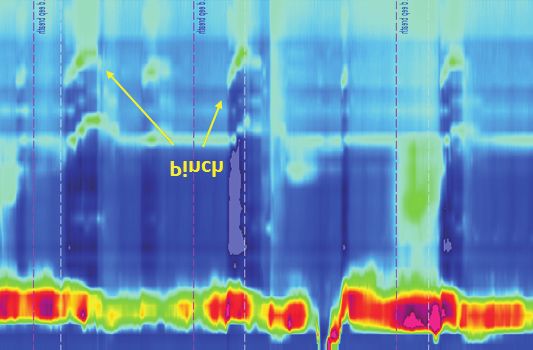
Esophageal Hiatus in Paraesophageal HH

     Esophageal peristalsis function was normal in all types of              Financial support: This work was supported by a NIH Grant
HHs with no difference in the contraction amplitude and DCI.                 (DK060733).
The above is surprising because sliding HH is commonly associ-
ated with reflux disease and hypotensive peristalsis. Since ours was         Conflicts of interest: None.
a retrospective study, it is possible that surgeons excluded patients        Author contributions: Ravinder K Mittal: conceived the proj-
with abnormal peristalsis for surgical correction of HH. There was           ect, analyzed data, and wrote manuscript; Dushyant Kumar: data
a difference in the LES relaxation between types of HH; normal               analysis, figure preparation, and manuscript editing; Ali Zifan: data
in sliding or type 1 HH but impaired in patients with types 2 and            analysis and figure preparation; Gary Ghahremani: read radiologi-
3 HH. One may argue that the latter could be due to compression              cal studies; David C Kunkel: manuscript writing; and Santiago
of the LES and distal esophagus by the stomach, since the latter             Horgan: data acquisition.
is located along the side of the esophagus in paraesophageal HH.
However, we did not observe increase in the esophageal pressure
(in between swallows) in these patients which would argue against            References
stomach compression of the esophagus as the reason. Impaired                  1. Nicholson F. Diphragmatic hernia. Ann Surg 1952;136:174-182.
LES relaxation resulted in high residual LES pressure during                  2. Jones FA. Diagnosis of hiatus hernia. Proc R Soc Med 1952;45:277-
swallows and higher intrabolus pressure. It is interesting that dys-             279.
phagia is more prevalent in types 2 and 3 HH patients compared to             3. Kahrilas PJ, Kim HC, Pandolfino JE. Approaches to the diagnosis and
                                                                                 grading of hiatal hernia. Best Pract Res Clin Gastroenterol 2008;22:601-
type 1 HH and it is possible that impaired LES relaxation plays a
                                                                                 616.
role in the dysphagia symptom. In a recent study, other authors also          4. Collis JL, Kelly TD, Willey AM. Anatomy of the crura of the diaphragm
found increased intrabolus pressure with swallows in patients with               and the surgery of hiatus hernia. Thorax 1954;9:175-189.
paraesophageal hernia.20                                                      5. Listerud MB, Harkins HN. Variations in the muscular anatomy of the
     In conclusion, our study is a comprehensive, cross sectional                esophageal hiatus: based on dissections of two hundred and four fresh
study of patients with paraesophageal and sliding HH. Our intent                 cadavers. West J Surg Obstet Gynecol 1959;67:110-112; discussion 112-
                                                                                 113.
was to determine if there are unique features in the anatomy of
                                                                              6. Costa MM, Pires-Neto MA. Anatomical investigation of the esophageal
the diaphragmatic hiatus, crural diaphragm function, LES, and                    and aortic hiatuses: physiologic, clinical and surgical considerations. Anat
esophageal motor function in these patients. The esophagus hiatus                Sci Int 2004;79:21-31.
is enlarged in all types of hernia as compared to normal subjects,            7. Callen PW, Filly RA, Korobkin M. Computed tomographic evaluation
however, there is no difference among the 3 hernia subtypes. To our              of the diaphragmatic crura. Radiology 1978;126:413-416.
surprise, we found that the LES in paraesophageal HH type 2 may               8. Mittal RK, Zifan A, Kumar D, Ledgerwood-Lee M, Ruppert E,
                                                                                 Ghahremani G. Functional morphology of the lower esophageal sphincter
also slide in and out of the hiatus. The LES relaxation of patients
                                                                                 and crural diaphragm determined by three-dimensional high-resolution
with paraesophageal HH is impaired, resulting in an increased                    esophago-gastric junction pressure profile and CT imaging. Am J Physi-
intrabolus pressure. The latter is seen in association with impaired             ol Gastrointest Liver Physiol 2017;313:G212-G219.
LES relaxation, an entity termed as EGJ outflow obstruction in                9. Kahrilas PJ, Peters JH. Evaluation of the esophagogastric junction using
the Chicago classification. Dysphagia is an important symptom in                 high resolution manometry and esophageal pressure topography. Neuro-
patients with outflow obstruction, which may be the reason for dys-              gastroenterol Motil 2012;24(suppl 1):11-19.
                                                                             10. Weitzendorfer M, Köhler G, Antoniou SA, et al. Preoperative diagnosis
phagia in these patients. There are several limitations of our study to
                                                                                 of hiatal hernia: barium swallow X-ray, high-resolution manometry, or
keep in mind: (1) our study was a retrospective study and therefore              endoscopy? Eur Surg 2017;49:210-217.
does not have a perfect study design to answer all the questions we          11. Zifan A, Kumar D, Cheng LK, Mittal RK. Three-dimensional myoar-
raised, (2) number of subjects that have CT scans available for the              chitecture of the lower esophageal phincter and esophageal hiatus using
analysis were relatively small, and (3) HH size was not measured at              optical sectioning microscopy. Sci Rep 2017;7:13188.
the time of surgery, which many considered to be more accurate. In           12. Marchand P. The anatomy of esophageal hiatus of the diaphragm and
                                                                                 the pathogenesis of hiatus herniation. J Thorac Surg 1959;37:81-92.
spite of the above, we feel that our study does provide some novel
                                                                             13. Edmundowicz SA, Clouse RE. Shortening of the esophagus in response
information worthy of further prospective inquiries.                             to swallowing. Am J Physiol 1991;260(3 Pt 1):G512-G516.
                                                                             14. Pandolfino JE, Zhang QG, Ghosh SK, Han A, Boniquit C, Kahrilas PJ.
                                                                                 Transient lower esophageal sphincter relaxations and reflux: mechanistic

                                                        Vol. 26, No. 1 January, 2020 (51-60)                                                             59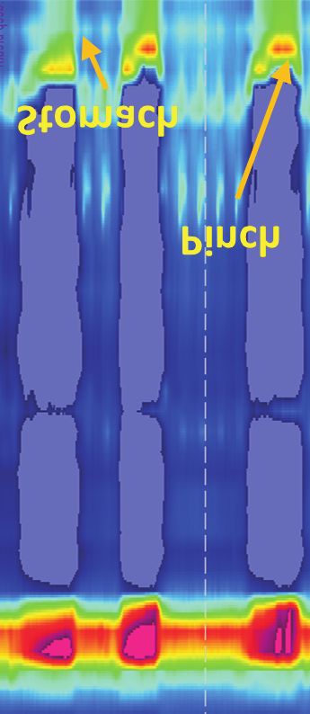
Dushyant Kumar, et al

    analysis using concurrent fluoroscopy and high-resolution manometry.             via capsaicin-sensitive neurokinin neurons. Gut 2007;56:1347-1352.
    Gastroenterology 2006;131:1725-1733.                                         18. Dunne DP, Paterson WG. Acid-induced esophageal shortening in hu-
15. Weber C, Davis CS, Shankaran V, Fisichella PM. Hiatal hernias: a re-             mans: a cause of hiatus hernia? Can J Gastroenterol 2000;14:847-850.
    view of the pathophysiologic theories and implication for research. Surg     19. Kääriäinen M. Diagnosis of reflux esophagitis. With special reference to
    Endosc 2011;25:3149-3153.                                                        double contrast radiography. Ann Clin Res 1985;17(suppl 4):1-43.
16. Mittal RK, Balaban DH. The esophagogastric junction. N Engl J Med            20. Rengarajan A, Arguero MJ, Kadirkamanathan SS, et al. Impact of para-
    1997;336:924-932.                                                                esophageal hernia on esophageal and esophagogastric junction (EGJ)
17. Paterson WG, Miller DV, Dilworth N, Assini JB, Lourenssen S,                     motor physiology. Gastroenterology 2017;152:S705-S706.
    Blennerhassett MG. Intraluminal acid induces oesophageal shortening

60                                                     Journal of Neurogastroenterology and Motility
You can also read