Development of the teeth, cervical vertebrae, hand and wrist combined for the estimation of the biological age
←
→
Page content transcription
If your browser does not render page correctly, please read the page content below
Research, Society and Development, v. 10, n. 3, e8510312948, 2021
            (CC BY 4.0) | ISSN 2525-3409 | DOI: http://dx.doi.org/10.33448/rsd-v10i3.12948

Development of the teeth, cervical vertebrae, hand and wrist combined for the
estimation of the biological age
Desenvolvimento dos dentes, vértebras cervicais, mão e punho combinados para estimativa da
idade biológica
Desarrollo de dientes, vértebras cervicales, mano y muñeca combinados para estimar la edad
biológica

                                                  Received: 02/14/2021 | Reviewed: 02/21/2021 | Accept: 02/27/2021 | Published: 07/03/2021

                                                                                                    Marisa de Matos Ferraz Pêgo
                                                                                           ORCID: https://orcid.org/0000-0001-5984-146X
                                                                                                    São Leopoldo Mandic College, Brazil
                                                                                                       E-mail: marisamfpego@gmail.com
                                                                                                   Paola Fernanda Leal Corazza
                                                                                           ORCID: https://orcid.org/0000-0002-8639-8392
                                                                                                       APCD College of Dentistry, Brazil
                                                                                                    E-mail: paola_corazza@hotmail.com
                                                                                                        Fernando Martins Baeder
                                                                                           ORCID: https://orcid.org/0000-0001-7101-5689
                                                                                                       Cruzeiro do Sul University, Brazil
                                                                                                     E-mail: fernandobaeder@uol.com.br
                                                                                                               Daniel Furtado Silva
                                                                                           ORCID: https://orcid.org/0000-0003-3319-2996
                                                                                                     Federal University of Paraíba, Brazil
                                                                                                           E-mail: furtado.ds@gmail.com
                                                                                            Ana Carolina Lyra de Albuquerque
                                                                                           ORCID: https://orcid.org/0000-0002-6532-5020
                                                                                                     Federal University of Paraíba, Brazil
                                                                                                             E-mail: lina.lyra@gmail.com
                                                                                                      José Luiz Cintra Junqueira
                                                                                           ORCID: https://orcid.org/0000-0001-6788-4021
                                                                                                   São Leopoldo Mandic College, Brazil
                                                                                                       E-mail: joseluiz@slmandic.edu.br
                                                                                                        Francine Kühl Panzarella
                                                                                           ORCID: https://orcid.org/0000-0002-5650-7711
                                                                                                   São Leopoldo Mandic College, Brazil
                                                                                                 E-mail: francine.panzarella@gmail.com

      Abstract
      This study aims to perform age estimation using three different parameters from dental and skeletal development. The
      sample consisted of 98 dental records of patients aged from 10 to 16 years old, containing the chronological age and a
      set of radiographs (panoramic, lateral cephalometric and carpal radiographs) taken in the same day. The biological age
      was assessed through the dental development from panoramic radiographs according to the Nicodemo's method. The
      stages of dental development were registered and imported in CRONOL software (UNESP, São Paulo, Brazil), which
      provided the estimated dental age. The lateral cephalometric radiographs were analyzed to assess the development of
      the vertebrae C2, C3 and C4. And carpal radiographs were evaluated according to Fishman's method. Shapiro-Wilk
      test was used to verify the normality of the chronological and estimated age. T-test for unpaired samples was used to
      compare the normal data. Chi-square test was used to analyze the age in function of sex. Moderate and strong
      correlations were found between the chronological and biological (estimated) ages for all the methods. Statistically
      significant differences between the development of males and females were not observed (p>0.05). A linear regression
      formula was designed to allow age estimates statistically more accurate (p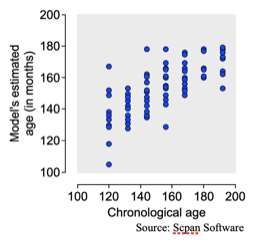
Research, Society and Development, v. 10, n. 3, e8510312948, 2021
               (CC BY 4.0) | ISSN 2525-3409 | DOI: http://dx.doi.org/10.33448/rsd-v10i3.12948

         Resumo
         Este estudo tem como objetivo realizar a estimativa da idade utilizando três diferentes parâmetros de desenvolvimento
         dentário e esquelético. A amostra foi composta por 98 documentações de pacientes com idade entre 10 e 16 anos,
         contendo a idade cronológica e radiografias (panorâmica, cefalométrica lateral e radiografia carpal) realizadas no
         mesmo dia. A idade biológica foi avaliada através do desenvolvimento dentário a partir de radiografias panorâmicas
         segundo o método de Nicodemo. As etapas de desenvolvimento dentário foram registradas e importadas no software
         CRONOL (UNESP, São Paulo, Brasil), fornecendo a idade dentária estimada. As radiografias cefalométricas laterais
         foram analisadas para avaliar o desenvolvimento das vértebras C2, C3 e C4. E as radiografias do carpo foram
         avaliadas por meio do método de Fishman. O teste de Shapiro-Wilk foi utilizado para verificar a normalidade da idade
         cronológica e estimada. O teste t para amostras desemparelhadas foi usado para comparar os dados normais. O teste
         qui-quadrado foi utilizado para analisar a idade em função do sexo. Correlações moderadas e fortes foram encontradas
         entre as idades cronológica e biológica (estimada) para todos os métodos. Não foram observadas diferenças
         estatisticamente significativas no desenvolvimento dos gêneros masculino e feminino (p> 0,05). Uma fórmula de
         regressão linear foi projetada para permitir estimativas de idade estatisticamente mais precisas (p  0.05). Se diseñó una fórmula de regresión lineal para permitir estimaciones de
         edad estadísticamente más precisas (p
Research, Society and Development, v. 10, n. 3, e8510312948, 2021
(CC BY 4.0) | ISSN 2525-3409 | DOI: http://dx.doi.org/10.33448/rsd-v10i3.12948
of malocclusions and to the management of functional and aesthetics disorders. Usually, dental and skeletal radiographs are
taken from orthodontic patients for treatment planning and follow-up. From a forensic scope, these radiographs are source of
multiple age information that may be combined to improve the accuracy in age estimation.
The present study aims to perform age estimation using three different parameters that include dental and skeletal
development registered in panoramic, lateral cephalometric and carpal radiographs. Next, the biological age estimated through
teeth and bones will be compared to the chronological age for the investigation of performance (accuracy). Finally, a formula
combining the three parameters will be designed from linear regressions models and will be tested in the sample. The age
estimation outcomes obtained with the formula will be compared to the chronological for a second assessment of performance.
All the analyses will be performed with the total sample and stratified by sex.
2. Methodology
This is a cross-sectional observational research with a correlational approach and statistical comparative procedure
(Pereira et al., 2018). The present research was conducted after the approval of the local Committee of Ethics in Research
under the protocol #1.303.139.
Dental records (n=121) were selected from the Department of Radiology of a private university in Brazil. The
inclusion criteria established consisted of dental records of Caucasian patients containing their date of birth and sex; they
should belong to patients aged between 10 and 16 years old; and they should contain a set of panoramic, lateral cephalometric
and carpal radiographs of the patient. The exclusion criteria consisted of patients that underwent previous orthodontic
treatment or the extraction of permanent teeth; trauma in the face, cervical vertebrae or hands; and medical history of systemic
diseases that influence on dental and bone development (Vieira et al., 2009; Araújo et al., 2010). After inclusion and exclusion
criteria, 98 dental records (48 females and 50 males) remained eligible. All the radiographs stored in the dental records were
analyzed by a single trained examiner blind for the age and sex of the patients.
The biological age was initially assessed in panoramic radiographs extracted from the dental records. In these
radiographs the developmental stages of the permanent teeth were classified according to the method of Nicodemo, Miranda &
Rangel (1974). The teeth chosen for classification according to this method were the canines, first and second premolars and
first and second molars (Moraes, Aragão & Heck 1998). The stages considered for each tooth were imported in CRONOL
software package (UNESP, São Paulo, Brazil), available in: http://ict.unesp.br/#!/departamentos-de-ensino/diagnostico-e-
cirurgia/disc-radiologia/. The software estimates the mean age of the patient taking into account the stages of all the teeth
analyzed previously. In order to make the outcomes also compatible with the clinical routine in Dentistry, the developmental
stage of the mandibular left second molar was also classified. The stage was implemented in Scpan software package (UNESP,
São Paulo, Brazil), available in: http://ict.unesp.br/#!/departamentos-de-ensino/diagnostico-e-cirurgia/disc-radiologia/. The
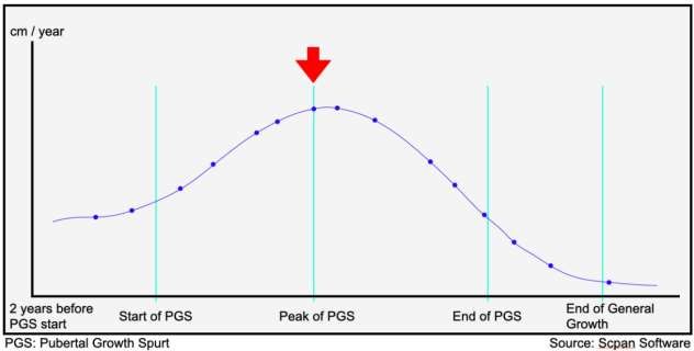
software reports the relation between the dental stage and the pubertal growth spurt (Moraes et al., 2008).
The cervical vertebrae C2, C3 and C4 were assessed in the lateral cephalometric radiographs and were classified
according to the method of Hassel & Farman (1995). This method is founded on the morphology of the cervical vertebrae,
which is classified in 6 developmental stages. The stages correspond to developmental initiation, acceleration, transition,
deceleration, maturation and finalization.
The development of the hand and wrist was assessed in carpal radiographs according to the method of Fishman
(1982), from which 4 stages were used to classify the development of the first (thumb), third and fifth fingers and the radius.
The developmental stages were observed in the epiphyses and diaphyses of the hand and wrist bones, as well in the
mineralization of the sesamoid.
3Research, Society and Development, v. 10, n. 3, e8510312948, 2021
(CC BY 4.0) | ISSN 2525-3409 | DOI: http://dx.doi.org/10.33448/rsd-v10i3.12948
2.1 Statistical analysis
Shapiro-wilk test was used to assess the normality of chronological and estimated dental ages, while the
homocedasticity was assessed with F test. Intraclass correlation coefficient was used to assess the agreement between the
chronological and dental ages. Mann-Whitney test was used to assess the agreement between the chronological age and the
estimated ages from cervical and carpal bones.
Chi-square test was used to investigate the influence of sex in the developmental stages of the teeth, cervical vertebrae
and hand and wrist bones.
Pearson’s and Spearman’s correlation coefficient were used to associate the chronological and estimated ages.
Forward linear regression analysis was used to investigate the level of dependence between the chronological age and the other
variables considered in the present study. Mann-Whitney test was used to investigate the relationship between the pubertal
growth spurt and the chronological age.
The statistical tests were performed with GraphPad Prism 6.0 (GraphPad Software Inc., La Jolla, CA, USA), BioEstat
5.0 (Instituto de Desenvolvimento Sustentável Mamirauá, Tefé, AM, Brazil) and SPSS 2.0 (IBM Corp., Armonk, NY, USA)
software packages considering a significance level of 5%.
3. Results
Weak concordance between the chronological and estimated age was observed both for the total sample (ICC = 0.263)
and for the sample stratified on sex (males: ICC = 0.206; females: ICC = 0.310). The absolute errors between the dental and
chronological age ranged between +30 (overestimation) and -53 (underestimation) months. In general, underestimations were
observed with the method of Nicodemo et al. (1974). Statistically significant differences were not observed between males and
females (p>0.05) (Table 1).
Table 1. Mean and standard deviation of the chronological and dental ages and their difference distributed based on sex.
Variable F+M (n=98) F (n = 48) M (n = 50)
CA 153.3 (±21.0) 149.8 (±20.9) 156.7 (±20.8)
DA 136.6 (±10.6) 135.5 (±11.2) 137.6 (±10.0)
Difference between CA and DA 16.7 (±16.3) 14.3 (±16.7) 19.1 (±15.7)
Error between CA and DA From +30 to +53 From +30 to -50 From +9 to -53
CA: chronological age; DA: dental age; F: females; M: males; Age expressed in months.
Source: Authors.
Statistically significant differences (p9 was the most prevalent (p=0.0076) in females compared to males (Table 2).
4Research, Society and Development, v. 10, n. 3, e8510312948, 2021
(CC BY 4.0) | ISSN 2525-3409 | DOI: http://dx.doi.org/10.33448/rsd-v10i3.12948
Table 2. Absolute (n) and relative (%) frequency of the sample distributed based on sex according to the different methods
used for age estimation.
Methods Stages F(n=48) M (n=50) p
ascending curve 10 (20.8%) 2 (4%)
peak 21 (43.8%) 9 (18%)
DA < 0.05
descending curve 17 (35.4%) 21 (42%)
end of PGS - 18 (36%)
1-3 18 (37.6%) 26 (52%)
CVA 0.2499
4-6 30 (62.6%) 24 (48%)
1-3 2 (4.2%) 7 (14%)
2-6 8(16.7) 17 (34%)
HWA < 0.05
7-9 14(29.2%) 14(28%)
>9 24 (50%) 12 (24%)
DA: dental age; CVA: cervical vertebrae age; HWA: hand and wrist age; F: females; M: males; PGS: pubertal growth spurt ; p: p-value out
of Chi-square test considering a significance level of 0.05.
Source: Authors.
In order to investigate the performance of the method for estimating the chronological age was assessed and compared
with the age estimates of each method (Table 3). Moderate (rS ou rP between 0.4 and 0.6) or strong (rS ou rP or 0.6 and 0)
correlations were found between all the involved variables (pResearch, Society and Development, v. 10, n. 3, e8510312948, 2021
(CC BY 4.0) | ISSN 2525-3409 | DOI: http://dx.doi.org/10.33448/rsd-v10i3.12948
Table 3. Correlation between chronological and estimated ages distributed based on sex.
Correlation outcomes
Sex Method
CA DA HWA
DA* 0.64
Females
+
Males HWA** 0.55 0.6
CVA** 0.53 0.62 0.75
DA 0.6
Females
HWA 0.7 0.63
CVA 0.6 0.51 0.7
DA 0.68
Males
HWA 0.6 0.71
CVA 0.53 0.75 0.74
CA: chronological age; DA: dental age; CVA: cervical vertebrae age; HWA: hand and wrist age; *Pearson’s
correlation; ** Spearman’s correlation.
Source: Authors.
The forward linear regression analysis resulted in a linear model (Table 4), which reliability (Table 5) and
performance were optimal (Table 6). The best model combined the following information: “Dental age” (p=0.0003), “Hand
and wrist classification” (p=0.0001) and sex (females=0, males=1, p=0.0019). The development of the cervical vertebrae did
not contribute significantly to the model (p=0.2840). The final formula is described as follows: Chronological age = 27.26 +
(0.72 × Nicodemo’s dental age) + (2.93 × Fishman’s hand and wrist stage) + (11.09 × Sex). Nearly 50% of the age is explained
by the variables and the accuracy rate reaches 71.4%. In other words, the formula has better performance for estimating the age
compared to a random chance of age estimation (pResearch, Society and Development, v. 10, n. 3, e8510312948, 2021
(CC BY 4.0) | ISSN 2525-3409 | DOI: http://dx.doi.org/10.33448/rsd-v10i3.12948
Table 4. Linear regression model obtained with the variables dental age, hand and wrist classification, cervical vertebrae
classification and sex for the estimation of age.
CI 95% of B
B SE Beta T-test p Min. Max.
Constant 27.26 22.93 - 1.19 0.24 -18.3 72.8
DA 0.7236 0.2081 0.36 3.68 0.0003 0.33 1.11
HWA 2.93 0.73 0.42 4.0 0.0001 1.48 4.39
Sex 11.09 3.48 0.27 3.18 0.0019 4.17 18.0
DA: Dental age; HWA: hand and wrist classification; SE: standard error; CI: confidence interval; Min.: minimum; Max.:
maximum.
Source: Authors.
Table 5. Reliability of the regression model.
R R2 Adjusted R2 SE
0.714 0.51 0.49 14.9
R: Determination coefficient; SE: standard error expressed in months.
Source: Authors.
7Research, Society and Development, v. 10, n. 3, e8510312948, 2021
(CC BY 4.0) | ISSN 2525-3409 | DOI: http://dx.doi.org/10.33448/rsd-v10i3.12948
Table 6. ANOVA outcome from the obtained regression model.
SS DF MS F P
Regression model 21786.7 3 7262.2 32.5Research, Society and Development, v. 10, n. 3, e8510312948, 2021
(CC BY 4.0) | ISSN 2525-3409 | DOI: http://dx.doi.org/10.33448/rsd-v10i3.12948
2017; Camacho-Basallo et al., 2017). Nevertheless, Perinetti et al. (2012) reported that the dental calcification was only usefull
for diagnosing pre-pubertal growth phase.
Statistically significant differences between the chronological age and the age estimated through the dental
development (Nicodemo et al., 1974) were not detected for both the males and females sampled in the present study. Similarly
to the previous scientific literature (Graziosi et al., 1999; Silva et al., 2005), the estimated age was lower than the chronological
age (underestimations) in the total sample and in the sample stratified by sex. Opposite outcomes (overestimations) were found
by Moraes et al. (2007) and Topolski et al. (2014). More specifically, a difference of 14.3 months between the estimated and
chronological ages was observed in females, while in males the difference reached 19.1 months. According to Moraes et al.
(2007), these outcomes are not common, but do not necessarily indicate an abnormal development of the individuals sampled
in the present study, since skeletal growth has periods of acceleration and maturation, and may not be directly associated with
chronological age.
In Table 1, the developmental stages of the teeth, cervical vertebrae and hand and wrist are distributed in function of
sex. Specific differences were observed in the distribution of the stages according to the age estimation method (based on
dental or skeletal parameters). Considering specifically the dental development, the most prevalent phase of the pubertal
growth spurt was the ascending curve in females, while in males was the descending curve. In these phases, the mandibular
second molar presents complete root development (with open or closed apices). This phenomenon corroborates the study of
Moraes et al. (1998) and indicates a developmental delay in females compared to males.
A few numbers of individuals represented each of the stages of cervical vertebrae development. For that reason, the
individuals were grouped based on a range of stages. Statistically significant differences were not observed between these
groups. A similar approach was used for the developmental stages of the hand and wrist. However, statistically significant
differences were observed in females for the group with stages above 9. From a biological point of view, this outcome
confirms the normal development of females, which remain above stage 9 for a longer period than males. Clinically, this
outcome points towards the early development of females, in which an insignificant pubertal growth is expected.
Many studies reported correlation between chronological age and skeletal maturation (Camacho-Basallo et al., 2017;
Alkhal, Wong & Rabie, 2008; Uysal et al., 2006), when considering the development of vertebral with hand and wrist stages
this association is more reliable in women than in men (Lamparski 1972; San Román et al., 2002; Uysal et al., 2006; Camacho-
Basallo et al., 2017). The study of Camacho-Basallo et al. (2017) highlighted the strong correlation between second molars and
females.
In order to test the methods based on their performance, the present study compared the chronological and estimated
ages. Moderate and strong correlations between ages were observed (Table 3). It indicates that all the methods investigated in
the present study may be used for age estimation based on dental and skeletal (cervical vertebrae with hand and wrist)
parameters. Similar outcomes were observed by Soegiharto, Cunningham and Moles (2008), Manhães Júnior (2006), Suma et
al., (2011), Kumar et al. (2012) and Maló et al. (2014). Moreover, these methods performed accurate in females and males.
This finding suggests that the relation between chronological and estimated ages is not affected by sex.
Like in this study, Lecca-Morales and Carruitero (2017) assessed the relationship with dental calcification and
pubertal growth peak stages and reported a high correlation between the mandibular second molar calcification and hand and
wrist maturation in both sex. However, the correlation of each phase of the pubertal growth spurt (ascending, peak and
descending) has not been tested. Therefore, the three parameters (teeth, cervical vertebrae and hand and wrist) were combined
in a linear regression analysis. Out of the three parameters, the development of the teeth and hand-wrist played a major part
and contributed to a new regression formula. The formula performed within an accuracy rate of 71.4% to estimate the age
using the developmental information from the teeth and hand and wrist. The accuracy rate observed with the formula reflected
9Research, Society and Development, v. 10, n. 3, e8510312948, 2021
(CC BY 4.0) | ISSN 2525-3409 | DOI: http://dx.doi.org/10.33448/rsd-v10i3.12948
the scientific literature (Carvalho et al., 2010; Camariere et al., 2015), which suggests that optimal outcomes are achieved with
the combination of dental and skeletal parameters.
It is important to note that studies in the scientific literature (Carneiro et al., 2010; Gundim et al., 2014; Karaday et al.,
2014) claim differences in dental mineralization timing among different populations. In order to overcome this potential bias,
the method of Nicodemo et al. (1974) was chosen – especially because these authors sampled Brazilian individuals as the
present study also did. This method was previously applied in Brazilians and it is currently considered practical and reliable
(Carvalho et al., 2010; Pessamiglio, 2011; Franco et al., 2020).
Methodological concerns on the limitations inherent to the combination between teeth and bones may justified mainly
by the sample size and sample age range. On the other hand, few studies combining age parameters are found in the scientific
literature. More specifically, the present study is the only combining the method of Nicodemo et al. (1974) and Fishman et al.
(1995) in the same formula. Despite the proper association with age, both methods may not be defined as ideal or perfect. In
this context, ideal is to combine age information to achieve optimal age estimations.
5. Conclusion
The three methods used in the present study tend to underestimate the age of females and males. The combination of
dental and skeletal parameters in a linear regression formula contributed to accurate age estimations. Age estimations stratified
by sex did not result different between females and males.
Additional studies remain necessary to assess the performance of the new formula within other populations and its
reproducibility.
References
Alkhal, H. A., Wong, R. W. & Rabie, A. B. (2008). Correlation between chronological age, cervical vertebral maturation and Fishman's skeletal maturity
indicators in southern Chinese. The Angle orthodontist, 78(4), 591–6.
Araújo, A. M. M., Pontual, M. L. A., França, K. P., Beltrão, R. V., & Pontual, A. A. (2010). Association between mineralization of third molars and
chronological age in a Brazilian sample. Rev Odonto Ciênc, 25(4), 391-4.
Bagherpour, A., Pousti, M. & Adelianfar, E. (2014). Hand skeletal maturity and its correlation with mandibular dental development. J Clin Exp Dent, 6(3),
e275-9.
Caldas, M. P., Ambrosano, G. M. B. & Haiter Neto, F. (2010). Computer-assisted analysis of cervical vertebral bone age using cephalometric radiographs in
Brazilian subjects. Braz Oral Res, 24(1), 120–6.
Camacho-Basallo, P., Yáñez-Vico, R. M., Solano-Reina, E. & Iglesias-Linares, A. (2017). Five radiographic methods for assessing skeletal maturity in a
Spanish population: is there a correlation? Acta Odontol Scand, 75(2), 106-12.
Cameriere, R., Flores-Mir, C., Mauricio, F. & Ferrante, L. (2007). Effects of nutrition on timing of mineralization in teeth in a Peruvian sample by the
Cameriere and Demirjian methods. Ann Hum Biol, 34(5), 547-56.
Carneiro, A. P. C., Guimarães, J. A. T. L., Silva, R.M., Santiago, A. P. C. S. & Laureano Filho, J. R. (2010). Chronological table of third molar mineralization
in a survey in the state of Alagoas, Brazil. Braz J Oral Sci, 9(4), 488-92.
Carvalho, A. C. A., Simões, C. C., Pinho, C., Freitas-Oliveira, L. S. A., Crusoé-Rebello, I. & Campos, P. S. F. (2010). Método de Análise da maturação óssea
e estimativa de idade. Rev. Ciências Med. Biolog, 9(supl. 1), 95-103.
Damian, M. F., Woitchunas, F. E., Cericato, G. O., Cechinato, F., Moro, G., Massochin, M. E. & Castoldi, F. L. (2006). Análise da confiabilidade e da
correlação de dois índices da maturação esquelética: índice carpal e índice vertebral. Rev Dental Press Ortodon Ortop Facial, 11(5), 110-2.
Duarte, H. E. M., Viek, R., Siqueira, D. F. & Sannomiya, E. K. (2008). Avaliação das idades dentária e óssea em indivíduos situados antes do surto do
crescimento puberal. Ortodontia SPO, 41(2), 95-100.
Eklöf, O. & Ringertz, H. (1967). A method for assessment of skeletal maturity. Ann Radiol, 10, 330-6.
Eto, L. F. & Mazzieiro, E. T. (2005). Avaliação da correlação entre os estágios de mineralização dos dentes inferiores e a idade esquelética observados sob o
gráfico de crescimento puberal. Rev Dent Press Ortodon Ortop Facial, 10(2), 75-86.
Fishman, L. S. (1982). Radiographic evaluation of skeletal maturation. A clinically oriented method based on hand-wrist films. Angle Orthod, 52(2), 88-112.
10Research, Society and Development, v. 10, n. 3, e8510312948, 2021
(CC BY 4.0) | ISSN 2525-3409 | DOI: http://dx.doi.org/10.33448/rsd-v10i3.12948
Franco, A., Vidigal, M. T. C., Oliveira, M. N., Nascimento, C. T. J. S., Silva, R. F. & Paranhos, L. R. (2020). Evidence-based mapping of third molar
techniques for age estimation applied to Brazilian adolescents – a systematic review. Research, Society and Development, 9(10), e9339109395.
Grave, K. C. & Brown, T. (1976). Skeletal ossification and the adolescent growth spurt. Am J Orthod, 69, 611-9.
Graziosi, M. A. O. C., Nicodemo, R. A., Moraes, L. C. & Carvalho, I. M. M. (1999). Estudo radiográfico da cronologia de mineralização dentária, em
portadores de fendas labiais e/ou palatinas - análise comparativa com a tabela da cronologia de mineralização dentária de Nicodemo, Moraes e Medici Filho.
Braz Dent Sci, 2(1), 7-15.
Greulich, W. W. & Pyle, S.I. (1959). Radiographic atlas of skeletal development of the hand and wrist. Stanford University Press.
Gundim, A. C., Sousa, A. P., Silva, J. C., Oliveira, R., Yamamoto-Silva, F. P. & Silva, B. S. F. (2014). Third molars stage of mineralization and its relation to
chronological age: Midwest Brazil sample. Rev Odontol UNESP, 43(5), 294-8.
Hassel, B. & Farm, A. G. (1995). Skeletal maturation evaluation using cervical vertebrae. Am J Orthod Dentofacial Orthop, 107(1), 58-66.
Karaday, B., Afsin, H., Ozaslan, A. & Karaday, S. (2014). Development of dental charts according to tooth development and eruption for Turkish children and
Young adults. Imaging SciDent, 44(2), 103-13.
Khan, R. M. S. & Ijaz, A. (2011). Correlation of dental calcification and skeletal maturity indicators. Annals, 17(1), 22-6.
Kumar, S., Singla, A, Sharma, R., Virdi, M. S., Anupam, A. & Mittal, B. (2012). Skeletal maturation evaluation using mandibular second molar calcification
stages. Angle Orthod, 82(3), 501-6.
Lamparski, D. G. (1972). Skeletal age assessment utilizing cervical vertebrae. Thesis (Master of Dental Science). University of Pittsburgh, Pittsburgh.
Lara, T. S., Bertoz, F. A., Santos, E. C. A. & Bertoz, A. P. M. (2008). Morfologia das terceiras e quartas vértebras cervicais representativas do surto de
crescimento puberal. Rev Dental Press Ortodon Ortop Facial, 13(6), 66-76.
Lecca-Morales, R. M. & Carruitero, M. J. (2017). Relationship between dental calcification and skeletal maturation in a Peruvian sample. Dental Press J
Orthod, 22(3), 89-96.
Maló, L., Lima, S., Teixeira, V., Canava, F. & Alves, S. (2014). Maturação esquelética numa população portuguesa – comparação entre maturação da mão e
punho e vértebras cervicais. Rev Port Estomatol Med Dent Cir Maxilof, 55(2), 102-9.
Manhães Júnior, L. R. C. (2006). Correlação entre a maturação óssea das vértebras cervicais com a maturação óssea de mão e punho e com a mineralização
dentária pelo método radiográfico. Tese (doutorado). Universidade Estadual Paulista, Faculdade de Odontologia, São José dos Campos.
Martins, J. C. R. & Sakima, T. (1977). Considerações sobre a previsão do surto de crescimento puberal. Ortodontia, 10(3), 164-70.
Mohammed, R. B., Kalyan, V. S., Tircouveluri, S., Vegesna, G. C., Chirla, A. & Varma, D. M. (2014). The reliability of Fishman method of skeletal
maturation for age estimation in children of South Indian population. J Nat Sci Biol Med, 5(2), 297–302.
Molina, A., Bravo, M., Fonseca, G. M., Márquez-Grant, N. & Martín-de-Las-Heras, S. (2021). Dental age estimation based on pulp chamber/crown volume
ratio measured on CBCT images in a Spanish population. International journal of legal medicine, 135(1), 359–364.
Moraes, S. H., Aragão, E. M. & Heck, A. R. (1998). Radiologia em endodontia. In: Berger, C R. (Coord.), Endodontia (pp.183-192). Pancast.
Moraes, M. E. L., Bastos, M. S., Santos, L.R.A., Castilho, J. C. M., Moraes, L. C. & Medici-Filho, E. (2007). Dental age in patients with Down syndrome.
Braz Oral Res, 21(3), 259-64.
Moraes, M. E. L., Takeshita, W. M., Soares, M.G., Moraes, L. C. & Médice Filho, E. (2008). Apresentação do software Scpan para avaliação do Surto de
Crescimento Puberal. Ortodontia SPO, 41(3), 179-83.
Moscatiello, V. A. M., Lederman, H., Moscatiello, R. A., Faltin Júnior, K. & Moscatiello, R. M. (2008). Cervical vertebral maturation and the correlation
between the skeletal age of the hand-wrist as orthodontic treatment indicators. Rev Dent Press Ortodon Ortop Facial, 13(4), 92-100.
Nicodemo, R. A., Miranda, P. & Rangel, F. J. C. (1974). Freqüência de perdas dos primeiros molares permanentes entre Nísseis [Frequency of loss of
Japanese first permanent molar teeth]. Rev Fac Odontol Sao Jose Dos Campos, 3(1), 73-7.
Pereira, A. S., Shitsuka, D. M., Parreira, F. J. & Shitsuka, R. (2018). Metodologia da pesquisa científica. UFSM.
Perinetti, G., Contardo, L., Gabrieli, P., Baccetti, T. & Di Lenarda, R. (2012). Diagnostic performance of dental maturity for identification of skeletal
maturation phase. Eur J Orthod, 34(4), 487-92.
Pessamiglio, J. F. (2011). Estimativa de idade em crianças através da arcada dentária. Trabalho de Conclusão de Curso de Graduação. Universidade Federal do
Rio Grande do Sul, Porto Alegre.
Pinchi, V., De Luca, F., Focardi, M., Pradella, F., Vitale, G., Ricciardi, F. & Norelli, G. A. (2016). Combining dental and skeletal evidence in age
classification: pilot study in a sample of Italian sub-adults. Leg Med, 20, 75-79.
Pineau, J. C. (2020). Age Estimation of Teenage Boys During Puberty. The American journal of forensic medicine and pathology, 41(3), 188–193.
Sachan, K., Sharma, V.P. & Tandon, P. (2011). A correlative study of dental age and skeletal maturation. Indian J Dent Res, 22(6), 882.
11Research, Society and Development, v. 10, n. 3, e8510312948, 2021
(CC BY 4.0) | ISSN 2525-3409 | DOI: http://dx.doi.org/10.33448/rsd-v10i3.12948
San Román, P., Palma, J. C., Oteo, M. D. & Nevado, E. (2002). Skeletal maturation determined by cervical vertebrae development. European journal of
orthodontics, 24(3), 303–311.
Santos, E. C. A., Bertoz, F. A., Arantes, F. M. & Reis, P. M. P. (2005). Avaliação da reprodutibilidade do método de determinação da maturação esquelética
por meio de vértebras cervicais. Rev Dental Press Ortodon Ortop Facial, 10(2), 62-8.
Soegiharto, B. M., Cunningham, S. J. & Moles, D. R. (2008). Skeletal maturation in Indonesian and white children assessed with hand-wrist and cervical
vertebrae methods. Am J Orthod Dentofacial Orthop, 134(2), 217-26.
Suma, G. N., Rao, B. B., Annigeri, R. G., Rao, D. J. K. & Goel, S. (2011). Radiographic correlation of dental and skeletal age: third molar, an age indicator. J
Forensic Dent Sci, 3(1), 14-8.
Tanner, J. M. & Whitehouse, R. H. (1959). Standards for skeletal maturation. International Children’s Center.
Topolsik, F., Souza, R. B., Franco, A., Cuoghi, A. O., Assunção, L. R. S. & Fernandes, A. (2014). Dental development of children and adolescents with cleft
lip and palate. Braz J Oral Sci, 13(4), 319-24.
Uysal, T., Ramoglu, S. I., Basciftci, F. A. & Sari, Z. (2006). Chronologic age and skeletal maturation of the cervical vertebrae and hand-wrist: is there a
relationship? Am J Orthod Dentofacial Orthop, 130(5), 622-8.
Vieira, C. L., Oliveira, A. E. F., Ribeiro, C. C. C. & Lima, A.A.S.J. (2009). Relação entre os índices de maturação das vértebras cervicais e os estágios de
calcificação dentária. Rev Press Ortodon Facial,14(2), 45-53.
12You can also read