What's New in the 2020 Update to the - CAP/ASCO ER/PR American Society of Clinical Oncology (ASCO) / CAP Guideline Update Webinar
←
→
Page content transcription
If your browser does not render page correctly, please read the page content below
What’s New in the 2020
                                     Update to the
                                     CAP/ASCO ER/PR
                                     Testing Guidelines in
                                     Breast Cancer?
                                     American Society of Clinical
                                     Oncology (ASCO) / CAP Guideline
                                     Update Webinar

                                     Kimberly H Allison, MD, FCAP   February 26th, 2020

© College of American Pathologists
Webinar Host

• This series is sponsored by
      the Personalized Healthcare
      Committee (PHC).
• Today’s webinar host is Jason
      Rosenbaum, MD

© College of American Pathologists   26 February 2020   2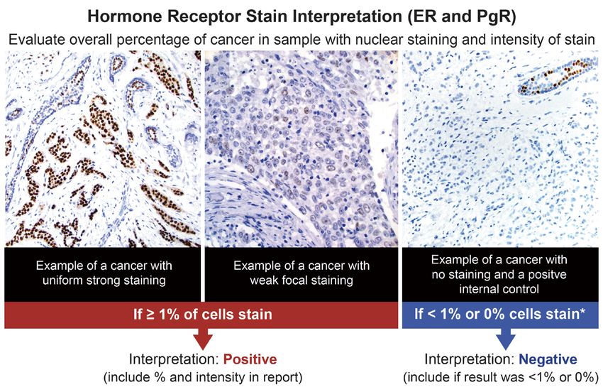
Housekeeping

    • This presentation will be recorded. The recording and PDF
          will go out to all registrants in one week

    • All lines are muted during the presentation

    • Please send in your questions as you think of them via the
          “Question Box” in your control panel

© College of American Pathologists                          26 February 2020   3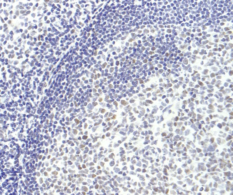
Kimberly Allison, MD, FCAP

 • Director of Breast Pathology and
   Professor of Pathology at Stanford
   University of Medicine.

 • Has a special interest in development
   of high-quality diagnostic standards
   and is active in setting practice
   guidelines and patient communication.

 • Actively involved in resident/fellow
   training as Director of the Stanford
   Breast Pathology Fellowship and
   Residency Director for the Department
   of Pathology.

                                           26 February 2020   4
© College of American Pathologists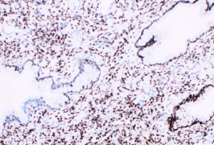
Disclaimer
    • The CAP does not permit reproduction of any substantial
          portion of the material in this Webinar without its written
          authorization. The CAP hereby authorizes attendees of the CAP
          Webinar to use the PDF presentation solely for educational
          purposes within their own institutions. The CAP prohibits use
          of the material in the Webinar – and any unauthorized use of
          the CAP’s name or logo – in connection with promotional
          efforts by marketers of laboratory equipment, reagents,
          materials, or services.

© College of American Pathologists                                  26 February 2020   5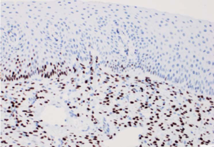
Disclaimer, continued
    • Opinions expressed by the speaker are the speaker’s own and
          do not necessarily reflect an endorsement by the CAP of any
          organizations, equipment, reagents, materials, or services used
          by participating laboratories.

© College of American Pathologists                               26 February 2020   6
Disclosures:

 • Scientific Advisory Board of Mammotome, Inc

© College of American Pathologists               26 February 2020   7
Learning Objectives

 • Identify new aspects of the CAP/ASCO ER/PR testing in
       breast cancer guideline updates that affect hormone
       receptor standard testing operating procedures,
       interpretation and reporting for invasive breast cancer and
       ductal carcinoma in situ.
 • Review how to apply the new recommendations in specific
       ER/PR testing patient care scenarios.

                    Also: To Answer your Frequently Asked Questions!

© College of American Pathologists                                     26 February 2020   8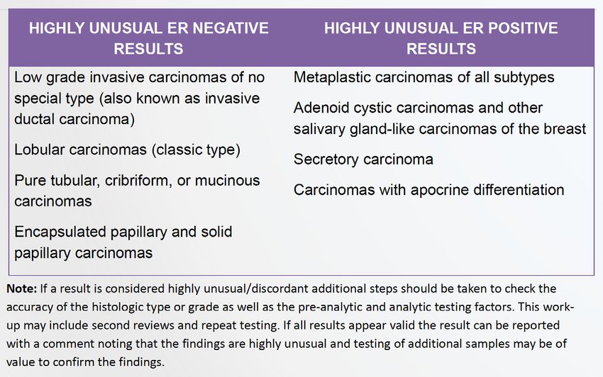
Why Update Now?
• CAP and ASCO agreed to partner to develop
  guidelines starting with HER2 testing in breast
  cancer in 2007
• After this successful venture, the ER/PgR
  guideline was jointly published in 2010                              Dr. Elizabeth
  o 2,800+ unique citations in publications from more than 99           Hammond
       different countries.
  o Now 1,400+ labs participate in CAP PT for ER/PgR

• Two updates to HER2 testing guidelines (updates
  in 2013 &2018)
• 10 years since initial ER/PgR guideline (2010)
  o More recent data on 1-10%/”low positive” ER cases                  Dr. Antonio
                                                                          Wolff
  o Is 1% threshold for positive still the most clinically relevant?
 © College of American Pathologists                                      26 February 2020   9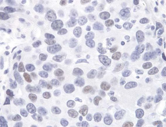
Guidelines in Breast Cancer are Living
  Documents
                                                    Big Questions, Setting First
                                                           Standards
• First focused on big                Pathologist
                                       feedback
 questions and standards for
 all cases                            New data
                                                              Fine tuning
• Subsequent updates based
                                       Experts
 on new data, feedback
• Fine tuning, often focused            Industry
                                       feedback                   Fine
 on less common scenarios                                        tuning
                                      Regulatory
                                       agencies

 © College of American Pathologists                                  26 February 2020   10
Has there been improvement
in ER/PgR testing since the
2010 Guidelines?
Where and how can additional improvements be made?
© College of American Pathologists 26 February 2020 11ER/PgR Testing in 2010
• No single national/international
guideline
• Only some countries had QA/PT
systems tracking data (UK-NEQAS,
Australia, NordicQC)
• Canadian ER testing controversy
(~40% false negative rate)
• Variability between local vs central
labs: Tables 3-6 from 2010 guidelines
• Issues:
o Long ischemic times, fixation variable
o Variability in thresholds used for positive
o Lack of appropriate internal or external positive
controls
o Labs without sufficient expertise in IHC methods,
QA or validation
© College of American Pathologists 26 February 2020 122010 Guidelines Set New Standards
• Issues Addressed:
o Long ischemic times, fixation variable recommendations made to standardize
and track/report
o Variability in thresholds used for positive standard set at 1% for positive
o Lack of appropriate internal or external positive controls required reporting of
and recommendations for rejection/retesting
o Labs without sufficient expertise in IHC methods, QA or validation CAP
accreditation requirements set for Validation and Proficiency Testing,
CanadianIQc, etc.
© College of American Pathologists 26 February 2020 13ER/PgR Testing since 2010: How are we doing?
• Look at Clinical Trial Data on Local vs Central Test Results:
o Local testing was routine per local lab standards without anticipation of central
re-testing (not a “test”) and on whole sections
o ER false negatives still a problem in “triple negative trials”
o Drawbacks: Trial population dependent/case selection, blocks/sample may be
different local vs central
• Look at Proficiency Testing and EQA Programs:
o Can control for more pre-analytical variables and do deeper dive into frequency
of analytical and post-analytical variability/errors and their causes
– Some use consensus and some use reference standard
o Dramatic increase in number of labs doing PT (ex. CAP now 1500+ labs)
o Drawbacks: Case selection for high agreement (ex. Weak positives often thrown
out due to failure to achieve 80% agreement for CAP PT) and known test
environment
© College of American Pathologists 26 February 2020 14NordiQC Trends Towards
Higher Pass Rates
Causes of improvement: Harmonization of use of Causes of improvement: Careful calibration of titer
optimized protocol setting, Fewer labs using 1D5, and HIER time
HIER in alkaline buffer Persistent issues: False positives w 1E2 (strong
Persistent issues: False negatives in cases with staining of tonsil B cells)
weaker staining but 10-80% positive Recommended controls: cervix for + (not post-
Recommended controls: cervix for strong +, tonsil menopause), tonsil for negative
for weak +, ER- breast cancer for negative control
www.nordiqc.org
© College of American Pathologists 26 February 2020 15• CAP PT 80 cores: 56 were scored similarly by 3 most common antibodies (1D5, 6F11, SP1) • ER: 92.5% graded, of intended positives with variability 17% were SP1+/1D5- or 6F11 – (more false neg ER for labs that don’t use SP1) • PR: 78.8% graded, of intended negatives with variability often near 1% threshold, frequent 1E2+/- by other clones (“false +” PR with 1E2?) © College of American Pathologists 26 February 2020 16
Current issues (“Fine-Tuning”):
• Variability still exists but is improved need for continued
regulation and guideline recommendations, importance of
publishing antibody-specific results + ideal methods
o Need to focus on avoiding false negatives for ER (weak intensity cases particularly
sensitive)
o Need to focus on avoiding false positives for PgR (careful titration with appropriate
controls)
• 1-10% ER cases uncommon (not in PT sets) but can cause
variability between labs (especially false negs)
o Is benefit of hormonal-Rx significant?
o Should they be treated with ER negative treatment algorithms?
o Do they have the prognosis of ER positive cancers?
o Is variability in this range avoidable? What are ways to improve reproducibility?
• PR and heterogeneity of expression (challenging on
proficiency testing)
© College of American Pathologists 26 February 2020 17Key guideline questions: 1. What is the optimum quality assurance, tissue handling, scoring system and reporting for Threshold determining potential benefit from endocrine question therapy? 2. What additional strategies can promote optimal How to avoid performance, interpretation, and reporting of IHC false assays, particularly in cases with low ER negative/low expression? 3. Are other ER expression assays acceptable for identifying patients likely to benefit from endocrine therapy? 4. Should DCIS be routinely tested for hormone receptors? © College of American Pathologists 26 February 2020 18
Setting Thresholds for Biomarkers
   • Dependent on what trying to prognosticate vs predict:
        o Best predictive threshold will depend on risk/benefit in giving drug

   • There will usually be a grey zone near the threshold
        o More variability in test results
        o Less clear clinical implications

                                  Increasing expression of biomarker
Multiple current uses of ER/PR Testing
1. Determining potential benefit from Test validated for as
endocrine therapies a predictive
biomarker =
2. Overall treatment pathways determined by Guideline’s focus
ER+ vs ER- (ex. NCCN guidelines)
Is the 1%
3. Surrogates for intrinsic/molecular subtype
threshold
determination (along with HER2) valid for all
4. Prognostic role (ex. AJCC prognostic uses?
subgroups)
5. Metastatic setting: ER+ vs ER- treatments
6. Diagnostic testing (is metastatic cancer
breast?)
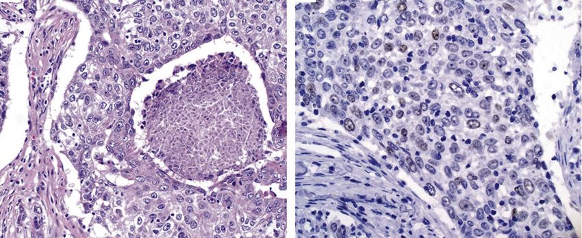
© College of American Pathologists 26 February 2020 20Allred study: Showing best predictive threshold?
• All patients received
endocrine therapy
• Actually only Prognostic…
• Samples were not
standard
Harvey et al JCO 1999
© College of American Pathologists 26 February 2020 21Clinical Trial data: Best predictive threshold?
• Limited clinical data on
threshold – mostly based
on LBA data
• 20 trials with over 200,000
women-years of follow-up Lancet 2011; 378 771-84
• Points to 10 fmol ER/mg
at best threshold.
o 10-19 fmol ER/mg had
recurrence reduced by 1/3 with
5 yrs Tam
Correlates best with 1% by IHC
© College of American Pathologists 26 February 2020 22Imperfect Clinical Trials
Data…but not likely to repeat
clinical trials on endocrine
therapy
Low risk drug with potentially high benefit…
© College of American Pathologists 26 February 2020 23What do we know about ER Low Positive
Cancers?
• Heterogeneous group & rare (2-3%)
• Often “basal-like” features (histology,
response to neoadjuvant
chemotherapy and molecular profiles)
o Don’t want to exclude these patients from “triple
negative” trials…?
• Potential benefit from endocrine
therapy (although less than stronger
positive):
May still need to be considered positive for at
least at trial of endocrine therapy but intent not to
be used to treat similar to other strong ER+
cancers…..
© College of American Pathologists 26 February 2020 24ER: What threshold for IHC+?
1% 10%
0 - < 1% 1-10% 11-100%
Worse prognosis Better prognosis
Basal or HER2-E Luminal (some HER2-E)
No Endocrine Benefit from Endocrine RX (50-66% reduction in recurrences, 30-40%
RX Benefit
reduction in mortality)
o To segregate out who will definitely NOT benefit from endocrine therapy? > 1% vs < 1% or
0%
o To select who is highly likely to benefit from endocrine therapy? Don’t want to exclude pts
from possible benefit in relatively low risk drug
o To determine overall treatment pathway? Use ~10%?
o To determine intrinsic/biologic subtype of breast cancer? Use ~10%?
o To determine overall prognostic groups? Use ~10%? EBCTCG (2015).
https://doi.org/10.1016/s0140 -
6736(15)61074 -1
© College of American Pathologists 26 February 2020 25UPDATED JANUARY 2020 Recommendation 1.1. Optimal algorithm for ER/PgR testing • Samples with 1-100% of tumor nuclei positive for ER or PgR are interpreted as positive. • For reporting of ER (not PgR), if 1-10% of tumor cell nuclei are immunoreactive, the sample should be reported as ER Low Positive with a recommended comment. • A sample is considered negative for ER or PgR if
New Low Positive ER Category and
 Recommended Reporting Comment:
 • ER : LOW POSITIVE (1-10%), SEE COMMENT

                                     Allison KH, Hammond MEH, Dowsett M, et al: J Clin Oncol doi:
                                                                            10.1200/JCO.19.02309
                                              Arch Pathol Lab Med doi: 10.5858/arpa.2019-0904-SA

© College of American Pathologists                                       26 February 2020   27
Recommendation 1.1., continued A sample may be deemed uninterpretable for ER or PgR if the sample is inadequate (insufficient cancer or severe artifacts present, as determined at the discretion of the pathologist), if external and internal controls (if present) do not stain appropriately, or if pre-analytical variables have interfered with the assay’s accuracy (see manuscript Figures 1-4). Clinicians should be aware of and able to discuss with patients the limited data on ER-low positive cases and issues with test results that are close to a positive threshold. Strong Recommendation © College of American Pathologists 26 February 2020 28
Example Case:
• Grade 3 invasive ductal carcinoma, LN
neg
• Core Biopsy outside read by image
analysis : ER 2%
• Core biopsy by our review: ER 10%, 1+
• Excision at Stanford: ER 20%, 1-2+
• Sent for Oncotype DX:
o High RS (54; 34% recur )
Cases close to threshold for positive
are more likely to have different
results by different assays, methods
or samples.
Any positive result is treatable but
need to acknowledge data limited.
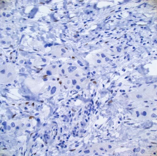
© College of American Pathologists 26 February 2020 29Example Case: 35 y/o female with Grade 3 IDC and
the following ER stain you estimate to be 1-10%
positive (1+)
What do you do next?
© College of American Pathologists 26 February 2020 30Recommendation 2.3 (NEW) Laboratories should establish and follow an SOP stating the steps the laboratory takes to confirm or adjudicate ER results for cases with weak stain intensity or
FIGURE 1

                                      For cases with < 10% or
                                           weak staining:
                                     1.Take steps to
                                       confirm/adjudicate
                                       result per your lab’s
                                       SOP.
                                     2.Correlate with histology.

                                        Allison KH, Hammond MEH, Dowsett M, et al: J Clin Oncol doi:
                                                                               10.1200/JCO.19.02309
                                                 Arch Pathol Lab Med doi: 10.5858/arpa.2019-0904-SA

© College of American Pathologists                                           26 February 2020   32
EXAMPLE SOP
Each lab can use their own
(base on lab specific
issues/data and good
practice)
Allison KH, Hammond MEH, Dowsett M, et al: J Clin Oncol doi: 10.1200/JCO.19.02309
Arch Pathol Lab Med doi: 10.5858/arpa.2019-0904-SA
© College of American Pathologists 26 February 2020 33 Evidence stain worked on
sample tested
If close to threshold for
positive second review to
make sure interpretation
reproducible in your lab
Allison KH, Hammond MEH, Dowsett M, et al: J Clin Oncol doi:
10.1200/JCO.19.02309
Arch Pathol Lab Med doi: 10.5858/arpa.2019-0904-SA
© College of American Pathologists 26 February 2020 34 Check if external
controls appropriate.
Check pre-analytic
variables
Report with additional
comment about
Allison KH, Hammond MEH, Dowsett M, et al: J Clin Oncol doi: controls
10.1200/JCO.19.02309
Arch Pathol Lab Med doi: 10.5858/arpa.2019-0904-SA
(recommended)
© College of American Pathologists 26 February 2020 35New Recommendations on Internal Control
Reporting (Recommendation 2.4):
• The status of internal controls should also be reported for
cases with 0-10% staining (with a special comment for those
lacking internal controls). See Table 2.
Allison KH, Hammond MEH, Dowsett M, et al: J Clin Oncol doi: 10.1200/JCO.19.02309
Arch Pathol Lab Med doi: 10.5858/arpa.2019-0904-SA
© College of American Pathologists 26 February 2020 36Recommendation 1.5.
Optimal internal QA procedures Updated
Standardized operating procedures (SOPs)
should be used that include routine use of
external control materials with each batch of
testing and routine evaluation of internal
normal epithelial elements or the inclusion of
normal breast sections (or other appropriate
control) on each tested slide, wherever
possible. External controls should include
negative and positive samples as well as
samples with lower percentages of ER
expression (such as tonsil). On-slide controls
are recommended. Allison KH, Hammond MEH, Dowsett M, et al: J Clin Oncol doi:
10.1200/JCO.19.02309
Arch Pathol Lab Med doi: 10.5858/arpa.2019-0904-SA
© College of American Pathologists 26 February 2020 37External Controls: Include a spectrum of ER expression, on-slide TMAs or similar preferred
Ex. Cancer
Strong positive (>95%, 3+) control. cases used Low positive (1-10%, 1+) control.
a external
controls
Moderate intensity positive (80%, 2+) control. Negative (0%) control.
Allison KH, Hammond MEH, Dowsett M, et al: J Clin Oncol doi:
10.1200/JCO.19.02309
Arch Pathol Lab Med doi: 10.5858/arpa.2019-0904-SA
© College of American Pathologists 26 February 2020 38TONSIL: An Excellent External Control For Low ER Positive
and PgR Negative
ER ER: Weak positive staining PgR: No staining
A B C
Tonsil is an excellent external control to monitor the analytical sensitivity for ER.
Dispersed germinal center cells and the squamous epithelium should be ER positive
but the B-cells in the mantle zones should be ER negative (as shown in panels A at 5x
and panel B at 20x). Tonsil is an appropriate negative control for PgR. In contrast to
ER, no nuclear PgR staining should be seen. Weak positive PgR staining in tonsil
should result in work-up to determine if assay drift has occurred.
Allison KH, Hammond MEH, Dowsett M, et al: J Clin Oncol doi:
10.1200/JCO.19.02309
Arch Pathol Lab Med doi: 10.5858/arpa.2019-0904-SA
© College of American Pathologists 26 February 2020 39Example case: External tonsil control for PgR stain reviewed Tonsil staining for PgR when should be negative…. Need to re-titer assay? Drift occurring? © College of American Pathologists 26 February 2020 40
Cervix as an external control

• PgR should variably stain the basal layer of the squamous
 mucosa (good for low limit of detection control)
• PgR should also stain endocervical columnar epithelium (with
 some variability)
• ER should stain almost all endocervical columnar epithelial cells
• Note: May be less robust staining in cervical tissue from
 postmenopausal women
  © College of American Pathologists                     26 February 2020   41
Double check stain worked (repeat test)
Check pre-analytic variables
May need to report as “indeterminate”
with recommendations for additional
samples if pre-analytic issues identified
Allison KH, Hammond MEH, Dowsett M, et al: J Clin Oncol doi:
10.1200/JCO.19.02309
Arch Pathol Lab Med doi: 10.5858/arpa.2019-0904-SA
© College of American Pathologists 26 February 2020 42FAQ: What if my case was decalcified?
• Recommend separating grossly: Van Es. EDTA Aceti HCl/For
AJSP.
o bony fragments decal c mic
2019;43:1355
–60
o non-bony fragments NO DECAL
ER % -0.5% -2.5% -21%
o Helpful for FISH & molecular change
ER false 0 0 42%
• Validate your lab’s decal/FFPE/Ab, or neg
o CAP disclaimer “This assay has not been validated PR % -1.5% -0.5% -14.5%
change
on decalcified tissues. Results should be
PR false 0 0 33%
interpreted with caution given the possibility of neg
false negative results on decalcified specimens.” HER2 -0.3 -0.3 -0.8
change
• Also: Most cyto fixatives alcohol based ISH failure 1/16 15/16 all
o Many labs use formalin-only for suspected breast
metastasis, or
o Validate your lab’s cyto fix/cell block/Ab See also: Clark. AIMM. 2019;27:223-30
Schrivjer. Mod Pathol. 2016;29:1460-70
Gertych. Diagn Pathol. 2014;9:213
Maclary. AIMM. 2017;25:144–149
© College of American Pathologists 26 February 2020 43Ensure reproducible, not a false negative
>10% staining 1-10% staining
Confirm result
Interpretation: Positive Report as ER Low Positive
(include % and intensity) w/comment
Allison KH, Hammond MEH, Dowsett M, et al: J Clin Oncol doi:
10.1200/JCO.19.02309
Arch Pathol Lab Med doi: 10.5858/arpa.2019-0904-SA
© College of American Pathologists 26 February 2020 44Stanford Practice: Data used to establish an SOP
• Test set of 30 cases reported as ER
Negative (0 or 10%) were
identified.
• 5 breast pathologists who perform ER
interpretations scored/interpreted
each case
• Agreement was very high for > 10%
• Agreement was high for < 1% (best for
0%)
ER 1-10%
ER 10%
Positive (1-10%)
• Decided our SOP should include
ER 1-10% second pathologist review for cases
ER >10%
ERExample case: You are reviewing as a second opinion a case with the following diagnosis from the original lab: DIAGNOSIS: INVASIVE LOBULAR CARCINOMA • ER negative (0%) with positive internal controls • PR negative (0%) with positive internal controls • HER2 positive (3+) by IHC Revised Diagnosis: Invasive Pleomorphic Lobular Carcinoma © College of American Pathologists 26 February 2020 46
Recommendation 2.2.

                                         Updated

   Interpretation of any ER result should include evaluation
   of the concordance with the histologic findings of each
   case. Clinicians should also be aware of when results are
   highly unusual/discordant and work with pathologists to
   attempt to resolve or explain atypical reported findings
   (see manuscript Table 3 as an aid in this process).

   Strong Recommendation

© College of American Pathologists                       26 February 2020   47
Also these should
be HER2 Negative
Allison KH, Hammond MEH, Dowsett M, et al: J Clin Oncol doi:
10.1200/JCO.19.02309
Arch Pathol Lab Med doi: 10.5858/arpa.2019-0904-SA
© College of American Pathologists 26 February 2020 48Grey Zones in Dual Probe HER2 ISH Test
Interpretation: 2018 Update Summary
Most cases
Grey Zones and Borderline
Results: Confirmation,
correlation and explanation
Required comments
REFERENCE:
Wolff AC, e. J Clin Oncol 2018; 36: 2105–22.
WHO 5th edition Tumours of the Breast 2019
© College of American Pathologists 26 February 2020 49Borderline or Unusual ER or HER2
Results: Summary
ER 1-10% or weak HER2 Unusual ISH
staining Groups
CONFIRMATION: CONFIRMATION:
Per SOP of Lab IHC and Recounts
CORRELATION (with CORRELATION (with IHC and
histology) histology)
EXPLANATION:
Required report comments EXPLANATION:
Required report comments
© College of American Pathologists 26 February 2020 50Should we still do PgR Testing??
• Yes, PR testing is still useful
o Biology suggests that it is important in modulating ER
o Marker of prognosis in multiple settings
o IHC subtyping / defining TNBC for many clinical trials; new AJCC staging
o To identify possible false-negative ER tests, as quality measure
• The utility of PgR testing continues to be largely prognostic in the ER
positive invasive cancer population, but testing using similar
principles to ER testing is still recommended for invasive cancers.
• No new evidence that PR+ vs PR- (however defined) is predictive
marker for ET vs no or choice of ET; consistent that higher ER+/PR+
(eg ≥50%) is more ET-responsive
• No data for ER-/PR-/HER2- vs ER-/HER2- to define TNBC
• No new data on utility in DCIS
© College of American Pathologists 26 February 2020 51Polling questions will now show
up on your screen
© College of American Pathologists 26 February 2020 52FAQ: Do you have to report “Low Positive” for PgR 1-10%? A. Yes B. No C. I don’t know © College of American Pathologists 26 February 2020 53
FAQ: Do you have to report “Low Positive” for
 PgR 1-10%?
 A. Yes
 B. No (Optional to do so - but if you do – do not include the
           comment intended for Low Positive ER)
 C. I don’t know

© College of American Pathologists                      26 February 2020   54
Recommendation 4: DCIS
Updated
ER testing in cases of newly diagnosed DCIS
(without associated invasion) is recommended to
determine potential benefit of endocrine therapies to
reduce risk of future breast cancer. PgR testing is
considered optional.
Moderate Recommendation
© College of American Pathologists 26 February 2020 55FAQ: Do you have to report “Low Positive” for DCIS with ER between 1-10%? A. Yes B. No C. I don’t know © College of American Pathologists 26 February 2020 56
FAQ: Do you have to report “Low Positive” for
 DCIS with ER between 1-10%?
 A. Yes
 B. No (Optional to do so - but if you do – do not include the
           comment intended for Low Positive ER)
 C. I don’t know

© College of American Pathologists                      26 February 2020   57
FAQ: Do we still need to report stain intensity for ER and PgR? A. No B. Yes C. I don’t know © College of American Pathologists 26 February 2020 58
FAQ: Do we still need to report stain intensity?

 A. No
 B. Yes (Stain intensity helps determine how well the assay
           worked and may be important biologically, but only the %
           of cells staining determines if the result is positive or
           negative)
 C. I don’t know

© College of American Pathologists                               26 February 2020   59
FAQ: What if I use the Allred Score or H-score?
That is fine. But….
Report needs to include both percent and intensity overall
raw results
Low positive ER needs to be defined as 1-10% positive for
consistency across labs.
© College of American Pathologists 26 February 2020 60FAQ: Does the actual formalin time and cold ischemia time need to be included in the template/original pathology report? Need to have documented: Time the tissue is removed from the patient The time it is placed in fixative The cold ischemia time The duration of fixation The fixative type These can be recorded in the pathology report or in another suitable location that is available for review. Including the specific times in the pathology report is at the laboratory's discretion (note: the CAP Laboratory Accreditation Program requires accredited laboratories to specify the type of fixative used and the cold ischemia time in all ER, PgR and HER2 reports). The laboratory is also responsible for determining if the cold ischemia and fixation times meet the requirements specified in the latest version of the ASCO/CAP ER/PgR testing guidelines. © College of American Pathologists 26 February 2020 61
Recommendation 1.6. Optimal external
 proficiency assessment
                                                                  Updated

 The laboratory performing ER and PgR testing must
 participate in external proficiency testing or alternative
 performance assessment as required by its accrediting
 organization.
                                     Recommendation was updated to
                                     remove information about what
                                     constitutes satisfactory proficiency
 Strong Recommendation               assessment. Laboratories are
                                     instructed to follow the requirements of
                                     their accrediting organization.

© College of American Pathologists                                     26 February 2020   62
Recommendation 1.7. Optimal laboratory
accreditation
• On-site inspection every other year should be
undertaken with annual requirement for self-
inspection.
• Moderate Recommendation
• Statement Reaffirmed
© College of American Pathologists 26 February 2020 63Initial test validation
• There will be an upcoming CAP guideline on principles of
predictive IHC test initial validation
o Recommendation 1.5., continued
o Regular, ongoing assay reassessment should be done at least semiannually
(as described in Fitzgibbons et al). Revalidation is needed whenever there is
a significant change to the test system (Torlakovic et al).
o Ongoing competency assessment and education of pathologists is required.
o Strong Recommendation
© College of American Pathologists 26 February 2020 64Recommendation 3.
Updated
Validated IHC is the recommended standard test for
predicting benefit from endocrine therapy. No other
assay types are recommended as the primary
screening test for this purpose.
No study has included
Strong Recommendation treatment in + vs - to
examine if predictive of
endocrine benefit
© College of American Pathologists 26 February 2020 65mRNA methods may be be less sensitive than IHC in detecting low level ER expression • Cancers with 1-9% ER staining by IHC had features overlapping with ER
Summary of Major Impact of 2020 Updates to
 ER/PgR IHC Guidelines:
 • New ER Low Positive reporting category for invasive cases
       with 1-10% staining
 • Need for lab specific SOP to ensure reproducibility/validity
       of invasive cases with
CAP’s Precision Medicine Webpage
• The webpage includes brief, relevant articles by CAP members that enable the
reader to gain a better understanding of a particular area of precision medicine.
o Examples include pharmacogenetics, immune response genes, and the latest in the molecular drivers of
cancer.
o Access them www.cap.org >
Member Resources > Precision Medicine
© College of American Pathologists 26 February 2020 68Short Presentations on Emerging Concepts
(SPECS)
• Pathology SPECs are:
– Short PowerPoints, created for pathologists
– Focused on diseases where molecular tests
play a key role in patient management
• Recent topics include:
– Microbiome
– Biomarkers in Lung Cancer
– MDS
– Other emerging topics
• Access them at www.cap.org >
Resources and Publications
© College of American Pathologists 26 February 2020 69CAP’s Pathology Resource Guide: Precision Medicine
• The CAP has created the Pathology Resource Guides to assist
pathologists in understanding key emerging technologies.
o Printed guides are now available for members ($39) and non-members ($69)
o The digital copy of the Resource Guides are a complimentary member benefit
o Access them www.cap.org > Resources and Publications
© 2018 College of American Pathologists. All rights reserved.
© College of American Pathologists 26 February 2020 70THANK YOU!
Thank you for attending our webinar,
“What’s New in the 2020 Update to the CAP/ASCO ER/PgR Testing
Guidelines in Breast Cancer?
by Kimberly Allison, MD, FCAP
For comments about this webinar or suggestions for upcoming
webinars, please contact phcwebinars@cap.org.
NOTE: There is no CME/CE credit available for today’s free
webinar. The PDF of the presentation will be sent out in a
week.
© College of American Pathologists 26 February 2020 71© College of American Pathologists
You can also read