The new WHO classification of gastrointestinal neuroendocrine tumors and immunohistochemical expression of somatostatin receptor 2 and 5
←
→
Page content transcription
If your browser does not render page correctly, please read the page content below
EXPERIMENTAL AND THERAPEUTIC MEDICINE 22: 1179, 2021

                    The new WHO classification of gastrointestinal
                   neuroendocrine tumors and immunohistochemical
                      expression of somatostatin receptor 2 and 5
                          OANA POPA1,2*, SORINA MARIA TABAN1*, STELIAN PANTEA3,
               ANDREI DOREL PLOPEANU1,4, ROBERT ALEXANDRU BARNA1,5, MARIOARA CORNIANU1,
                           ANCA‑ARIANA PASCU5 and ALIS LILIANA CARMEN DEMA1*

    1
     Department of Microscopic Morphology‑Pathology, ANAPATMOL Research Center, ‘Victor Babeș’ University of Medicine
    and Pharmacy of Timisoara, 300041 Timisoara; 2Endocrinology Clinic, ‘Pius Brînzeu’ County Emergency Clinical Hospital,
        300723 Timisoara; 3Surgical Emergency Clinic, ‘Victor Babeș’ University of Medicine and Pharmacy of Timisoara,
     300041 Timisoara; 4Anatomic Pathology Service, ‘Pius Brînzeu’ County Emergency Clinical Hospital, 300723 Timisoara;
                       5
                         Department of Internal Medicine II‑Discipline of Gastroenterology and Hepatology,
                 ‘Victor Babeș’ University of Medicine and Pharmacy of Timisoara, 300041 Timisoara, Romania

                                           Received June 2, 2021; Accepted July 2, 2021

                                                    DOI: 10.3892/etm.2021.10613

Abstract. The 2019 World Health Organization (WHO) classifi‑          neuroendocrine tumors were more common SSTR2‑positive
cation of gastrointestinal tumors defines well‑differentiated grade   in comparison with G3 carcinomas (P
2                                 POPA et al: SSTRs IN DIGESTIVE NEUROENDOCRINE NEOPLASMS

tract and a second category for neuroendocrine carcinomas               Currently, only a few studies have followed immunohis‑
(NECs) that are poorly differentiated and, although they posi‑      tochemical (IHC) SSTR expression in gastrointestinal NENs,
tive for most neuroendocrine markers, have a poor prognosis         although immunohistochemistry allows precise cellular
and evolution (3).                                                  localization of SSTRs. In this study, the aims were to evaluate
    The actual classification and the tumor grading algorithm       the IHC expression of SSTR2 and SSTR5 in gastrointestinal
are very similar to the one discussed in the 2017 WHO clas‑         NENs, MiNENs and AGCCs, as well as to correlate the
sification of pancreatic NENs. Therefore, a new category            expression of these markers with clinical and morphological
was generated, the well‑differentiated grade 3 NETs of the          factors that impact the overall prognosis, outcome and treat‑
digestive system. The distinction between NETs and NECs             ment of the patients.
with large cells (LCNEC) and small cells (SCNEC) consists
of different morphological characteristics (1,4). The pres‑         Patients and methods
ence of both components, low and high grade, in a unique
tumor is a strong argument in favor of naming NETs as               Patient data. The retrospective study included 76 patients with
grade 3 well‑differentiated NETs. Mixed tumors, in which            gastrointestinal NENs confirmed by histology and immuno‑
each component, neuroendocrine and non‑neuroendocrine,              histochemistry at the Pathology Laboratory of Timis County
represents more than 30 percent of the tumor cells, are             Emergency Clinical Hospital (Timisoara, Romania) from
termed mixed neuroendocrine‑non‑neuroendocrine tumors               January 2008 to December 2018. The cases were selected
or MiNENs (2).                                                      according to histopathological diagnosis and tissue material
    Over the last 2‑3 years, genetic studies have shown that        available for pathological evaluation and IHC reactions. In
the genetic mutations in neuroendocrine tumor cells with            5 cases, the examined tissue material was not sufficient to
extra‑pancreatic origin (especially in those of the gastro‑         perform the required number of sections for IHC investiga‑
intestinal tract) are very similar to those of the pancreas. In     tion. Our study batch consisted of 52 gastrointestinal tumors
gastrointestinal NECs, TP53 and RB1 mutations are frequently        (endoscopic biopsies, specimens of polypectomy and surgical
encountered, similar to pancreatic and pulmonary NECs, but          samples) and 19 cases of liver metastases in the absence of
absent in NETs. MEN1, DAXX and ATRX mutations are char‑             evident primary disease. The median age of the patients
acteristic of well‑differentiated NETs (5,6).                       (37 men and 34 women) was 59.9 years. Patient clinical and
    Aside from the morphological and molecular aspects, G3          pathological characteristics are summarized in Table I.
NETs and NECs also differ from a clinical point of view.                The study was conducted in accordance with Declaration
Platinum‑containing chemotherapy is successfully used in            of Helsinki, in compliance with good clinical practice, and was
NECs, sometimes with noteworthy results in SCNECs. Despite          approved by the Ethics Committee of ‘Pius Brinzeu’ Emergency
this aspect, the observation was stated that some patients do       Clinical County Hospital and Victor Babes University of
not respond to this therapy but have longer survival and better     Medicine and Pharmacy Timisoara (no. 20 b/2015 extended
outcome than patients that were responsive to platinum‑based        in 2019). Written informed consent was obtained from each
chemotherapy. This subgroup of patients was subsequently            patient included in our study.
diagnosed with G3 NETs (1).
    Gastroenteropancreatic NENs are characterized by the            Histological and IHC interpretation. Tumors were reclassi‑
overexpression of somatostatin receptors (SSTRs) 2 and 5, a         fied according to the 2019 WHO classification (Table II). IHC
family of G protein‑coupled receptors present in neuroendo‑         stains were performed in all 71 cases. The specimens were
crine cells (7,8). Somatostatin is a cyclic neuropeptide that       fixed in 10% neutral‑buffered formalin for a maximum of 24 h
is ubiquitously expressed in humans, acting as an inhibitor         at room temperature, paraffin‑embedded and sectioned at 3‑ to
of exocrine and endocrine secretions on target organs. It           4‑µm. IHC staining was performed with a Leica Bond‑Max,
exerts its biological effects by binding to five specific           which is an automatic and continuous access slide‑staining
high‑affinity receptors on the cell surface (9). Somatostatin       system that simultaneously processes IHC protocols, using
then activates the second messenger system with a wide              a Bond Polymer Refine Detection Kit (Leica Biosystems
range of actions: inhibition of adenylate cyclase, activation       Newcastle). Ki67 (clone MM1, catalog no. PA0118), CgA (clone
of calcium channels, stimulation of phosphotyrosine phos‑           5H7, catalog no. PA0515), synaptophysin (Syn) (clone 27G12,
phatase or MAPK kinase activity (10). The expression of             catalog no. PA0299) and p53 (clone DO‑7, catalog no. PA0057)
these markers is the foundation for somatostatin analogue           antibodies from Leica with ready‑to‑use (RTU) kits following
therapy.                                                            the manufacturer protocols were used. For the IHC detection
    Adenocarcinoma ex‑goblet cell carcinoid (AGCC), a term          of SSTR2 and SSTR5 antibodies, the following protocol
proposed by Klimstra et al (1) and Tang et al (11) or mixed         was performed: tissues were deparaffinized and pre‑treated
goblet cell carcinoid‑adenocarcinoma (12) is an enigmatic           with the Epitope Retrieval Solution 1 at 98˚C for 20 min.
entity, an amphicrine tumor with glandular/mucinous and             Specimens were then incubated with the primary antibody for
neuroendocrine differentiation (at least, focal differentiation).   30 min at a dilution of 1:150 for SSTR2 (clone UMB1, Abcam,
Tang et al revealed that these tumors are adenocarcinomas or        catalog no. ab134152) and 1:125 for SSTR5 (clone UMB4,
AGCC, but not NECs (11). It seems that focal immunoreac‑            Abcam, catalog no. ab109495), followed by visualization with
tions to chromogranin A (CgA) and other neuroendocrine              a Leica Bond Polymer Refine Detection kit for 20 min at room
markers support this hypothesis (12‑14). In the current WHO         temperature. Finally, the sections were washed in water and
classification, these neoplasms are classified as goblet cell       counterstained with hematoxylin. Appropriate negative and
adenocarcinomas (15).                                               positive controls were generated with satisfactory staining.
EXPERIMENTAL AND THERAPEUTIC MEDICINE 22: 1179, 2021 3
Table I. Clinicopathological characteristics of the patients with cells were present, all tumor cells were counted. Tumors with
gastrointestinal NENs, MiNENs and AGCCs. a mitotic rate >20% and a Ki‑67 proliferation index of >20%
were IHC evaluated for the expression of p53. The immuno‑
Clinicopathological characteristics No. % expression was considered positive if intense nuclear staining
was present in >25% of the tumor cells. Strongly positive p53
Sex was considered abnormal and indicated mutations in the TP53
Male 37 52.1 gene (16,17).
Female 34 47.9 The expression of SSTR2 was evaluated according
Age at diagnosis (years) to the system proposed by Volante et al (18). Therefore,
20%. The
bodies. The proliferation index Ki‑67 represents the percentage histological examination of the NECs revealed solid sheets
of cells with nuclear expression of a total of 500 tumoral cells or trabeculae of large cells with pale eosinophilic cytoplasm,
in hot‑spots. In biopsies where only a small number of tumor vesicular, pleomorphic nuclei with large nucleoli and a high4 POPA et al: SSTRs IN DIGESTIVE NEUROENDOCRINE NEOPLASMS Table II. Classification and grading criteria for NENs of the gastrointestinal tract (1). Terminology Differentiation Grade Mitotic ratea Ki‑67 indexb NET, G1 Well‑differentiated Low 20% SCNEC Poorly differentiated High >20 >20% LCNEC >20 >20% MiNEN Well or poorly differentiated Variable Variable Variable a Mitotic rate, the number of mitosis/2 mm2. bKi‑67 index, counting ≥500 cells in the regions of highest labelling (hot‑spots) which are identified at scanning magnification. NENs, neuroendocrine neoplasms; NET, neuroendocrine tumor; NEC, neuroendocrine carcinoma; SCNEC, small cell neuroendocrine carcinoma; LCNEC, large cell neuroendocrine carcinoma; MiNEN, mixed neuroendocrine non‑neuroendocrine neoplasm. Table III. The correlation between SSTR2/SSTR5 expression and clinicomorphological factors. Clinicopathological factors No. SSTR2+ cases n (%) SSTR5+ cases n (%) Tumor location Stomach 10 8 (80) 3 (30) Duodenum 2 0 (0) 0 (0) Small intestine 10 10 (100) 5 (50) Appendix 6 6 (100) 1 (16.7) Right colon 11 5 (45.5) 3 (27.3) Left colon (including rectum) 13 7 (53.8) 5 (38.5) Hepatic metastases 19 11 (57.9) 3 (15.8) Tumor grading G1 28 27 (96.4) 10 (35.7) G2 21 5 (71.4) 7 (33.3) G3 22 5 (22.7) 3 (13.6) Tumor type NET 52 39 (75) 17 (32.7) NET G1 27 26 (96.3) 10 (37) NET G2 18 12 (66.7) 6 (33.3) NET G3 7 1 (14.3) 1 (14.3) NEC 12 4 (33.3) 2 (16.7) MiNEN 3 2 (66.7) 1 (33.3) AGCCs 4 AGCC G1 + G2 2 2 (100) 0 (0) AGCC G3 2 0 (0) 0 (0) Tumor stage I 7 7 (100) 3 (42.9) II 9 9 (100) 4 (44.4) III 23 13 (56.5) 8 (34.8) IV 22 12 (54.5) 3 (13.6) SSTR, somatostatin receptor; G, grade; NET, neuroendocrine tumor; NEC, neuroendocrine carcinoma; SCNEC, small cell neuroendocrine carcinoma; LCNEC, large cell neuroendocrine carcinoma; MiNEN, mixed neuroendocrine non‑neuroendocrine neoplasm; AGCC, adenocar‑ cinoma ex‑goblet cell carcinoid. mitotic rate (LCNEC) or areas of small cells with scant well‑differentiated G3 NETs are difficult to identify on cytoplasm and hyperchromatic nuclei (SCNEC), tumor histology alone, distinct areas of organoid pattern of tumor necrosis and occasional desmoplastic stroma. Although cells and foci of tumor cells with relatively monomorphic
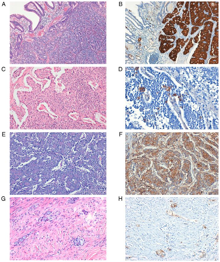
EXPERIMENTAL AND THERAPEUTIC MEDICINE 22: 1179, 2021                                                        5

Figure 1. Expression of SSTR2 in NETs. (A) Well‑differentiated NET,             Figure 2. Expression of SSTR5 in NETs. (A) Well‑differentiated NET, G1.
G2. (B) Strong SSTR2 positive staining (score 3), magnification x10.            (B) SSTR5‑positive staining, magnification x20. (C) Well‑differentiated
(C) Well‑differentiated NET, G2. (D) SSTR2 positive staining (score 2),         NET, G2 with microcalcifications. (D) SSTR5‑positive staining,
magnification x40. (E) LCNEC. (F) Strong SSTR2‑positive staining                magnification x20. (E) Well‑differentiated NET, G3. (F) Weak
(score 3), magnification x20. (G) Appendiceal AGCC. (H) SSTR2‑positive          SSTR5‑positive staining, magnification x40. (G) LCNEC. (H) Strong
staining (score 3), x20 magnification. SSTR, somatostatin receptor; G, grade;   SSTR5‑positive staining, x20 magnification. SSTR, somatostatin receptor;
NET, neuroendocrine tumor; LCNEC, large cell neuroendocrine carcinoma;          G, grade; NET, neuroendocrine tumor; LCNEC, large cell neuroendocrine
AGCC, adenocarcinoma ex‑goblet cell carcinoid.                                  carcinoma.

nuclei are highly suggestive of this type of tumor. To correctly                were more frequently found in the small intestine (100% of
diagnose and classify these tumors, immunoreaction for p53                      the cases), appendix (100% of the cases) and the stomach
was performed. Tumors with >25% intensely positive cells                        (66,66% of the cases). In addition, 50% of the tumors in
for the above‑mentioned marker were classified as G3 NEC.                       the small intestine were also positive for SSTR5. Well‑ and
Two cases of MiNEN were identified in the left colon and                        moderately differentiated tumors exhibited significantly more
one with a gastric location. According to the 2019 WHO                          common SSTR2 in comparison with G3 tumors (P
6                                      POPA et al: SSTRs IN DIGESTIVE NEUROENDOCRINE NEOPLASMS

Table IV. SSTR2 and SSTR5 expression and clinicopathological characteristics.

Clinicopathological
characteristic                   No.      SSTR2+ cases n (%)          χ2 value      P‑value       STR5+ cases n (%)           χ2 value       P‑value

Tumor location
 Stomach + duodenum 12  8 (66.66)  1.252  0.869   3 (25)    Non‑valid
 Small intestine    10 10 (100)			                5 (50)
 Appendix            6  6 (100)			                1 (16.7)
 Colon              24 12 (50)			                 8 (33.33)
 Hepatic metastases 19 11 (57.9)			               3 (15.8)
Tumor grade
 G1                 28 27 (96.4)  30.27
EXPERIMENTAL AND THERAPEUTIC MEDICINE 22: 1179, 2021                                           7

    NETs located in the stomach, duodenum (excepting                least one neuroendocrine marker (predominantly focal), the
gastrinomas), pancreas and rectum, with sizes ≤10 mm and            latest WHO classification lists these tumors as carcinomas
considered G1 in the WHO classification, are considered by          and not NETs (33). Scientific data are scarce despite some
some authors ‘early’ NETs. These patients have an excellent         studies proving the severe outcome of patients with AGCCs.
prognosis. Endoscopic mucosal or submucosal resection is            According to NANETS 2020 consensus guidelines (34) in
recommendable ‘early NETS’ (some authors include tumors             limited disease, appendectomy is advised for tumors
8 POPA et al: SSTRs IN DIGESTIVE NEUROENDOCRINE NEOPLASMS
Funding 0000‑0002‑0027‑1674; Alis Liliana Carmen Dema: ORCID:
0000‑0003‑0767‑2718.
No funding is received.
References
Availability of data and materials
1. Klimstra DS, Kloppel G, La Rosa S and Rindi G: Digestive System
All data generated or analyzed during this study are included Tumours. In: WHO Classification of Tumours. 5th edition. Vol 1.
IARC, Lyon, 2019.
in this published article. 2. Rindi G, Klimstra DS, Abedi‑A rdekani B, Asa SL,
Bosman FT, Brambilla E, Busam KJ, de Krijger RR, Dietel M,
Authors' contributions El‑Naggar AK, et al: A common classification framework for
neuroendocrine neoplasms: An international agency for research
on cancer (IARC) and World Health Organization (WHO) expert
OP contributed to the conception of the study, collected, consensus proposal. Mod Pathol 31: 1770‑1786, 2018.
analyzed and interpreted data from the literature and critically 3. Klöppel G: Neuroendocrine neoplasms: Dichotomy, origin and
classifications. Visc Med 33: 324‑330, 2017.
revised the manuscript. SMT contributed to the conception of 4. Cockburn A and Rege TA: Gastrointestinal neuroendocrine lesions.
the study, performed the literature research, drafted the manu‑ In: Fenoglio‑Preiser' Gastrointestinal Pathology. 4th edition.
script and is responsible for confirming the authenticity of all Wolters Kluwer, Alphen aan den Rijn, pp3300‑3512, 2017.
5. Mafficini A and Scarpa A: Genetics and epigenetics of gastro‑
the raw data. SP contributed to the conception of the study, enteropancreatic neuroendocrine neoplasms. Endocr Rev 40:
performed the literature research, drafted the manuscript and 506‑536, 2019.
is responsible for confirming the authenticity of all the raw 6. Crona J and Skogseid B: GEP‑nets update: Genetics of neuroen‑
docrine tumors. Eur J Endocrinol 174: R275‑R290, 2016.
data. ADP contributed to the interpretation of the data from the 7. Cakir M, Dworakowska D and Grossman A: Somatostatin
literature, collected, analyzed and interpretated the data corre‑ receptor biology in neuroendocrine and pituitary tumours: Part
sponding to the patient and critically revised the manuscript. 1‑Molecular pathways. J Cell Mol Med 14: 2570‑2584, 2010.
8. Klomp MJ, Dalm SU, de Jong M, Feelders RA, Hofland J and
RAB contributed to the interpretation of the data from the Hofland LJ: Epigenetic regulation of somatostatin and soma‑
literature and critically revised the manuscript. MC collected, tostatin receptors in neuroendocrine tumors and other types of
analyzed and interpretated the data corresponding to the cancer. Rev Endocr Metab Disord: Oct 21, 2020 (Epub ahead of
print).
patient and critically revised the manuscript. AAP performed 9. Barbieri F, Bajetto A, Pattarozzi A, Gatti M, Würth R, Thellung S,
the literature research, selected the included studies, analyzed Corsaro A, Villa V, Nizzari M and Florio T: Peptide receptor
and interpretated the data and drafted the manuscript. ALCD targeting in cancer: The somatostatin paradigm. Int J Pept 2013:
926295, 2013.
contributed to the conception of the study, performed the 10. Hankus J and Tomaszewska R: Neuroendocrine neoplasms and
literature research, selected the included studies, analyzed and somatostatin receptor subtypes expression. Nucl Med Rev Cent
interpretated the data and drafted the manuscript. All authors East Eur 19: 111‑117, 2016.
11. Tang LH, Shia J, Soslow RA, Dhall D, Wong WD, O'Reilly E,
read and approved the final manuscript. Qin J, Paty P, Weiser MR, Guillem J, et al: Pathologic classifica‑
tion and clinical behavior of the spectrum of goblet cell carcinoid
Ethics approval and consent to participate tumors of the appendix. Am J Surg Pathol 32: 1429‑1443, 2008.
12. Taggart MW, Abraham SC, Overman MJ, Mansfield PF and
Rashid A: Goblet cell carcinoid tumor, mixed goblet cell carci‑
The study was conducted in accordance with Declaration of noid‑adenocarcinoma, and adenocarcinoma of the appendix:
Helsinki, in compliance with good clinical practice, and was Comparison of clinicopathologic features and prognosis. Arch
Pathol Lab Med 139: 782‑790, 2015.
approved by the Ethics Committee of ‘Pius Brinzeu’ Emergency 13. Hristov AC, Young RH, Vang R, Yemelyanova AV, Seidman JD
Clinical County Hospital and Victor Babes University of and Ronnett BM: Ovarian metastases of appendiceal tumors with
Medicine and Pharmacy Timisoara (no. 20 b/2015 extended goblet cell carcinoidlike and signet ring cell patterns: A report of
30 cases. Am J Surg Pathol 31: 1502‑1511, 2007.
in 2019). Written informed consent was obtained from each 14. Reid MD, Basturk O, Shaib WL, Xue Y, Balci S, Choi HJ,
patient included in our study. Akkas G, Memis B, Robinson BS, El‑Rayes BF, et al:
Adenocarcinoma ex‑goblet cell carcinoid (appendiceal‑type
crypt cell adenocarcinoma) is a morphologically distinct entity
Patient consent for publication with highly aggressive behavior and frequent association with
peritoneal/intra‑abdominal dissemination: An analysis of
Not applicable. 77 cases. Mod Pathol 29: 1243‑1253, 2016.
15. Misdraji J, Carr NJ and Pai Rk: Appendiceal goblet cell adenocar‑
cinoma. In: WHO Classification of Tumours. 5th edition. Vol 1.
Competing interests IARC, Lyon, pp149‑151, 2019.
16. Murnyák B and Hortobágyi T: Immunohistochemical correlates
of TP53 somatic mutations in cancer. Oncotarget 7: 64910‑64920,
The authors declare that they have no conflicts of interest or 2016.
competing interests. 17. Nielsen K, Binderup T, Langer SW, Kjaer A, Knigge P,
Grøndahl V, Melchior L, Federspiel B and Knigge U: P53,
Somatostatin receptor 2a and Chromogranin A immunostaining
Authors' information as prognostic markers in high grade gastroenteropancreatic
neuroendocrine neoplasms. BMC Cancer 20: 27, 2020.
Oana Popa: ORCID: 0000‑0003‑3883‑2438; Sorina Maria 18. Volante M, Brizzi MP, Faggiano A, La Rosa S, Rapa I, Ferrero A,
Mansueto G, Righi L, Garancini S, Capella C, et al: Somatostatin
Taban: ORCID: 0000‑0002‑3971‑2756; Stelian Pantea: receptor type 2A immunohistochemistry in neuroendocrine
ORCID: 0000‑0002‑6048‑6909; Andrei Dorel Plopeanu: tumors: A proposal of scoring system correlated with soma‑
ORCID: 0 0 0 0 ‑ 0 0 02‑ 490 0 ‑ 4809; Rober t Alexandr u tostatin receptor scintigraphy. Mod Pathol 20: 1172‑1182, 2007.
19. Vinik AI and Chaya C: Clinical presentation and diagnosis of
Barna: ORCID: 0000‑0003‑4634‑969X; Marioara Cornianu: neuroendocrine tumors. Hematol Oncol Clin North Am 30:
ORCID: 0000‑0001‑5675‑5339; Anca‑Ariana Pascu: ORCID: 21‑48, 2016.EXPERIMENTAL AND THERAPEUTIC MEDICINE 22: 1179, 2021 9
20. Wang R, Zheng‑Pywell R, Chen HA, Bibb JA, Chen H and Rose JB: 33. Nagtegaal ID, Klimstra DS and Washington MK: Tumours of
Management of gastrointestinal neuroendocrine tumors. Clin Med the appendix. Digestive System Tumours. WHO Classification of
Insights Endocrinol Diabetes 12: 1179551419884058, 2019. Tumours, 5th Edition, Edited by WHO Classification of Tumours
21. Ito T, Sasano H, Tanaka M, Osamura RY, Sasaki I, Kimura W, Editorial Board, 135‑156, 2019.
Takano K, Obara T, Ishibashi M, Nakao K, et al: Epidemiological 34. Kunz PL, Reidy‑Lagunes D, Anthony LB, Bertino EM,
study of gastroenteropancreatic neuroendocrine tumors in Japan. Brendtro K, Chan JA, Chen H, Jensen RT, Kim MK, Klimstra DS,
J Gastroenterol 45: 234‑243, 2010. et al: Consensus guidelines for the management and treatment of
22. Dasari A, Shen C, Halperin D, Zhao B, Zhou S, Xu Y, Shih T neuroendocrine tumors. Pancreas 42: 557‑577, 2013.
and Yao JC: Trends in the incidence, prevalence, and survival 35. Amin MB, Edge S, Greene F, Byrd DR, Brookland RK,
outcomes in patients with neuroendocrine tumors in the united Washington MK, Gershenwald JE, Compton CC, Hess KR,
states. JAMA Oncol 3: 1335‑1342, 2017. Sullivan DC, et al (eds): AJCC Cancer Staging Manual (8th edition).
23. Hofland J, Kaltsas G and de Herder WW: Advances in the diag‑ Springer International Publishing, New York, NY, 2017.
nosis and management of well‑differentiated neuroendocrine 36. Papotti M, Bongiovanni M, Volante M, Allìa E, Landolfi S,
neoplasms. Endocr Rev 41: 371‑403, 2020. Helboe L, Schindler M, Cole SL and Bussolati G: Expression
24. Scherübl H and Cadiot G: Early gastroenteropancreatic neuro‑ of somatostatin receptor types 1‑5 in 81 cases of gastrointestinal
endocrine tumors: Endoscopic therapy and surveillance. Visc and pancreatic endocrine tumors. A correlative immunohisto‑
Med 33: 332‑338, 2017. chemical and reverse‑transcriptase polymerase chain reaction
25. Lewis MA and Hobday TJ: Treatment of neuroendocrine tumor analysis. Virchows Arch 440: 461‑475, 2002.
liver metastases. Int J Hepatol 2012: 973946, 2012. 37. van Adrichem RC, Kamp K, van Deurzen CH, Biermann K,
26. Hendifar AE, Ramirez RA, Anthony LB and Liu E: Current Feelders RA, Franssen GJ, Kwekkeboom DJ, Hofland LJ and
practices and novel techniques in the diagnosis and management de Herder WW: Is there an additional value of using soma‑
of neuroendocrine tumors of unknown primary. Pancreas 48: tostatin receptor subtype 2a immunohistochemistry compared
1111‑1118, 2019. to somatostatin receptor scintigraphy uptake in predicting
27. Calabrò D, Argalia G and Ambrosini V: Role of PET/CT and gastroenteropancreatic neuroendocrine tumor response?
therapy management of pancreatic neuroendocrine tumors. Neuroendocrinology 103: 560‑566, 2016.
Diagnostics (Basel) 10: 1059, 2020. 38. Okuwaki K, Kida M, Mikami T, Yamauchi H, Imaizumi H,
28. Bartsch DK and Scherübl H: Neuroendocrine tumors of the Miyazawa S, Iwai T, Takezawa M, Saegusa M, Watanabe M, et al:
gastrointestinal tract. Visc Med 33: 321‑322, 2017. Clinicopathologic characteristics of pancreatic neuroendo‑
29. Righi L, Volante M, Tavaglione V, Billè A, Daniele L, Angusti T, crine tumors and relation of somatostatin receptor type 2A to
Inzani F, Pelosi G, Rindi G and Papotti M: Somatostatin receptor outcomes. Cancer 119: 4094‑4102, 2013.
tissue distribution in lung neuroendocrine tumours: A clinico‑ 39. Wang Y, Wang W, Jin K, Fang C, Lin Y, Xue L, Feng S, Zhou Z,
pathologic and immunohistochemical study of 218 ‘clinically Shao C, Chen M, et al: Somatostatin receptor expression indicates
aggressive’ cases. Ann Oncol 21: 548‑555, 2010. improved prognosis in gastroenteropancreatic neuroendocrine
30. Srirajaskanthan R, Watkins J, Marelli L, Khan K and Caplin ME: neoplasm, and octreotide long‑acting release is effective and
Expression of somatostatin and dopamine 2 receptors in neuro‑ safe in Chinese patients with advanced gastroenteropancreatic
endocrine tumours and the potential role for new biotherapies. neuroendocrine tumors. Oncol Lett 13: 1165‑1174, 2017.
Neuroendocrinology 89: 308‑314, 2009.
31. Parghane RV, Ostwal V, Ramaswamy A, Bhandare M, Chaudhari V,
Talole S, Shrikhande SV and Basu S: Long‑term outcome of
'Sandwich' chemo‑PRRT: A novel treatment strategy for metastatic This work is licensed under a Creative Commons
neuroendocrine tumors with both FDG‑ and SSTR‑avid aggressive Attribution-NonCommercial-NoDerivatives 4.0
disease. Eur J Nucl Med Mol Imaging 48: 913‑923, 2021. International (CC BY-NC-ND 4.0) License.
32. Basu S, Parghane RV, Kamaldeep and Chakrabarty S: Peptide
receptor radionuclide therapy of neuroendocrine tumors. Semin
Nucl Med 50: 447‑464, 2020.You can also read