Hamster IFN-γ+CD4+ and IL-4+CD4+ T cell responses against leptospires are significantly higher than those of mice
←
→
Page content transcription
If your browser does not render page correctly, please read the page content below
ORIGINAL ARTICLE
                  Asian Pacific Journal of
                  Allergy and Immunology

  Hamster IFN-γ+CD4+ and IL-4+CD4+ T cell responses against
    leptospires are significantly higher than those of mice
        Yaowarin Nakornpakdee,1,3 Rasana W Sermswan,2,3 Santi Maneewatchararangsri,4 Surasakdi Wongratanacheewin,1,3

    Abstract

    Background: Leptospirosis is a bacterial disease caused by the Leptospira interrogans. The hamster is considered a
    susceptible host while the mouse is resistant. The knowledge of hamster T cell immunity is limited compared to the mouse.
    The reason why the hamster and the mouse give different responses to leptospires remains unclear.

    Objective: To determine the differential responses of CD4+ T cells between hamsters and mice using Leptospira interrogans
    as an infectious model.

    Methods: The CD4+ T-cell reactivity and their intracellular cytokine responses after infection with live L.interrogans
    serovar Autumnalis or leptospiral antigens, or injection with recombinant LipL32 protein (rLipL32) were elucidated. For
    secondary immune responses, mononuclear cells were re-stimulated with leptospiral crude antigens (LAg) or rLipL32.
    Intracellular cytokines and CD4+ T cells were determined using flow cytometry.

    Results: There were no significant differences between the percentages of hamster and mouse CD4+ and CD25+CD4+ T
    cell responses to live bacteria. Mouse CD4+ (24.50±1.98%) and CD25+CD4+ T cells (3.83±0.88) responded significantly
    higher than those of hamster (15.07±2.82% and 2.00±0.37%) when infected and re-stimulated with LAg. The numbers of
    IFN-γ and IL-4 producing cells in hamsters at 1.76±0.10% and 0.82±0.25% for IFN-γ+CD4+ and IL-4+CD4+ T cells were
    significantly higher than those in resistant mice at 0.10±0.02% and 0.23±0.03% for IFN-γ+CD4+ and IL-4+CD4+ T cells.

    Conclusion: Hamsters responded significantly higher in secondary stimulation especially in the levels of the IFN-γ+ and
    IL-4+CD4+ T cells. The mechanisms of this dissimilarity remain to be elucidated.

    Keywords: Leptospirosis, LipL32, L.interrogans serovar Autumnalis, T cell response, CD4

    From:                                                                  Corresponding author:
    1
      Department of Microbiology,                                          Surasakdi Wongratanacheewin
    2
      Department of Biochemistry, Faculty of Medicine, Khon Kaen           Department of Microbiology, Faculty of Medicine, Khon Kaen
      University, Khon Kaen, 40002, Thailand                               University, Khon Kaen, 40002, Thailand
    3
      Melioidosis Research Center, Khon Kaen University, Khon Kaen,        Email: sura_wng@kku.ac.th
      40002, Thailand
    4
      Department of Molecular Tropical Medicine and Genetics, Faculty of
      Tropical Medicine, Mahidol University, Bangkok, 10400, Thailand

Introduction
    Leptospirosis is a worldwide zoonotic disease caused by                especially in pathogenesis and protection, the variances in
the pathogenic Leptospira genus and there is variable host                 these models were therefore studied. The animal models with
susceptibility toward pathogenic Leptospira strains. The                   different susceptibilities to leptospires may help discovering
commonly used animal models for leptospirosis studies are                  the crucial factors for survival of the infection. The Syrian
hamsters and guinea pigs while mice and rats are generally                 hamster is highly susceptible to many organisms and has
resistant to leptospirosis and are often found to be reservoirs            been used as an excellent experimental model for several
of the bacteria.1 Although the clinical aspects and progression            infectious diseases caused by microorganisms, such as
of the disease are well understood, knowledge of host factors              Treponema pallidum,3 Leishmania spp.,4 Opisthorchis viverrini,5
which determine the outcome of infection is limited.2 As the               and Leptospira interrogans.6 It is still unclear, however,
hamsters and mice give different responses to leptospires                  why the hamster is extremely susceptible to such infections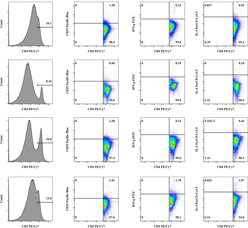
Asian Pac J Allergy Immunol DOI 10.12932/AP-130917-0158

and gives different outcomes to leptospirosis compared to the       CA, USA). The sterility of proteins was confirmed by absence
well characterized mouse model. In the mouse model, the             of bacterial growth on Luria Bertani (LB) agar plates at 37°C
interaction between host and pathogens can induce chemokine         and EMJH media at 30°C. The contaminated endotoxins were
expression and different levels of host susceptibility can give     determined by the Limulus amebocyte lysate (LAL) assay
differential chemokine profiles. BALB/c mice are considered         using Pierce LAL Chromogenic Endotoxin Quantitation Kits
as the most resistant mouse model against leptospirosis and         (Thermo Fisher Scientific, MA, USA). The proteins were kept at
gave the highest level of chemokine expression compared             -20 °C until used.
to C3H/HeJ and C3H/HePas which are sensitive and have
intermediate susceptibility.7 Due to the highest resistance level   2. Recombinant LipL32 protein (rLipL32)
in BALB/c mouse, it was accordingly selected as the model               rLipL32 was produced from BL21(DE3) E.coli carrying
to compare with hamsters which are susceptible to this              the recombinant lipl32-pET23a(+) plasmid as described pre-
pathogen. Furthermore, previous studies reported that antigen       viously with modifications.11,12 Briefly, the transformed E.coli
-presenting cells (APCs) present the processed leptospiral          was grown in LB with 100 µg/ml ampicillin (LB-A) at 37 °C
antigens to CD4+ T cells through MHC Class II molecules,            with shaking at 200 rpm. The rLipL32 protein expression was
leading to their activation and production of cytokines such        induced by 0.2 mM IPTG at 37 °C for 3 hours.
as IL-4 and IFN-γ to support the role of B cells in protection          The His6-tagged rLipL32 was purified from crude solu-
against leptospires.8,9 Therefore, the responses of CD4+ T-cell     bilized protein prepared from bacterial inclusion bodies by
subsets and their intracellular cytokines, IFN-γ and IL-4,          a Ni-NTA affinity column (GE Healthcare, Uppsala, Sweden)
were studied between susceptible hamsters and resistant             under a denaturing condition. The rLipL32 was concentrated
BALB/c mice infected with virulent L.interrogans serovar            and its buffer was exchanged to RPMI1640 plain medium
Autumnalis or the recombinant LipL32 protein (rLipL32) and          (Gibco, Thermo Fisher Scientific, MA, USA) using a 3 kDa
were compared by flow cytometry in this study. In addition,         cut-off Amicon Ultra-tubes (Merck Millipore, County Cork,
the differences in responses of mice and hamsters to                Ireland) at 4 °C, followed by filtration with a 0.2 µm filter
L.interrogans might also reflect the dissimilarities between        membrane (Whatman, Buckinghamshire, England). Protein
hamster and mouse immunities.                                       concentration was measured by a BCA protein assay kit (Ther-
                                                                    mo Fisher Scientific, MA, USA) and the aliquots of proteins
                                                                    were stored at -20 °C.
Methods                                                                 The rLipL32 protein was analyzed by reverse phase nano
Animals and ethics                                                  -liquid chromatography (Dionex, Surrey, UK) coupled with
    Outbred 4 week old female Syrian golden hamsters ob-            MicroToF Q II mass spectrometry (Bruker, Bremen, Germany)
tained from the Animal Laboratory Breeding Unit, Faculty of         and the mass spectrometric result was identified using the
Medicine, Khon Kaen University and four-week-old inbred             MASCOT search engine 2.2 (Matrix Science, Ltd.). Protein
female BALB/c mice purchased from Nomura Siam Inter-                purity of rLipL32 protein was verified under 13% SDS-PAGE
national Co. Ltd. were used in this study. All animals were         and colloidal Coomassie Briliant Blue G-250 stain. Antigenic
maintained in the animal care unit at Faculty of Medicine,          specificity of rLipL32 was confirmed by Western blotting using
Khon Kaen University. All experiments were approved by the          anti-6x His antibody “(R&D Systems, MN, USA)”. The protein
Animal Ethics Committee of Khon Kaen University (No. AEK            sterility and contaminated endotoxins in the rLipL32 were
KU 6/2558 and AEKKU-NELAC 3/2558, No. 0514.1.75/1) and              determined as the same in LAg preparation.
performed in accordance with institutional guidelines.
                                                                    Leptospira infection and rLipL32 injection
Antigen Preparation                                                 1. Live L.interrogans serovar Autumnalis infection
1. Leptospiral crude antigens (LAg)                                     Hamsters and BALB/c mice were divided into three groups,
    LAg was prepared as described.10 Briefly, L.interrogans se-     3 per group, including a non-injected group as a normal
rovar Autumnalis UI13372 was cultured in Leptospira medium          control, an EMJH-injected, and a 102 live L.interrogans serovar
Ellinghausen–McCullough–Johnson–Harris (EMJH) (Becton,              Autumnalis-infected group. Hamsters and BALB/c mice were
Dickinson and Company, Maryland, USA) at 30 °C for 7-10             injected intraperitoneally with EMJH or 102 live L.interrogans
days to yield a cell density of 108 cells/ml. Bacteria were         serovar Autumnalis on day 0. After 10 days of infection, all
harvested by centrifugation at 10,000 ×g for 10 minutes and         animals were sacrificed and spleens were collected.
killed with 0.5 mg/l sodium azide for 30 minutes. The bacteria
were washed twice in 0.01 M phosphate-buffered saline (PBS),        2. rLipL32 injection
pH 7.4, resuspended in PBS, and frozen at -20 °C for 7 days.            Hamsters and BALB/c mice were divided into four groups,
They were centrifuged at 10,000 ×g for 30 minutes at 4 °C           3 per group, including a non-injected group as a normal
after being thawed. The pellets were washed two times with          control, an RPMI1640-injected, a TiterMax gold adjuvant
PBS, resuspended in PBS, and sonicated on ice at 20 kHz (High       (Sigma-Aldrich, USA) injected group and a 20 µg of rLipL32
intensity ultrasonic processor model VC/VCX 750, Sonics) for 3      emulsified in adjuvant-injected group. Hamsters and BALB/c
periods of 3 minutes each.                                          mice were injected intraperitoneally with RPMI1640, TiterMax
    LAg was filtered with 0.2 µm pore size filter membranes         gold adjuvant, or rLipL32 on day 0. The same antigens were
(Whatman, Buckinghamshire, England) and the protein con-            subcutaneously injected at multiple sites on the backs of the
centrations were determined using Bradford reagents (Bio-rad,       hamsters and BALB/c mice on days 7, 14, and 21. Spleens were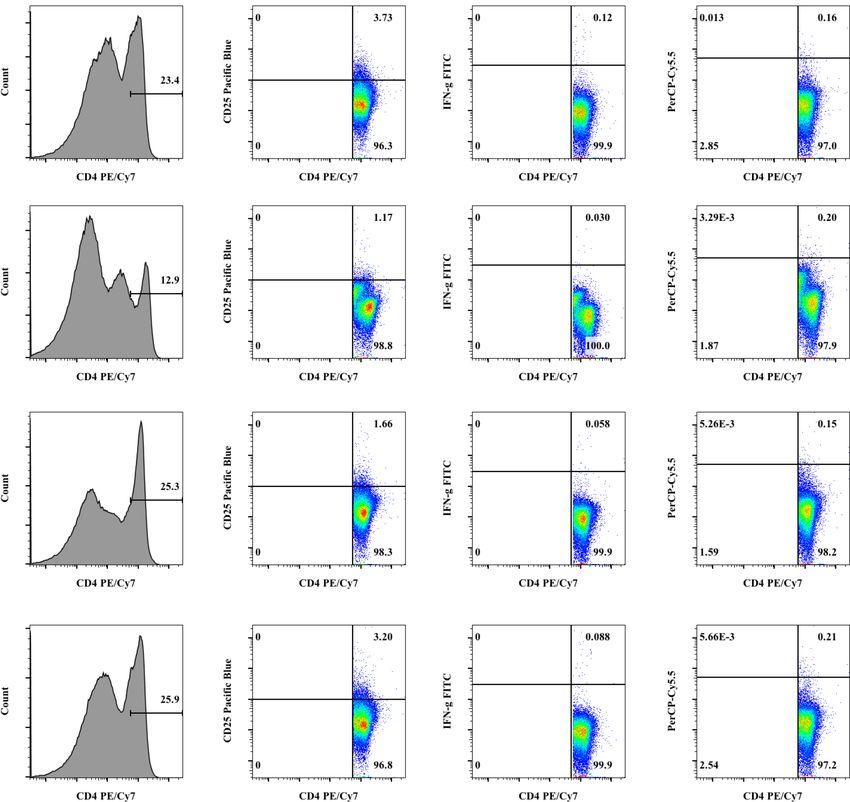
CD4+ T cell responses against leptospires

collected 3 days after the last injection.                         350 ×g for 5 minutes at 4 °C. The culture cells were then washed
                                                                   in fluorescence-activated cell sorting (FACS) staining buffer
Flow cytometric analysis                                           (PBS, 5% FBS, and 0.1% sodium azide) and resuspended in 50
Fluorescent antibodies                                             µl of FACS staining buffer containing an optimal concentra-
    Anti-mouse antibodies used in this study were CD4-PE/          tion of the desired fluorescent antibodies and 2% of normal rat
Cy7 (GK1.5), CD25-Pacific blue (PC61), IFN-γ-FITC (XMG             serum as Fc receptor blocking. Cells were washed with FACS
1.2), and IL-4-PerCP/Cy5.5 (11B11). Isotypic controls were rat     staining buffer after being incubated for 30 minutes at room
IgG2b-PE/Cy7 (RTK4530), rat IgG1-Pacific blue (RTK2071),           temperature. Intracellular staining was subsequently performed.
rat IgG1-FITC (RTK2071), and rat IgG1-PerCP/Cy5.5 (RTK
2071). All antibodies were purchased from Biolegend.                   Intracellular cytokine staining
                                                                       To analyze intracellular cytokine production, the cell
Cell stimulation and surface immunofluorescence staining           surface marker stained cells were fixed with Cytofix/Cytoperm
    Splenic mononuclear cells were isolated from all experi-       solution (BD Biosciences, CA, USA) for 20 minutes at 4 °C
mental animals with Ficoll-Paque solution (GE Healthcare,          and washed twice with Perm/Wash solution (BD Biosciences,
Uppsala, Sweden) according to the manufacturer’s protocols.        CA, USA). Fixed and permeabilized cells were thoroughly
Splenic mononuclear cells were sequentially stained with           resuspended in 50 µl of Perm/Wash solution containing an
fluorescent antibodies and analyzed by flow cytometry.             optimal concentration of anti-IFN-γ-FITC (XMG1.2), anti
Besides flow cytometric analysis, hamster and mouse splenic        -IL-4-PerCP/Cy5.5 (11B11), or isotype control antibody (Bio-
mononuclear cells derived from live serovar Autumnalis             legend, CA, USA) and incubated at 4 °C for 1 hour in the dark.
infections were stimulated with RPMI1640 alone as an               The cells were then washed twice with Perm/Wash solution and
unstimulated control or with 20 µg/ml of LAg at 24 and 48          resuspended in FACS staining buffer prior to flow cytometric
hours. Those derived from rLipL32 injections were stimu-           analysis.
lated with RPMI1640 alone as an unstimulated control or 20             All samples were analyzed on a FACS Canto II flow
µg of rLipL32 at 48 hours. In brief, splenic mononuclear cells     cytometer (BD Biosciences, CA, USA) and data were analyzed
were suspended in complete RPMI1640 medium (RPMI1640               by FlowJo software 10.2 (FlowJo LLC, OR, USA). Lymphocytes
medium supplemented with 10% FBS, 100 U/mL Penicillin              were gated based on an FSC-SSC gate. The stained anti-CD4
and 100 μg/mL Streptomycin). One million cells were cultured       mAb areas were subsequently gated and defined as percentages
with or without mentioned antigens in 48-well plates and           of CD4+ T cells of lymphocytes. Further gating adjustments
5 µg/ml of Brefeldin A (Biolegend, CA, USA) as a protein           were performed based on the expressions of CD25, IFN-γ,
transport inhibitor was added into the cultures 12 hours           and IL-4 (Figure S1-S4). Percentages of each cell subpopula-
before harvesting. The stimulated cells were centrifuged at        tion were calculated. Isotype controls of each antibody were
                                                                   included in each staining protocol.

                                                                                          C: Non-injected group
                                                                                          L: 102 Live L.interrogans-injected group
                                                                                          LAg 24: In vitro re-stimulation with LAg for 24
                                                                                          hours
                                                                                          LAg 48: In vitro re-stimulation with LAg for 48
                                                                                          group

C

C-LAg 24

Figure S1. Flow cytometry gating strategy. Hamster cells derived from live L.interrogans serovar Autumnalis infection and LAg
re-stimulation for 24 and 48 hours were stained with indicated antibodies and examined by flow cytometry. FSC-W, FSC-H,
SSC-W and SSC-H were used to gate out doublet cells after applying the FSC-SSC gate. After selection for CD4+ cells, further gating
adjustments were performed based on the expressions of CD25, IFN-γ, and IL-4.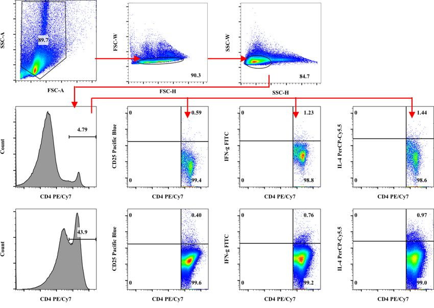
Asian Pac J Allergy Immunol DOI 10.12932/AP-130917-0158
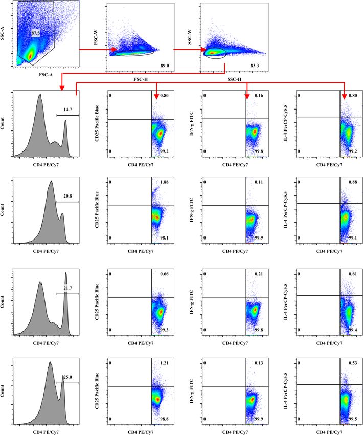
C-LAg 48
L
L-LAg 24
L-LAg 48
Figure S1. (Continued) Flow cytometry gating strategy.
C: Non-injected group
L: 102 Live L.interrogans-injected group
LAg 24: In vitro re-stimulation with LAg for 24
hours
LAg 48: In vitro re-stimulation with LAg for 48
group
C
C-LAg 24
Figure S2. Flow cytometry gating strategy. Mouse cells derived from live L.interrogans serovar Autumnalis infection and LAg
re-stimulation for 24 and 48 hours were stained with indicated antibodies and examined by flow cytometry. FSC-W, FSC-H,
SSC-W and SSC-H were used to gate out doublet cells after applying the FSC-SSC gate. After selection for CD4+ cells, further gating
adjustments were performed based on the expressions of CD25, IFN-γ, and IL-4.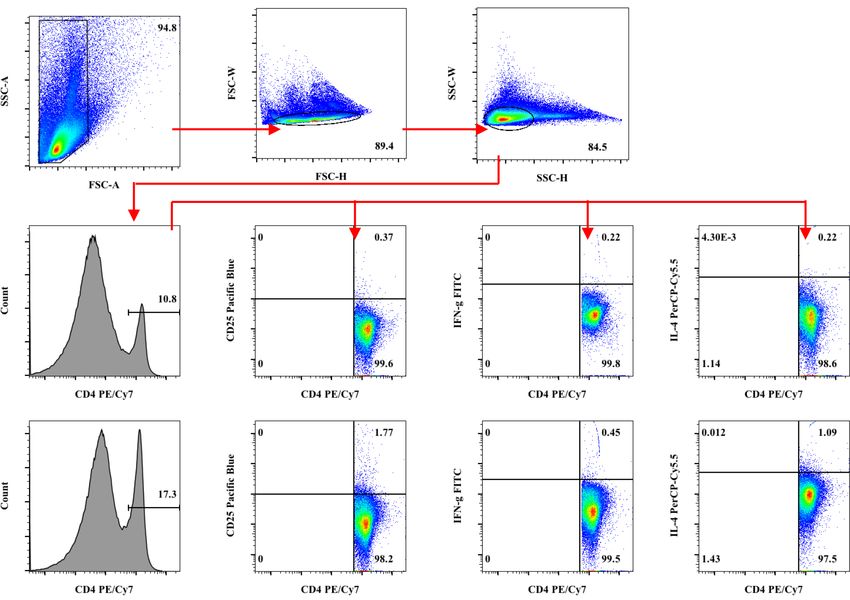
CD4+ T cell responses against leptospires

C-LAg 48

L

L-LAg 24

L-LAg 48

Figure S2. (Continued) Flow cytometry gating strategy.

                                                                                                   C: Non-injected group
                                                                                                   rLipL32: rLipL32-injected group
                                                                                                   rLipL32 48: in vitro re-stimulation with
                                                                                                   rLipL32 for 48 hours

C

C-rLipL32 48

Figure S3. Flow cytometry gating strategy. Hamster cells derived from rLipL32 infection and rLipL32 re-stimulation for 48 hours
were stained with indicated antibodies and examined by flow cytometry. FSC-W, FSC-H, SSC-W and SSC-H were used to gate out
doublet cells after applying the FSC-SSC gate. After selection for CD4+ cells, further gating adjustments were performed based on the
expressions of CD25, IFN-γ, and IL-4.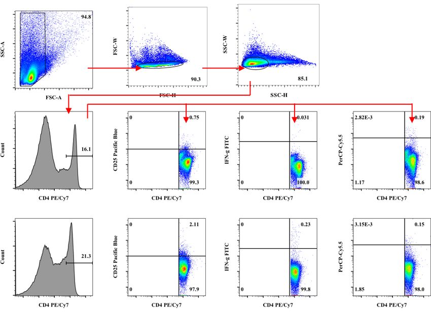
Asian Pac J Allergy Immunol DOI 10.12932/AP-130917-0158

rLipL32

rLipL32-rLipL32 48

Figure S3. (Continued) Flow cytometry gating strategy.

                                                                                                   C: Non-injected group
                                                                                                   rLipL32: rLipL32-injected group
                                                                                                   rLipL32 48: in vitro re-stimulation with
                                                                                                   rLipL32 for 48 hours

C

C-rLipL32 48

rLipL32

rLipL32-rLipL32 48

Figure S4. Flow cytometry gating strategy. Mouse cells derived from rLipL32 infection and rLipL32 re-stimulation for 48 hours
were stained with indicated antibodies and examined by flow cytometry. FSC-W, FSC-H, SSC-W and SSC-H were used to gate out
doublet cells after applying the FSC-SSC gate. After selection for CD4+ cells, further gating adjustments were performed based on the
expressions of CD25, IFN-γ, and IL-4.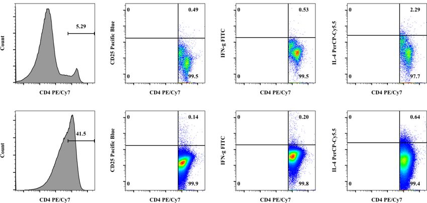
CD4+ T cell responses against leptospires

Statistical analysis                                                        hamsters and BALB/c mice with or without live L.interrogans
   Data are shown as means±standard deviations (SDs). The                   serovar Autumnalis infection were identified by flow cytome-
one-way ANOVA was used to analyze multiple groups and                       try. There were no significant differences between percentages
Student’s t-test was used to compare data between mouse and                 of hamster and mouse CD4+ and CD25+CD4+ T cells respond-
hamster in each parameter. The comparison data with P values                ing to live bacteria in both L.interrogans infected and control
Asian Pac J Allergy Immunol DOI 10.12932/AP-130917-0158

Figure 1. (Continued) Quantification of CD4+ T cells (A) CD25+CD4+ T cells (B) IFN-γ+CD4+ T cells (C) and IL-4+CD4+ T cells
(D) derived from Leptospira infection.

                                      Hamster     C: Non-injected group
                                                  rLipL32: rLipL32-injected group
                                      Mouse       rLipL32 48: in vitro re-stimulation with rLipL32 for 48 hours

Figure 2. Quantification of CD4+ T cells (A) CD25+CD4+ T cells (B) IFN-γ+CD4+ T cells (C) and IL-4+CD4+ T cells (D) derived
from rLipL32 injection. Hamsters and BALB/c mice were intraperitoneally injected with RPMI1640, Adjuvant (TiterMax gold
adjuvant), or 20 µg of rLipL32 on day 0. The non-injected group served as a control. The same antigens were subcutaneously
injected at multiple sites on the backs of hamsters and BALB/c mice on days 7, 14, and 21. Spleens were collected 3 days after the last
injection and splenic mononuclear cells were isolated. Splenic mononuclear cells were analyzed by flow cytometry and also cultured
with or without 20 μg/ml of rLipL32 for 48 hours. Cells were stained with anti-mouse (CD4, CD25, IFN-γ, and IL-4) mAbs. Data
are reported as means ± standard deviations for three animals per group. Statistically significant differences were evaluated using
one-way ANOVA and Student’s t-test. The asterisks (*) and (**) indicate statistical significance at p
CD4+ T cell responses against leptospires
Figure 2. (Continued) Quantification of CD4+ T cells (A) CD25+CD4+ T cells (B) IFN-γ+CD4+ T cells (C) and IL-4+CD4+ T cells
(D) derived from rLipL32 injection.
The IFN-γ+CD4+ and IL-4+CD4+ T cells of hamsters responded IL-4+CD4+ T cells than those of the mouse in the primary
significantly higher than those of mice response (Figure 2D). This result indicates the striking
In contrast to the percentage of CD4+ T cells, the percent- difference of both animal models to the common pathogenic
ages of IFN-γ+CD4+ and IL-4+CD4+ T cells in hamsters were leptospiral antigens. These data suggested that T cell responses
significantly higher than those in mice among all groups. elicited against Leptospira protein, LipL32, are different
L.interrogans serovar Autumnalis-infected hamsters with LAg between hamster and mouse models.
re-stimulation for 48 hours gave the strongest response of
IFN-γ+CD4+ T cells (1.76±0.10%). In vitro LAg re-stimulation Discussion
conditions showed significantly higher responses of IL-4+CD4+ The humoral-mediated immune response is known to be
T cells than conditions without LAg re-stimulation. This cir- a major immune system component against leptospirosis as
cumstance, however, occurred only in hamsters pre-infected leptospires are extracellular pathogens13 while the knowledge
with live L.interrogans (Figure 1C-D). This might indicate the of the T cell response to this disease remains poorly under-
hamster CD4+ T cells produced either IFN-γ or IL-4 differently stood. Several animal models have been used to elucidate host
from mice. immune responses and leptospirosis pathology. Hamsters,
guinea pigs, and gerbils are susceptible to leptospirosis while
Hamster CD4+ T cells responded against rLipL32 differently mice and rats are resistant.1 In order to discover the crucial
from the mice factors for the host defense mechanisms in survival to lepto-
As LipL32 is the common surface protein of pathogenic spirosis and provide more strategies to control this disease,
leptospire serovars, it was then used as the stimulation Leptospira-specific CD4+ T-cell subsets and the cytokine release
antigens in this study for the investigation of differential associated with different host susceptibilities to leptospires were
responses between hamsters and mice. Similar to live infec- analyzed between susceptible hamsters and resistant BALB/c
tions, the numbers of mouse CD4+ T cells (21.23±3.55%) mice. Although the Syrian hamster is highly susceptible to
were significantly higher than the hamsters (5.98±1.59%). many organisms and has been used as an excellent experimen-
Interestingly, after in vitro re-stimulation with rLipL32, ham- tal model for several infectious diseases, it remains unclear why
ster CD4+ T cells (45.90±5.80%) were significantly greater than the hamster is extremely susceptible to such infections and gives
mouse cells (26.05±1.06%) while the hamster CD25+CD4+ the different outcomes in leptospirosis compared to the well
T cells (0.22±0.11%) were significantly lower than mouse characterized mouse model. It might be because of limited
cells (1.02±0.21%) (Figure 2A-B). While the IFN-γ+CD4+ and availability of immunological reagents, specific monoclonal
IL-4+CD4+ T cells of both animals were comparable; LipL32 antibodies (mAbs), and molecular tools to study the immune
stimulated slightly higher, but not significantly, hamster system of this hamster model. The production and development
IFN-γ+CD4+ T cells (0.55±0.26% and 0.31±0.15% for LipL32 of new specific mAbs is time-consuming and expensive. Several
injections and injections with in vitro re-stimulation) than in commercially available anti-mouse mAbs including anti-mouse
the mouse (0.16±0.05% and 0.10±0.03% for LipL32 injections CD4 clone GK1.5,14 anti-rat CD8β clone 341,15 anti-mouse
and injections with in vitro re-stimulation) (Figure 2C). In CD25 clone PC61, anti-mouse IFN-γ clone XMG1.2, and IL-4
contrast, the hamster gave a significantly higher number of clone 11B115 are available which have previously been shown toAsian Pac J Allergy Immunol DOI 10.12932/AP-130917-0158 cross-react with hamsters and were thus used to determine Autumnalis-injected hamsters with LAg re-stimulation for the responses of CD4+ T-cell subsets and their intracellular 48 hours gave the greatest response of IFN-γ+CD4+ T cells cytokines, IFN-γ and IL-4, between leptospirosis susceptible among all samples (Figure 1C). This indicates that the primary hamsters and resistant BALB/c mice in this study. Although infection with L.interrogans serovar Autumnalis primes the the outbred hamsters were used to compare with the inbred populations of antigen-specific hamster CD4+ T cells resulting mice in this study, most of the available hamsters were in the high level of IFN-γ production when re-stimulated extensively line bred from the same mother and litters so that with LAg correlated with host susceptibilities to this infection. they could be closely related to inbred stock. Inbred hamsters The study of specific CD4+ T cell reactivity in various clinical are usually unhealthy with shorter life spans than those outcomes of leptospirosis patients reported that the response constantly outcrossed. Thus, a limitation regarding this point of IFN-γ+CD40L+CD4+ T cells derived from whole-blood spec- could not be excluded. The severity of outcomes in leptospiro- imens stimulated with the leptospiral antigen in vitro and was sis has been based considerably on the environment, pathogen correlated with the severity of leptospirosis in these patients.9 virulence, and host susceptibility.16 Host immune responses LipL32 is derived only from pathogenic strains of Leptospira are hypothesized to be the more significant ones to exhibit and is a well-known outer membrane protein.19 According to the dramatic symptoms of the disease than virulence of the this previous study, the in vivo gene expression of Leptospira pathogen.1 In this study, the ex vivo phenotypes of CD4+ T-cell LipL32 was quantified in blood of animal models with subsets were compared among different groups. The results different susceptibilities to leptospires; the susceptible Syrian demonstrated that there were no differences between the golden hamster and the resistant BALB/c mouse. Their results hamster or mouse models. This indicates similar CD4+ T-cell indicated that the lipl32 expression in hamsters was signifi- stimulation of leptospiral antigens in both animals. After in cantly higher than in mice.20 This result may correlate with vitro re-stimulation with rLipL32 for 48 hours, the responses the present data in which the responses of hamster LipL32 of the mouse CD4+ and CD4+CD25+ T cells were significantly -specific CD4+ T cells were higher than those of the mouse higher than those of the hamster. This might be due to model. Although the response of hamster CD4+ T cells was different secondary immune responses leading to the more dramatically increased in in vitro re-stimulation with rLipL32, rapid production of chemokines which are important for lower levels of IFN-γ and IL-4 producing CD4+ T cells were recruitment and activation of T cells in the resistant model detected. This might be because of the different stimulations of compared to susceptible models. Several studies compared the epitopes in mice and hamsters. immune responses of the host with different susceptibilities to leptospires. The immune responses of the susceptible Syrian Conclusion golden hamster were compared with the resistant Oncins Taken together, the results of the present study appear to be France 1 (OF1) mouse in terms of histological analysis, cyto- the first report demonstrating the different CD4+ T cells and kine mRNA expression, and the quantification of leptospire CD25+CD4+ T cells responses between hamster and mouse loads in target organs and blood. Severe outcomes such as models when infected with live Leptospira. Although there hemorrhage, inflammation, and augmentation of leptospire were a similar number of CD4+ T cells and CD25+CD4+ T burdens were found in hamster organs, while a rapid clear- cells in the primary response, the IFN-γ and IL-4 producing ance was observed in the mice resulting in limited changes cells were different especially when re-stimulated with LAg or in histological observations. The pro-inflammatory cytokines LipL32 antigens. The significantly higher levels of the IFN-γ+ TNF-α, IL-1β, cyclo-oxygenase-2, and IL-6 and anti-inflam- and IL-4+CD4+ T cells in hamsters might make them to be matory cytokine IL-10 were delayed and vast overexpression more susceptible of such infections. The mechanisms of this in the hamster occurred while rapid induction was found in phenomenon remain to be elucidated when reagents for mice. The same result was also observed for the chemokines, hamsters are more available. IP-10/CXCL10 and MIP-1α/CCL3. The rapid cytokine pro- duction and recruitment of immune cells, especially T cells, in resistant mice might be the important factor to rapidly Acknowledgements controlling leptospires and limiting pathological lesions.17 We would like to acknowledge financial supports from Although the numbers of mouse CD4+ T cells was higher Faculty of Medicine and Melioidosis Research Center, Khon than those of hamster CD4+ T cells, these cells produced low Kaen University. Yaowarin Nakornpakdee was supported by levels of IFN-γ and IL-4. The high production of hamster The Development and Promotion of Science and Technol- IFN-γ+CD4+ T cells may lead to the marked inflammation ogy Talents Project (DPST). We would like to acknowledge of infected hamsters causing animal death. This finding was Emeritus Professor James A. Will, University of Wisconsin also reported in previous data by the present authors18 when -Madison, under Publication Clinic, KKU, Thailand for heat-killed vaccine protected hamsters from leptospirosis with editing this manuscript and Assistant Professor Dr. Onrapak lower levels of IFN-γ+CD4+ hamster cells. Another explana- Reamtong, Department of Molecular Tropical Medicine and tion might be due to the various subpopulations of CD4+ T Genetics, Faculty of Tropical Medicine, Mahidol University for cells with distinct cytokine profiles between hamsters and mice analyzing rLipL32 peptides by mass spectrometry. giving the different responses. As the antibody used for deter- mination of the number of hamster CD4+ and CD4+CD25+ T Conflict of Interest cells were anti-mouse antibodies, therefore, the low reactivity to none hamster cells could not be excluded. The L.interrogans serovar
CD4+ T cell responses against leptospires

Source of Funding with Grant Number                                                8.    Faisal SM, McDonough SP, Chang Y. Leptospira: Invasion, Pathogenesis and
                                                                                         Persistence. In: Embers ME, editor. The Pathogenic Spirochetes: Strategies
   Faculty of Medicine, Melioidosis Research Center, Khon                                for Evasion of Host Immunity and Persistence. New York: Springer; 2012.
Kaen University, Higher Education Research Promotion                                     p. 143-72.
and National Research University Project of Thailand, CHE,                         9.    Volz MS, Moos V, Allers K, Luge E, Mayer-Scholl A, Nockler K, et al.
through the Health Cluster (SheP-GMS) and The Development                                Specific CD4+ T-Cell Reactivity and Cytokine Release in Different Clinical
                                                                                         Presentations of Leptospirosis. Clin Vaccine Immunol. 2015;22(12):1276-
and Promotion of Science and Technology Talents Project                                  84.
(DPST).                                                                            10.   Tansuphasiri U, Deepradit S, Phulsuksombati D, Tangkanakul W. Two
                                                                                         simple immunoassays using endemic leptospiral antigens for serodiagnosis
                                                                                         of human leptospirosis. Southeast Asian J Trop Med Public Health.
Author Contributions                                                                     2005;36:302-11.
•    Yaowarin Nakornpakdee did almost all experiments.                             11.   Maneewatch S, Sakolvaree Y, Saengjaruk P, Srimanote P, Tapchaisri P,
•    Rasana W Sermswan designed the leptospiral experiments.                             Tongtawe P, et al. Monoclonal antibodies to LipL32 protect against
                                                                                         heterologous Leptospira spp. challenge. Hybridoma (Larchmt). 2008;27:
•    Santi Maneewatchararangsri designed, cloned, and                                    453-65.
     expressed rLipL32.                                                            12.   Maneewatch S, Adisakwattana P, Chaisri U, Saengjaruk P, Srimanote P,
•    Surasakdi Wongratanacheewin designed all experiments                                Thanongsaksrikul J, et al. Therapeutic epitopes of Leptospira LipL32
     except leptospiral work, wrote grants, and wrote and edited                         protein and their characteristics. Protein Eng Des Sel. 2014;27:135-44.
                                                                                   13.   Fraga TR, Barbosa AS, Isaac L. Leptospirosis: aspects of innate immunity,
     the manuscript.                                                                     immunopathogenesis and immune evasion from the complement system.
                                                                                         Scand J Immunol. 2011;73(5):408-19.
                                                                                   14.   Dondji B, Bungiro RD, Harrison LM, Vermeire JJ, Bifulco C, McMahon
References                                                                               -Pratt D, et al. Role for nitric oxide in hookworm-associated immune
1.   da Silva JB, Ramos TM, de Franco M, Paiva D, Ho PL, Martins EA,
                                                                                         suppression. Infect Immun. 2008;76(6):2560-7.
     et al. Chemokines expression during Leptospira interrogans serovar
                                                                                   15.   Hammerbeck CD, Hooper JW. T cells are not required for pathogenesis
     Copenhageni infection in resistant BALB/c and susceptible C3H/HeJ
                                                                                         in the Syrian hamster model of hantavirus pulmonary syndrome. J Virol.
     mice. Microb Pathog. 2009;47(2):87-93.
                                                                                         2011;85(19):9929-44.
2.   Adler B. Pathogenesis of leptospirosis: cellular and molecular aspects. Vet
                                                                                   16.   Adler B. History of leptospirosis and leptospira. Curr Top Microbiol
     Microbiol. 2014;172:353-8.
                                                                                         Immunol. 2015;387:1-9.
3.   Adler J, Jarvis K, Mitten M, Shipkowitz NL, Gupta P, Clement J.
                                                                                   17.   Matsui M, Rouleau V, Bruyere-Ostells L, Goarant C. Gene expression
     Clarithromycin therapy of experimental Treponema pallidum infections
                                                                                         profiles of immune mediators and histopathological findings in animal
     in hamsters. Antimicrob Agents Chemother. 1993;37(4):864-7.
                                                                                         models of leptospirosis: comparison between susceptible hamsters and
4.   Rouault E, Lecoeur H, Meriem AB, Minoprio P, Goyard S, Lang T. Imaging
                                                                                         resistant mice. Infect Immun. 2011;79(11):4480-92.
     visceral leishmaniasis in real time with golden hamster model: Monitoring
                                                                                   18.   Srikram A, Wongratanacheewin S, Puapairoj A, Wuthiekanun V, Sermswan
     the parasite burden and hamster transcripts to further characterize the
                                                                                         RW. Analyses of vaccination protocols for Leptospira interrogans serovar
     immunological responses of the host. Parasitol Int. 2017;66(1):933-9.
                                                                                         Autumnalis in hamsters. Am J Trop Med Hyg. 2008;79:779-86.
5.   Kaewraemruaen C, Sermswan RW, Wongratanacheewin S. Induction
                                                                                   19.   Haake DA, Chao G, Zuerner RL, Barnett JK, Barnett D, Mazel M, et al. The
     of regulatory T cells by Opisthorchis viverrini. Parasite Immunol. 2016;
                                                                                         leptospiral major outer membrane protein LipL32 is a lipoprotein expressed
     38(11):688-97.
                                                                                         during mammalian infection. Infect Immun. 2000;68(4):2276-85.
6.   Gomes-Solecki M, Santecchia I, Werts C. Animal Models of Leptospirosis:
                                                                                   20.   Matsui M, Soupe ME, Becam J, Goarant C. Differential in vivo gene
     Of Mice and Hamsters. Front Immunol. 2017;8:58.
                                                                                         expression of major Leptospira proteins in resistant or susceptible animal
7.   Domingos RH, Pavanel EB, Nakajima E, Schons-Fonseca L, Da Costa
                                                                                         models. Appl Environ Microbiol. 2012;78(17):6372-6.
     RMA, De Franco M, et al. Resistance of mice to Leptospira infection and
     correlation with chemokine response. Immunobiology. 2017;222(11):1004-
     13.
You can also read