Tooth Resorption in Cats - Fraser Hale
←
→
Page content transcription
If your browser does not render page correctly, please read the page content below
Tooth Resorption in Cats
Introduction                                                lies against the tooth and alveolar bone. The
                                                            free-gingival margin is above the CEJ, but
It has been a little over eight years since I wrote
                                                            gingiva does not attach to enamel, so the gingiva
about tooth resorption in cats and while not a lot
                                                            above the CEJ is known as the free gingiva
has changed, in a practical sense, I think it is
                                                            (‘cause it ain’t attached) and the space between
time I update you on some more recent findings
                                                            gingiva and enamel is known as the gingival
and thoughts. By the way, there is also a paper
                                                            sulcus. From the CEJ to the crest of the alveolar
on tooth resorption in dogs which is something
                                                            bone, the gingiva should be firmly attached to
we are seeing with increasing frequency. I would
                                                            the root cementum in a tight collar completely
encourage you to review that paper as well:
                                                            encircling the tooth. It is this collar of gingival
www.toothvet.ca/PDFfiles/TRs_in_Dogs.pdf                    attachment that isolates the periodontal ligament
                                                            space from oral bacteria and acts as the barrier to
The nomenclature committee of the American
                                                            periodontal disease.
Veterinary Dental College has decided we
should abandon the old terms such as "neck
lesions", "feline odontoclastic resorptive lesions"
and such in favour of the very simple tooth
resorption which is abbreviated as TR. For more
on proper dental nomenclature, grades and stages
of pathology, please visit the AVDC website:
http://www.avdc.org/Nomenclature/Nomen-
Teeth.html#resorption
Over the past decade there have been a number
of papers presented and published examining the
problem of tooth resorption and though the body
of knowledge has been growing, the pieces of
the puzzle have not been adding up to a clear
understanding of the condition. Around the
beginning of this century there were a number of              Line drawing of the periodontal tissues.
new studies presented that showed some promise
of bringing the bits together so that a plausible           Within the alveolus, there is a space between the
picture almost came into focus.                             root and bone (the periodontal ligament space),
Anatomy Review                                              occupied by the periodontal ligament, which
                                                            attaches to the bone and cementum. The
Before I embark on this discussion, we need to              periodontal     ligament        space     appears
review a bit of anatomy to make sure we are all             radiographically as a fine radiolucent line
speaking the same language.                                 following the contour of the root.
The crown of the tooth is that part covered by              I need to quote an axiom that must be kept in
enamel and the root is covered by cementum.                 mind. A little knowledge is a dangerous thing.
Where the crown and root meet is the neck,                  What follows will be a condensed review of
cervix or cementoenamel junction (CEJ). The                 some relatively recent papers. Much of this
bulk of the tooth is composed of dentin and                 information needs further study before we can
inside the tooth is a hollow chamber containing             change our approach to treating tooth resorption.
the dental pulp.                                            I do not want you going off half-cocked.
Most of the root is situated in a depression in the         One more introductory note (which will be
bone known as the alveolus. The most coronal                repeated because it is soooo important).
portion of the root extends above the margin of
the alveolar bone. Gingiva is a tough tissue that

Hale Veterinary Clinic               toothvet@toothvet.ca      www.toothvet.ca          Local Calls: 519-822-8598
Fraser A. Hale, DVM, FAVD, Dipl AVDC Page 1                    August 2012         Long Distance: 1-866-866-8483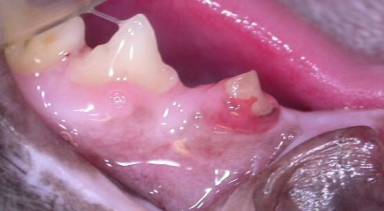
INTRA-ORAL DENTAL RADIOGRAPHY IS
ESSENTIAL    FOR THE   ACCURATE
ASSESSMENT AND PROPER TREATMENT
OF TOOTH RESORPTION, REGARDLESS
OF THE SPECIES.

I am not just saying this to encourage you to
refer (though that would be nice) – it just makes
sense. I doubt any of you would even consider
treating a fractured limb without diagnostic
radiographs. Nor would you do an exploratory                We can see the uniform density of the roots, the
laparotomy without radiographs. Without dental              pulp chamber within, the thin dark line of the
radiographs, you simply cannot know what you                periodontal ligament space and the normal
are getting into or when you have gotten out. So            variations in the density of the alveolar and
if you are not willing and able to do whole-                mandibular bone, including the lucency
mouth intra-oral dental radiographs on all of               associated with the mandibular canal.
your dental patients, then you should stop                  Below is a digital photograph of an old analog
offering dental services. Diagnosis first...then            radiograph of the left mandible of a cat with type
treatment.                                                  1 tooth resorption.
Types of Tooth Resorption
From the Nomenclature page at www.avdc.org:
On a radiograph of a tooth with type 1 (T1)
appearance,     a    focal  or    multifocal
radiolucency is present in the tooth with
otherwise normal radiopacity and normal
periodontal ligament space.
On a radiograph of a tooth with type 2 (T2)
appearance, there is narrowing or
disappearance of the periodontal ligament
space in at least some areas and decreased
radiopacity of part of the tooth.
On a radiograph of a tooth with type 3 (T3)
                                                            There is loss of bone and tooth structure, no new
appearance, features of both type 1 and
                                                            hard tissue is filling the defects and beyond the
type 2 are present in the same tooth. A tooth
                                                            areas of tissue loss, the bone, roots and
with this appearance has areas of normal
                                                            periodontal ligament space all look normal. The
and narrow or lost periodontal ligament
                                                            mesial root of the 4th premolar has been
space, and there is focal or multifocal
                                                            transected allowing bacteria into the endodontic
radiolucency in the tooth and decreased
                                                            system of this tooth resulting in septic pulp
radiopacity in other areas of the tooth.
                                                            necrosis. The infection has then oozed out
Here is a normal digital radiograph of the left             through the tip of the distal root resulting in
mandible of a cat with radiographically normal              apical periodontitis and bone loss (arrow).
teeth and surrounding tissues.
                                                            Type 1 lesions are associated with moderate to
                                                            severe gingivitis and/or periodontitis. They
                                                            typically start at the CEJ and can extend in all
                                                            directions from that starting point. The cause of
                                                            these lesions might well be local inflammatory
                                                            reaction due to gingivitis or periodontitis. It is

Hale Veterinary Clinic               toothvet@toothvet.ca      www.toothvet.ca         Local Calls: 519-822-8598
Fraser A. Hale, DVM, FAVD, Dipl AVDC Page 2                    August 2012        Long Distance: 1-866-866-8483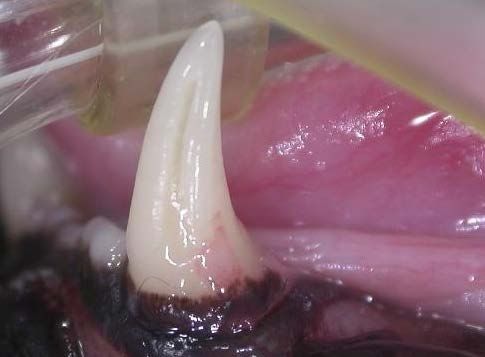
hard to say if the lesions are the cause of the
periodontitis or the result of it.
As type 1 lesions are often associated with
periodontitis and endodontic disease, these teeth
must be entirely extracted. It is not permissible
to leave any remnants of these roots behind. The
good news is that the presence of a periodontal
ligament (i.e., the lack of ankylosis) makes these
roots fairly easy to extract. The bad news is that
the resorptive process usually creates serious
weak spots on the roots, so the roots break and
then it is necessary to surgically remove the root
remnants. Having a pre-operative dental x-ray
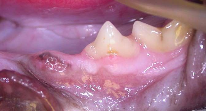
lets you know where the weak spots are. In the photo above, there is soft tissue creeping
up the crown of the lower left canine tooth and
Type 2 lesions are more frustrating to deal with.
this almost always indicates a resorptive lesion
These are the ones that we thought we might be
of some sort.
getting closer to understanding.
Below is a radiograph of healthy, normal
mandibular canine teeth.
Type 2 lesions are not generally associated with
endodontic disease or periodontitis, though there
may be localized gingivitis or granulation tissue The radiograph shows advanced type 2 tooth
in and around the lesion itself. These lesions resorption affecting both lower canine teeth. The
seem to be able to arise at any location on the right canine lesion (to your left) is all within the
root and are associated with extensive root confines of the alveolus but the one affecting the
resorption, loss of periodontal ligament and left canine tooth has extended above the alveolus
ankylosis. and onto the crown, as shown in the photograph.
New(ish) Histologic Studies
Steinberg looked at 80 clinically normal teeth
taken from cats that had at least one clinical TR
and found that 100% of these teeth had
microscopic evidence of resorption and these
lesions were found primarily in the non-cervical
Hale Veterinary Clinic toothvet@toothvet.ca www.toothvet.ca Local Calls: 519-822-8598
Fraser A. Hale, DVM, FAVD, Dipl AVDC Page 3 August 2012 Long Distance: 1-866-866-8483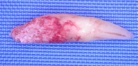
region of the root. This suggests that if these cats        extends through the gingival attachment and
live long enough, every single tooth will                   comes in contact with oral bacteria,
eventually succumb to resorptive lesions. Taken             inflammation develops as a result of the lesion,
further, it could be said that if they live long            not its cause.
enough, every cat will develop type 2 TRs.
                                                            New(ish) Biochemical Studies
Gorrel and Larsson also did a study looking for
                                                            Alex Reiter has done a study which looked like it
microscopic lesions on the roots of cat teeth.
                                                            might prove to be the key to unlocking the
They harvested 56 teeth that appeared clinically
                                                            mystery. He looked at the serum concentration
and radiographically free of TRs. Of these, 43
                                                            of calciotropic hormones in cats with TRs. What
teeth (group A) were taken from cats that had a
                                                            he found was that cats with TRs had
clinical TR on at least one tooth and 13 teeth
                                                            significantly higher serum levels of 25
(group B) were taken from cats with no clinical
                                                            hydroxyvitamin D than cats without TRs.
or radiographic evidence of TRs on any teeth.
Twenty six teeth from group A and one tooth                 As he points out, rather than asking “why do
from group B showed microscopic evidence of                 some cat teeth undergo resorption?”, we should
external root resorption.                                   ask “why do some cat’s teeth not undergo
                                                            resorption?”. After all, teeth are made of the
Among the salient observations of this study
                                                            same tissues as bone and it is constantly
were the following:
                                                            undergoing remodeling. Normally, the roots of
The cervical cementum on the roots of teeth                 permanent teeth are considered to be resistant to
from TR cats was thicker and more irregular                 resorption because of a protective organic
than that found on the roots of teeth taken from            matrix. If this matrix is lost or becomes calcified,
TR-free cats.                                               then odontoclasts (which are virtually identical
The periodontal ligament around non-ankylosed               in every way to osteotoclasts) can attack the root
resorptive lesions was narrow and the fibers                surface.
arranged vertically (rather than the normal                 Reiter proposes that hypercementosis, osteoid
horizontal or oblique arrangement). The                     production along the socket wall or gradual
ligament around ankylosed lesions was grossly               calcification of the periodontal ligament might
abnormal with edematous vascularized tissue.                be the trigger. He further postulates that this is
Teeth from TR cats were much more likely                    caused by hypervitaminosis D. Another study
(60%) to have microscopic resorptive lesions                found that excess administration of vitamin D or
than teeth from TR-free cats (8%). These                    its metabolites to experimental animals resulted
microscopic lesions were all located at the mid             in dental and periodontal changes very similar to
root or apical portion of the root and were not             those seen around teeth affected by TRs. Since
associated with inflammation.                               cats cannot manufacture vitamin D, they must
                                                            take it in from dietary sources.
Healed cemental lesions covered by intact
periodontal tissue was seen in some cases.                  Looking at the vitamin D content in canned cat
                                                            foods, Reiter found that 20 of 49 brands (41%)
Ankylosis was associated with formation of                  had in excess of 30 times the vitamin D
reparative bone-cementum tissue filling in the              requirements of 250IU/kg diet dry matter and
space vacated by the disappearing root tissue.              31% actually exceeded the AAFOO maximum
These findings suggest that type 2 lesions start            level of 10 000 IU/kg diet dry matter.
within the cementum of the root and in some                 Colin Harvey and others have done some work
cases they may heal spontaneously. The other                looking at a biphosphonate drug (alendronate)
big surprise was the finding that these early               that is used in humans to prevent osteoporosis.
lesions are not associated with inflammation. It            One study found that the drug bound very
seems that as long as the lesion remains on the             heavily to alveolar bone and root cementum –
root below the level of gingival attachment and             that’s good news as it means we can get this
protected from oral bacterial contamination,                drug right where we want it.
there is no inflammation. Once the lesion
Hale Veterinary Clinic               toothvet@toothvet.ca      www.toothvet.ca          Local Calls: 519-822-8598
Fraser A. Hale, DVM, FAVD, Dipl AVDC Page 4                    August 2012         Long Distance: 1-866-866-8483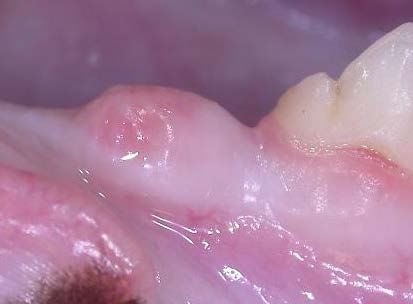
The next study looked at the effect this drug               cats with lower serum 25-OH-D concentrations.
might have on established type 2 lesions over               In conclusion, the hypothesis that higher serum
time. Using colony cats at research facilities, he          25-OH-D concentrations are associated with a
quantified the size of lesions found. The cats              higher prevalence of TR is not supported by this
then received alendronate for a year and the                study. J Vet Dent 27 (3); 142-147, 2010
lesions were re-measured. In the vast majority of
                                                            And, there are serious concerns in human
cases, the lesions were no larger a year later and
                                                            patients and warnings to veterinarians as well
in some cases they were actually smaller. In the
                                                            that bisphosphonates can cause a severe
control cats, the lesions increased in size as
                                                            osteonecrosis of the mandible in some patients.
predicted. These findings suggested that
                                                            So the use of these drugs in cats at this time
alendronate may someday be useful to prevent
                                                            cannot be justified.
the progression of very early lesions and even in
preventing them completely.                                 What does all this add up to?
However, more recently there was this:                      The two studies looking at Vitamin D and TR
                                                            found opposite results so all we can say on that
Tooth Resorption and Vitamin D3 Status in
                                                            score is that more study is indicated.
Cats Fed Premium Dry Diets
                                                            Type 1 lesions are possibly the result of gingival
Nicolas Girard, DVM; Eric Servet, Food Ing;
                                                            and periodontal inflammation and arise at the
Philippe Hennet, DVM; Vincent Biourge, DVM,
                                                            cervical region of the tooth. From there, they
PhD
                                                            may extend up the crown and/or down the root.
Summary: It has been suggested that tooth                   Prevention would be based on maintaining good
resorption (TR) in cats is associated with vitamin          oral hygiene. Treatment of detectable lesions is
D3 status. The purpose of this study was to                 complete extraction of the tooth and its roots
evaluate any correlation between serum 25-OH-               (leave nothing behind). On the other hand, these
D concentrations and the prevalence of TR. The              may be caused by the same process as causes
healthy adult domestic cats (n=64) of this study            Type 2 lesions but they take on a different
had been fed similar premium dry-expanded                   clinical presentation because their location
foods throughout their lives. Serum 25-OH-D                 allows them to be contaminated with oral
was measured, and cats received a single,                   bacteria very early in the process and so
complete     periodontal     examination,      with         inflammatory changes dominate the situation
periodontal probing of each tooth and                       (chicken or egg?).
exploration of the tooth surface using a dental
                                                            Type 2 lesions are a non-inflammatory
explorer. A complete set of 10 dental
                                                            resorption that begins within the socket or at
radiographs was taken for each cat. There were
                                                            least below the level of gingival attachment and
168 TRs diagnosed in 40 of 64 cats (85 were
                                                            may be triggered by vitamin D toxicity or not.
Type 1 TR and 83 were Type 2). The mean serum
                                                            Only after the lesion enlarges sufficiently to
25-OH-D concentration was 187.7 ± 87.3
                                                            break through the gingival attachment to become
nmol/L. The mean serum 25-OH-D in cats with
                                                            contaminated     with      oral   bacteria     does
one or more TR was 164.2 ± 78.8 nmol/L,
                                                            inflammation become a factor. Treatment is still
compared with 226.8 ± 88.2 nmol/L for those
                                                            extraction and in my view it should be complete
without TR (p = 0.14). The mean serum 25-OH-
                                                            removal of all dental tissues (more on this later).
D in the 13 cats with >5 TR was 131.2 ± 49.5
nmol/L, which was significantly less than in cats           The only way to distinguish between a type 1
with no TR (p < 0.05). There was no relationship            lesion and a type 2 lesion is with intra-oral
between TR Type and serum 25-OH-D. There                    dental radiography.
was no effect of age or sex on serum 25-OH-D.               For now, my feline discharge statement contains
On the contrary, variations in serum 25-OH-D                the following statement:
were observed according to the studied breeds.
There was no relationship between TR Type and               While we do not know the causes of tooth
serum 25-OH-D. TR prevalence was greater in                 resorption, we do know that cats that have
                                                            had some are likely to develop more. There
Hale Veterinary Clinic               toothvet@toothvet.ca      www.toothvet.ca          Local Calls: 519-822-8598
Fraser A. Hale, DVM, FAVD, Dipl AVDC Page 5                    August 2012         Long Distance: 1-866-866-8483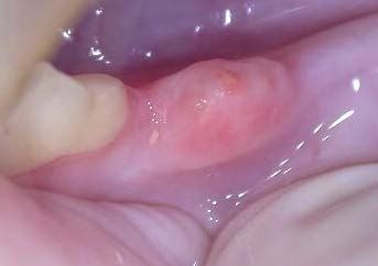
is no way to predict which teeth will be                    Here is the position statement from the American
affected next or when and we currently have                 Veterinary Dental College:
no recommendations for prevention of new
                                                            Feline tooth resorption typically originates in
lesions. All we can do is monitor for new
                                                            the cementum, may progress into root
problems and deal with them as they arise.
                                                            dentin, and then either progress through the
That is not a very comforting thought, but it is            root, into the crown, or both. Tooth
the best we have at the moment.                             resorption that can be identified on oral
                                                            examination is an indication for radiographic
More on the treatment of Type 2 TRs. Here is an
                                                            evaluation      and    treatment.      Intraoral
old reference that started a lot of problems.
                                                            radiography is required to properly evaluate
Crown amputation with intentional root                      this condition. Whole-mouth radiographs are
retention for advanced feline resorptive                    recommended to evaluate other teeth in the
lesions - A clinical study.                                 mouth. Complete extraction is the treatment
Gregg DuPont, DVM                                           of choice for teeth that have detectable
                                                            crown resorption but no radiographic
Summary: Whole tooth extraction is generally                evidence of root resorption. Teeth with
considered to be the treatment of choice for teeth          crown resorption but radiographic signs of
with advanced feline external odontoclastic                 advanced root resorption (and no concurrent
resorptive lesions. These teeth often have both a           periodontal disease, periapical periodontitis
weakened, brittle crown and radicular ankylosis.            or stomatitis) may be treated by subgingival
These two factors cause frustration and                     amputation. Either form of treatment should
sometimes complications during attempts at                  be followed by gingival closure. If there is
extraction. This study investigated the                     radiographic evidence of root resorption, but
alternative of intentionally leaving part or all of         no clinical resorption can be detected on
non-pathologic tooth roots in situ to prevent               oral examination, the tooth can be
iatrogenic trauma to the patient, loss of alveolar          "monitored" or preemptively extracted.
bone, and prolonged healing of surgical defects.            Restoration of these teeth is not
Fifty one roots from 23 teeth were radiographed             recommended.         Semiannual          dental
5-36 months following elective root retention;              examinations are recommended for all cats
continued resorption without surrounding bony               with previous diagnosis of tooth resorption.
reaction was seen in almost all cases. In one cat,          Radiography should be repeated annually or
the roots retained normal periodontal ligament              more frequently as dictated by the oral
one year later, and in another cat that developed           examination.
severe stomatitis, the intentionally retained roots
were extracted at the same time that the                    Adopted by the Board of Directors, April
remaining molar teeth were extracted. J Vet                 2006, revised April 2007
Dent 12 (1); 9- 14, 1995.                                   That statement is now five years old.
This paper is now 17 years old. Sadly, some of              I will be the first to admit that veterinary dentists
the most important features of this paper have              do not have all the answers with regard to TRs
been lost on the general veterinary population.             yet, however, we do always take dental
The paper suggested that in very particular                 radiographs as part of our evaluation of TRs and
circumstances, it might be permissible to leave             that is absolutely essential.
some root remnants in place when extracting
                                                            As I have stated, type 1 lesions should be
teeth with resorptive lesions. Unfortunately, this
                                                            completely extracted. Also, teeth with any
message got bastardized until the word on the
                                                            radiographic or clinical evidence of deep
street was that it was okay to just snap off the
                                                            periodontal or endodontic disease should be
crowns and leave the rest to sort itself out. This
                                                            extracted completely. Teeth in cats who are
gets back to the axiom about a little bit of
                                                            positive for FeLV, FIV or who have a history of
knowledge being dangerous.
                                                            Feline Chronic GingivioStomatitis should be
                                                            completely removed. Let me say that again. For

Hale Veterinary Clinic               toothvet@toothvet.ca       www.toothvet.ca          Local Calls: 519-822-8598
Fraser A. Hale, DVM, FAVD, Dipl AVDC Page 6                     August 2012         Long Distance: 1-866-866-8483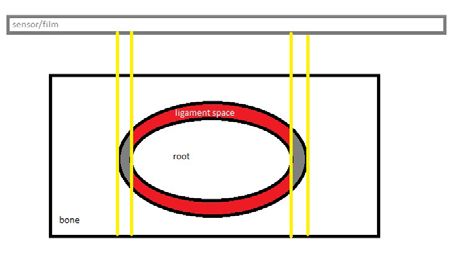
cats with FCGS, you must remove every scrap                 gingival flaps, remove all visible or
of every root of every tooth and you must                   radiographically detectable dental tissues,
confirm/document complete extraction with                   smooth the bone and suture the wound with 5-0
immediate post-operative radiographs.                       monofilament absorbable.
If a tooth has a type 2 lesion without periodontal          In the radiograph below, the left lower 3rd
or      endodontic      disease     (as    confirmed        premolar seems to be almost completely
radiographically) and the root appears to have              resorbed and replace by new hard tissue.
been largely replaced by new bone-cementum
tissue, then it might be permissible to leave a
little of this tissue in the socket. The theory goes
that the lesions are non-inflammatory and the
tissue is healing the defect. My worry is that the
lesion has been chronically exposed to oral
bacteria by the time the lesion is detected and so
I expect that bacterially induced inflammation
must extend at least part way down the “root”.
Therefore, my approach is still to remove all
identifiable dental tissue and then suture the
wound closed.

                                                            In the clinical photo of the area, the gingiva is
                                                            fistulated and inflamed over the remains of the
                                                            3rd premolar. So I reflected a flap and removed
                                                            all that remained of the 3rd premolar as shown in
                                                            the post-op radiograph.

Even those who advocate the more conservative
approach of “intentional root retention” agree
that removing just the crown is not enough. In
the radiograph above, we can see that there is
still some coronal enamel at the most distal
aspect of the tooth and there are also portions of
the root projecting above the alveolar crest.
Much of the root tissue has been replaced by                I do not consider this intentional root retention as
bone-cementum tissue and so the roots appear as             there was no root left - it had been completely
ghosts. The gingiva overlying this tooth typically          resorped and replaced. This is in contrast to the
would have a fistula and so there is ongoing                right side. Here are the images of the area.
bacterial invasion. Left on its own, the rest of
this tooth will likely resorb, but in the meantime,
there is an open and contaminated wound with
inflammation and pain. At the risk of being
repetitive, my approach would be to reflect
Hale Veterinary Clinic               toothvet@toothvet.ca      www.toothvet.ca          Local Calls: 519-822-8598
Fraser A. Hale, DVM, FAVD, Dipl AVDC Page 7                    August 2012         Long Distance: 1-866-866-8483
Summary: The aim of this retrospective study
was to follow the progression of radiographic
changes in intentionally retained roots of teeth
affected with tooth resorption type 2 in cats.
Emphasis was placed on assessment of degree of
resorption as well as the occurrence of
inflammatory changes in tooth roots. The results
confirm that crown amputation is an adequate
treatment in cat for teeth affected by type 2
resorption. J Vet Dent 29 (1); 20-26, 2012
Now, this paper purports to validate the practice
of intentional root retention, but I found the logic
used to reach that conclusion unconvincing. The
paper refers to lack of radiographic signs of
inflammation, but inflammation does not show
up on radiograph and neither does pain. I have
seen plenty of cats with persistent inflammation
around retained root remnants and so my
approach is, if I am removing any of the root, I
will remove ALL of the root. Since I can remove
In that pre-operative radiograph, I felt the tissue all of the root, why would I leave any behind to
where the root should be also looked like new potentially cause persistent pain? It might save a
hard tissue and so I removed the crown and part bit of time to leave some roots behind, but
way down into the socket, but I did not remove saving time is not the priority. Doing the job
the new hard tissue. Again, I do not consider this properly is the goal and in my view, that still
to be so-called crown amputation or intentional means removing all that remains of the tooth.
root retention as I removed everything that bore
any resemblance (visually or radiographically) to Here is a case where the lower 3rd premolars
dental tissue and what I left behind was the new were in advanced stages of tooth resorption but I
hard tissue that had replaced the resorbed root. could still tell the difference between tooth and
bone and so I completely removed these teeth.
First the photo, pre-op and post-op radiographs
of the right lower 3rd premolar (note the
inflammation of the soft tissue over the remains
of the tooth).
Here is a more recent paper on the issue that
seems to lend further support to the practice of
intentional root remnant retention:
Radiographic changes associated with tooth
resorption type 2 in cats.
Mihaljevic SY, Kernmaier A, Mertens-Jentsch S.
Hale Veterinary Clinic toothvet@toothvet.ca www.toothvet.ca Local Calls: 519-822-8598
Fraser A. Hale, DVM, FAVD, Dipl AVDC Page 8 August 2012 Long Distance: 1-866-866-8483And next the same images of the left 3rd
premolar.
More on Radiographic interpretation
Now, one of the criteria you will see for
intentional root retention is that if you cannot see
the periodontal ligament space on the
radiograph, then you can leave the roots in place.
I could not disagree with this terrible over
simplification more.
A radiograph is a two-dimensional
representation of a three dimensional group of
structures. On even the best radiograph, you can
only ever see a tiny percentage of the periodontal
ligament space. In the highly simplified
representation of the cross-section of an oval
root in an oval socket with a periodontal
ligament space in between them, there is a
radiograph film/sensor to the top of the image to
capture the radiation passed through the tissues.
The yellow lines represent some of the x-ray
beams. The radiographic shadow cast by this
section of jaw with the root in place will only
Hale Veterinary Clinic toothvet@toothvet.ca www.toothvet.ca Local Calls: 519-822-8598
Fraser A. Hale, DVM, FAVD, Dipl AVDC Page 9 August 2012 Long Distance: 1-866-866-8483show the periodontal ligament space in the area
between each pair of yellow lines (shaded grey).
None of the red periodontal ligament space is
going to be seen on the image because it is
hidden within the superimposed hard tissues of
the root and the bone. There might be some
ankylosis within the visible grey areas but if the
rest of the (red) periodontal ligament space is
normal, then the tooth should come out in one
piece without trouble. On the other hand, the
visible (grey) area might look fine on the
radiograph but the tooth might be a monster to Here is the extracted tooth, all in one piece.
extract due to ankylosis within the invisible (red)
areas. So, while radiographs are completely
essential, they are only part of the overall
picture.
And a radiograph of that extracted tooth.
I commonly find teeth that look on radiograph as
if they are ankylosed suggesting that there is no
chance I will be able to remove them intact, yet I
am still able to get them out entirely without
breaking any of the root(s).
Here is the pre-operative radiograph of a
resorbing right upper canine tooth in a cat. The It is also common to have teeth that look, on
periodontal ligament appears absent. radiograph, as if they should co-operate and
come out intact, yet they crumble to bits.
Conclucsion
How do I want you to manage TRs?
In my fantasy world, you would just refer them
all to me.
If you are going to treat cats with TRs here is
what you have to do:
Top of the next page is the post-operative Radiograph every tooth in the head (even those
radiograph showing that the entire canine tooth that appear to be missing) and examine each
was removed and the bone at the top of the tooth subgingivally with a dental explorer. If you
socket has been reduced in preparation for are not going to radiograph, then do not try to
wound closure. treat these cats, just refer them to someone who
will radiograph them.
Hale Veterinary Clinic toothvet@toothvet.ca www.toothvet.ca Local Calls: 519-822-8598
Fraser A. Hale, DVM, FAVD, Dipl AVDC Page 10 August 2012 Long Distance: 1-866-866-8483For type 1 lesions, extract the entire tooth and root(s), smooth off the alveolar bone and suture the wound closed with a fine, absorbable monofilament (I like 5-0 Monocryl™). For type 2 lesions, extract as much of the tooth as you can without causing excessive trauma (that is a judgment call), being sure to at least get everything above the alveolar crest and 1 – 2 millimeters into the socket before smoothing the bone and suturing as above. Personally, I will continue with my habit of removing all that remains of any tooth I am removing. Plan to re-examine and re-radiograph annually as cats that have had one lesion will almost certainly get others sooner or later. Send the animals home with analgesics (I like transdermal codeine for cats) for at least four days and put them on softened food for 14 days. Have the owners keep the pet quiet for a few days and tell them to not touch the mouth for 2 weeks. Consider sending home an E-collar to prevent pawing/rubbing at the face. Antibiotics are for the treatment of infection, not for the prevention of infection. When I do oral surgery, I remove the infected tissue and so only rarely (almost never) do I feel the need to send home antibiotics. There will continue to be confusion and frustration around tooth resorption until we know what causes it and can offer some solid recommendations for prevention. There will also continue to be controversy regarding the advisability of intentional root retention. Hopefully we will have some solid answers down the road, but in August of 2012, this is it. Hale Veterinary Clinic toothvet@toothvet.ca www.toothvet.ca Local Calls: 519-822-8598 Fraser A. Hale, DVM, FAVD, Dipl AVDC Page 11 August 2012 Long Distance: 1-866-866-8483
You can also read