Methylglyoxal, the Major Antibacterial Factor in Manuka Honey: An Alternative to Preserve Natural Cosmetics? - MDPI
←
→
Page content transcription
If your browser does not render page correctly, please read the page content below
cosmetics
Article
Methylglyoxal, the Major Antibacterial Factor in
Manuka Honey: An Alternative to Preserve
Natural Cosmetics?
Claudia Juliano *        and Giovanni Antonio Magrini
 Department of Chemistry and Pharmacy, University of Sassari, Via Muroni 23/A, 07100 Sassari, Italy;
 giovanniantonio.magrini@gmail.com
 * Correspondence: julianoc@uniss.it; Tel.: +39-7922-8729

 Received: 28 November 2018; Accepted: 20 December 2018; Published: 25 December 2018

 Abstract: Microbial safety is an essential prerequisite of cosmetics, and preservatives are required to
 prevent product spoilage and damage to consumers’ health. Consumer concern about the safety of
 some cosmetic ingredients and the increasing demand for more natural beauty products has driven
 cosmetic industries and formulators to find natural alternatives to replace synthetic preservatives
 currently used. In this study, methylglyoxal (MGO, the main factor responsible for the antimicrobial
 activity of manuka honey) was tested for antimicrobial activity against a panel of selected bacteria
 and mycetes by using conventional microbiological techniques (determination of M.I.C., time-kill
 assay), and its potential preservative in an O/W emulsion was investigated (challenge test). MGO
 showed a remarkable and fast antibacterial activity (M.I.C. values 0.150–0.310 mg/mL), while the
 inhibitory activity against fungi was less marked (M.I.C. values 1.25–10 mg/mL); chitosan has proven
 to be a synergist of antimicrobial effectiveness of MGO. Results of the challenge test showed that
 the addition of MGO to a cream formulation was efficient against microbial contamination. On the
 basis of our results, MGO appears to be a good candidate as a cosmetic preservative of natural origin;
 further studies are needed to confirm its applicability and its safety.

 Keywords: methylglyoxal; cosmetic preservatives; antibacterial activity; antifungal activity; chitosan

1. Introduction
     Microbial contamination and growth can occur in cosmetics and personal care products during the
manufacture, storage, and particularly during the period of use [1]. Multiplication of microorganisms
may cause spoilage of cosmetic formulations, but can also constitute a threat to consumer health,
especially when cosmetics are intended for use in areas of particular concern or when they are
used by young children or immunocompromised subjects. Therefore, preservatives that are able to
prevent microorganisms from growing play a crucial role, particularly in formulations containing
water. In the European Union, only preservatives listed in Annex V of Regulation (EC) [2] can be
used in cosmetics and personal care products. Unfortunately, exposure to some of these substances
can produce undesirable effects, such as skin irritation and sensitization [3], and some of them are
recognised as potential endocrine disruptors [4]. Moreover, a hypothetical connection between cosmetic
preservatives—namely parabens—and breast cancer was recently suggested [5,6], although this link
has been denied in a relatively recent review of literature [7]. These concerns and the rising consumer
demand for more natural cosmetics are driving the cosmetic industry to find natural, safer alternatives
to synthetic preservatives. In that context, a number of botanical extracts and essential oils have been
tested in vitro as potential cosmetic preservatives, sometimes with promising results [8–11]. As part of
the research about potential natural preservatives, a recent study [12] investigated the preservative

Cosmetics 2019, 6, 1; doi:10.3390/cosmetics6010001                           www.mdpi.com/journal/cosmetics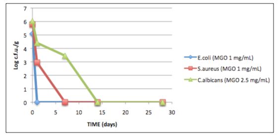
Cosmetics 2019, 6, 1                                                                                  2 of 8

efficacy of manuka honey, a New Zealand honey well known for its pronounced antimicrobial activity
also against clinical multi-resistant isolates [13–15], in an O/W emulsion. Manuka honey was proven
to be more effective than methylparaben against Pseudomonas aeruginosa and showed the same efficacy
of this synthetic compound against Escherichia coli; however, it did not reveal inhibitory activity
against fungal contamination [12]. Methylglyoxal (MGO), a 1,2-dicarbonyl compound present in
manuka honey in exceptionally high levels (38–761 mg/kg), is the main factor responsible for its
bactericidal activity [16,17]; however, neutralization of MGO reduced but did not completely eliminate
antibacterial activity, which proves the presence in manuka honey of other antibacterial factors [18].
The aim of the present investigation was to evaluate the potential of MGO as an alternative natural
preservative intended for cosmetics and personal care products, by using conventional microbiological
techniques (M.I.C. and M.B.C. evaluation, time-kill test, challenge test). Moreover, it is well known that
a combination of preservatives is often a good solution to improve the efficacy of a preserving system
and to reduce the side effects associated with the individual substances; in accordance with these
considerations, we also investigated an eventual synergistic interaction between MGO and chitosan,
a biopolymer obtained by partial deacetylation of chitin that possesses, among others, interesting
antimicrobial properties [19].

2. Materials and Methods

2.1. Materials
      Methylglyoxal (MGO) solution (40% w/vol in water) was supplied by Sigma Aldrich; before each
experiment, it was diluted tenfold in sterile, distilled, MilliQ water, and the resulting solution (4%) was
sterilized by filtration using sterile membrane filters (Sartorius, pore size 0.22 µm). Chitosan (molecular
weight 190–310 kDa; deacetylation degree 75–85%; viscosity Brookfield, 1% solution in acetic acid
200–800 cps; manufacturer values) was supplied by Aldrich (Milwaukee, WI). Its aqueous solution
was prepared by dissolving chitosan in hydrochloric acid 0.1 M at 1% w/vol and by evaporating the
resulting solution to dryness in a Rotavapor R110 (Buchi, Flawil, Switzerland) at 70 ◦ C under vacuum;
the residue was then re-dissolved in 100 mL of Milli-Q water and sterilised by filtration through
0.22 µm Sartorious filters. The pH of this solution, evaluated with a pH meter Hanna 8417, was 2.35.
      The test organisms used in this study were as follows: Escherichia coli (ATCC 8739), Staphylococcus
aureus (ATCC 6538), Pseudomonas aeruginosa (ATCC 9027), Streptococcus mutans (ATCC 35668), Candida
albicans (ATCC 10231), Rhodotorula mucilaginosa (ATCC 66034), Aspergillus brasiliensis (niger) (ATCC
16404) and Geotrichum candidum (ATCC 34614) (all purchased from Oxoid-Thermofisher Scientific,
Rodano, Italy), and three Candida spp. clinical strains (respectively isolated from a vaginal, rectal, and
pharyngeal swab, kindly supplied by Dr. M. Tidore, Laboratory of Clinical and Microbiological
Analysis of Policlinic Hospital of Sassari, Italy). Mueller Hinton Agar (MHA), Mueller Hinton
Broth (MHB), Sabouraud Liquid Medium (SLM), Sabouraud Dextrose Agar (SDA), Peptone Water
(PW), Blood Agar Base n◦ 2, sterile defibrinated horse blood, and phosphate-buffered saline tablets
(PBS, Dulbecco A, pH 7.3) were purchased from Oxoid-Thermofisher Scientific (Rodano, Italy).
Culture media, PBS, and other solutions were prepared with MilliQ water.

2.2. Antibacterial Activity of MGO
     The antibacterial activity of MGO was determined as Minimum Inhibitory Concentration (M.I.C.)
by using a broth microdilution test performed in 96-well microplates [20]. Twofold dilutions of the 4%
solution of MGO, ranging from 1% to 0.007%, were prepared in MHB; control wells contained only
liquid medium. All assays were performed at least in triplicate. Microplates were inoculated with
about 1 × 10⁴ bacteria/well and aerobically incubated at 35 ◦ C for 24 h. After incubation, plates were
visually checked for bacterial growth, and the M.I.C. of MGO was defined as the lowest concentration
at which no growth was observed. To determine the M.B.C. (Minimum Bactericidal Concentration),
aliquots of 2 µL of medium from each well with no visible growth were subcultured onto MHA plates
Cosmetics 2019, 6, 1 3 of 8
(or blood agar 5% for Streptococcus mutans), which were then incubated at 35 ◦ C for 24 h; M.B.C. was
defined as the lowest concentration at which no growth was detectable. Results are reported in Table 1.
2.3. Antifungal Activity of MGO
The antifungal activity of MGO was assessed on Candida albicans, Rhodotorula mucilaginosa,
Geotrichum candidum, and Aspergillus brasiliensis standard strains and on Candida spp. clinical isolates.
Fungi were grown on SDA plates at 35 ◦ C (Candida and Rhodotorula) or 25 ◦ C (other fungi), and
stock cultures were maintained at 4 ◦ C during all the experiments. M.I.C.s of MGO for Candida and
Rhodotorula strains were determined by using the same broth microdilution assay described in the
previous paragraph; twofold dilutions of the 4% solution of MGO, ranging from 1% to 0.007%, were
prepared in SLM. All assays were performed at least in triplicate. Microplates were inoculated with
about 1 × 10⁴ yeasts/well and aerobically incubated at 35 ◦ C for 24 h. After incubation, plates were
visually checked for fungal growth, and the M.I.C. of MGO was defined as the lowest concentration
at which no growth was observed. To determine the M.F.C. (Minimum Fungicidal Concentration),
aliquots of 2 µL of medium from each well with no visible growth were subcultured onto SDA plates,
which were then incubated at 35 ◦ C for 24 h; M.F.C. was defined as the lowest concentration at which
no growth was detectable. Results are reported in Table 1.
M.I.C.s and M.F.C.s of MGO against Aspergillus brasiliensis and Geotrichum candidum were
determined using an agar macrodilution method [21]. Twofold serial dilutions of MGO in SDA
were made in 5 mm Petri dishes (final volume 10 mL) in order to obtain final concentrations of
2 mg/mL, 1 mg/mL, 0.5 mg/mL, and 0.25 mg/mL. The experiments were all performed in triplicate.
Control plates containing only SDA were run simultaneously. The agar surface of the plates was then
inoculated into the center with 1–3 µL of a conidial or yeast cell suspension prepared in sterile distilled
water +0.05% Tween-80, containing 103 –104 conidia/cells. Plates, wrapped with Parafilm to maintain
the correct water activity in the medium, were inverted and incubated at room temperature (about
25 ◦ C). Five days later, plates were visually checked for fungal growth; results are reported in Table 1.
2.4. Killing Time Test
The antimicrobial activity of MGO was also characterized by a “killing time” assay performed
on Escherichia coli ATCC 8739, Staphylococcus aureus ATCC 6538, and C. albicans ATCC 10231; this test
evaluates the reduction of viable microorganism count when a standardized inoculum is incubated
with different MGO concentrations in a liquid medium that does not support cell growth. The assay
was performed in agreement with Juliano et al. [22]. Microorganisms in the logarithmic phase of
growth were centrifuged at 1500 rpm for 10 min, washed in PBS, and then re-suspended at a density of
5 × 10⁵ –1 × 10⁶ colony-forming units (cfu)/mL in appropriate volumes of PBS containing a suitable
concentration of MGO (equal or greater than M.B.C.). Control tubes (microorganisms suspended in
PBS) were included in each assay. Test tubes were incubated at 35 ◦ C. At time zero and at predetermined
intervals (30, 60, and 120 min), 0.5 mL of the suspensions were removed and subjected to serial tenfold
dilutions in PBS; aliquots of 0.5 mL of the appropriate dilutions were thoroughly mixed in Petri plates
(50 mm diameter) with molten SDA or MHA (45 ◦ C). Plates were then incubated for 24 h at 35 ◦ C; after
this time, the number of viable microorganisms at each time was evaluated by counting plates with
30–300 colonies.
2.5. Challenge Test
A challenge test consists of inoculating selected microorganisms (bacteria, fungi) individually into
a formulation and determining the log reduction of viable counts at prescribed time intervals in order
to evaluate the effectiveness of an antimicrobial preservative. In this investigation, several preliminary
challenge tests were performed according to European Pharmacopoeia [23]. The formulation
challenged in our experiments was an O/W emulgel composed of 87% (w/w) distilled sterile water,
10% sweet almond oil, and 3% Sepigel 305 (Farmalabor, Canosa di Puglia, Italy) as an emulsifier.Cosmetics 2019, 6, 1 4 of 8
To obtain the emulgel, Sepigel 305 was dispersed in water at room temperature, and the oil phase was
incorporated in the resulting suspension under mechanical agitation; the result was an emulsion of
creamy, gel-like consistency. Samples of emulgel of 50 g were directly prepared in the final containers.
Formulations (with suitable MGO concentrations and without MGO as control) were contaminated
with 105 –106 microorganisms/g. Immediately after inoculation, 1 g of each sample was transferred
under sterile conditions in a beaker with a magnetic stirring bar, brought to 10 mL with PW, and
thoroughly mixed at room temperature on a magnetic stirrer. Finally, tenfold dilutions of the resulting
suspension were prepared in saline and spread on plates of appropriate solid media (MHA for bacteria,
SDA for mycetes); plates were incubated at 37◦ C, and the number of colony-forming units (cfu) was
evaluated in each plate after 24 h. After the first count (zero time count), inoculated formulations were
maintained at 25◦ C and subjected to the same enumeration of viable microorganisms at time intervals
of 24 h, 7, 14, and 28 days. The evaluation of antimicrobial activity was expressed in terms of the log
reduction in the number of viable microorganisms compared to the initial inoculum.
2.6. Enhancement by Chitosan
The inhibitory activity of chitosan solution against Ps. aeruginosa, E. coli, and C. albicans strains was
previously evaluated as M.I.C. and M.B.C./M.F.C. by using the techniques described in the Sections 2.2
and 2.3. Chitosan solution was tested at twofold serial dilutions ranging from 2 to 0.0039 mg/mL
in MHB or SLM (pH value of these did not change after the addition of chitosan solution; data not
shown). Afterwards, M.I.C. and M.B.C./M.F.C. of MGO were evaluated as previously described in the
presence of concentrations of chitosan equal to one-half and one-quarter of its M.I.C. value.
3. Results
3.1. Antimicrobial Activity of MGO
A preliminary assessment of the antimicrobial activity of MGO was made with conventional
microbiological assays (M.I.C. and M.B.C. determination in liquid or solid medium). The analysis of
Table 1 points out that MGO presents a good antimicrobial activity against bacterial strains examined,
particularly against Gram+ microorganisms (S. aureus, Str. mutans), with M.I.C. values of 0.150 mg/mL,
while slightly higher concentrations were required to inhibit Gram− bacteria (E. coli, Ps. aeruginosa).
On the other hand, MGO was found to be rather less active against yeasts (M.I.C.s 0.63–1.25 mg/mL)
and against filamentous fungi (7.5–10 mg/mL); these results are in agreement with the lower activity
shown by manuka honey against fungi compared with its antibacterial activity [12].
Table 1. Inhibitory activity of MGO (expressed in mg/mL and as %w/vol) against bacterial
and fungal strains. M.I.C. = Minimum Inhibitory Concentration; M.B.C. = Minimum Bactericidal
Concentration; M.F.C. = Minimum Fungicidal Concentration. Results are the average of at least three
independent determinations.
Strain M.I.C. M.B.C./M.F.C.
Escherichia coli ATCC 8739 0.220 mg/mL (0.022%) 0.310 mg/mL (0.031%)
Pseudomonas aeruginosa ATCC 9027 0.310 mg/mL (0.031%) 0.310 mg/mL (0.031%)
Staphylococcus aureus ATCC 6538 0.150 mg/mL (0.015%) 0.310 mg/mL (0.031%)
Streptococcus mutans ATCC 35668 0.150 mg/mL (0.015%) 0.310 mg/mL (0.031%)
Candida albicans ATCC 10231 0.630 mg/mL (0.063%) 1.25 mg/mL (0.125%)
Candida spp. from rectal swab (1) 1.25 mg/mL (0.125%) 5 mg/mL (0.5%)
Candida spp. from pharyngeal swab (2) 1.25 mg/mL (0.125%) 5 mg/mL (0.5%)
Candida spp. from vaginal swab (3) 1.25 mg/mL (0.125%) 5 mg/mL (0.5%)
Rhodotorula mucilaginosa ATCC 66034 1.25 mg/mL (0.125%) 3.75 mg/mL (0.375%)
Aspergillus brasiliensis ATCC 16404 10 mg/mL (1%) 10 mg/mL (1%)
Geotrichum candidum ATCC 34614 7.5 mg/mL (0.75%) 7.5 mg/mL (0.75%)Candida spp. from vaginal swab (3) 1.25 mg/mL (0.125%) 5 mg/mL (0.5%)
Rhodotorula mucilaginosa ATCC 66034 1.25 mg/mL (0.125%) 3.75 mg/mL (0.375%)
Aspergillus brasiliensis ATCC 16404 10 mg/mL (1%) 10 mg/mL (1%)
Geotrichum
Cosmetics 2019, 6, 1 candidum ATCC 34614 7.5 mg/mL (0.75%) 7.5 mg/mL (0.75%) 5 of 8
3.2. Killing Time Test
3.2. Killing Time Test
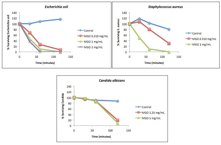
The results of this assay are summarized as shown in Figure 1. MGO was tested at
The results of this assay are summarized as shown in Figure 1. MGO was tested at concentrations
concentrations equal or greater than M.B.C. Time–kill curves show that MGO is able to reduce the
equal or greater than M.B.C. Time–kill curves show that MGO is able to reduce the viability of the
viability of the microbial population very quickly in a dose-dependent manner (Figure 1). After 60
microbial population very quickly in a dose-dependent manner (Figure 1). After 60 min of contact
minutes of contact with MGO 2 mg/mL, the viable count of E. coli was virtually zero, while with
with MGO 2 mg/mL, the viable count of E. coli was virtually zero, while with MGO 1 mg/mL, it took
MGO 1 mg/mL, it took 120 minutes to achieve the same result. After this time, MGO 0.310 mg/mL
120 min to achieve the same result. After this time, MGO 0.310 mg/mL did not kill all the bacteria, but
did not kill all the bacteria, but it reduced their number to a very large extent. S. aureus viable counts
it reduced their number to a very large extent. S. aureus viable counts were reduced at a slower rate
were reduced at a slower rate since MGO 1 mg/mL required 120 minutes to kill the standard
since MGO 1 mg/mL required 120 min to kill the standard inoculum; moreover, after 120 min, MGO
inoculum; moreover, after 120 minutes, MGO 0.310 mg/mL reduced bacterial viability by over 70%.
0.310 mg/mL reduced bacterial viability by over 70%. In the same test, after a 120 min contact with
In the same test, after a 120 minutes contact with MGO at 1.25 and 5 mg/mL, only 19% and 9% of C.
MGO at 1.25 and 5 mg/mL, only 19% and 9% of C. albicans survived, respectively; the rate of killing of
albicans survived, respectively; the rate of killing of yeasts was slower than that of bacteria, as
yeasts was slower than that of bacteria, as indicated by the lower slope of the curves.
indicated by the lower slope of the curves.
Figure 1. Time–kill curves of E. coli, S. aureus, and C. albicans in PBS in the presence of different
Figure 1. Time–kill of
concentrations curves
MGO.of E. coli, S. aureus, and C. albicans in PBS in the presence of different
concentrations of MGO.
3.3. Challenge Test
3.3. Challenge Test obtained in the challenge test showed that, as expected, control creams (without any
Results
preservative)
Results did not
obtained meet
in the the European
challenge Pharmacopoeia
test showed microbiological
that, as expected, controlstandards (data notany
creams (without shown).
The addition
preservative) did ofnot
1 mg/mL
meet the of MGO to thePharmacopoeia
European cosmetic emulsion allowed effective
microbiological control of
standards the bacterial
(data not
strains
shown). The tested
addition coli,
(E.of S. aureus)
1 mg/mL (Figure
of MGO 2). cosmetic
to the In fact, MGO led allowed
emulsion to the total eradication
effective controlofofbacterial
the
bacterial strains tested (E. coli, S. aureus) (Figure 2). In fact, MGO led to the total eradication of to
populations after 7 days of testing, meeting criterion A of European Pharmacopoeia referring
bacterial contamination of preparations for cutaneous application (this criterion is satisfied when the
total number of microorganisms is reduced in 2 to 3 logarithmic units between the second and the
seventh day, and an increase in the bacterial number does not occur to the end of the test, which is after
28 days) [23]. The same criterion referring to fungi is met when the log reduction of microorganisms
after 14 days is of two units and no growth occurs at the end of the test; therefore, MGO at 2.5 mg/mL
was also found to be effective against C. albicans (Figure 2).referring to bacterial contamination of preparations for cutaneous application (this criterion is
satisfied when the total number of microorganisms is reduced in 2 to 3 logarithmic units between the
second and the seventh day, and an increase in the bacterial number does not occur to the end of the
test, which is after 28 days) [23]. The same criterion referring to fungi is met when the log reduction
of microorganisms after 14 days is of two units and no growth occurs at the end of the test; therefore,
Cosmetics 2019, 6, 1 6 of 8
MGO at 2.5 mg/mL was also found to be effective against C. albicans (Figure 2).
Figure 2. Challenge test: growth inhibition of different microorganisms in a cosmetic formulation
Figure 2. Challenge test: growth inhibition of different microorganisms in a cosmetic formulation
containing MGO.
containing MGO.
3.4. Interaction of MGO with Chitosan
3.4. Interaction of MGO with Chitosan
When evaluated in the presence of sub-inhibitory concentrations of chitosan (1/4 and 1/2 of M.I.C.
When
value), M.I.C.evaluated
and M.B.C.in the presence
of MGO wereofreduced
sub-inhibitory concentrations of chitosan
in a concentration-dependent (1/4
manner and 2);
(Table 1/2this
of
M.I.C. value), M.I.C. and M.B.C. of MGO were reduced in a concentration-dependent manner
effect was particularly evident for E. coli (M.B.C. of MGO was reduced by a factor of ten in the presence (Table
2);
of athis effect was particularly
concentration of chitosanevident for1/2
equal to E. coli (M.B.C.
M.I.C.), of MGO
while in thewas
samereduced by a factor
condition, M.B.C.ofoften in the
Candida
presence of a concentration of chitosan equal to 1/2 M.I.C.), while in the same condition,
spp. strain 1 was reduced by four times. These results show that, in our experimental conditions, M.B.C. of
Candida spp. strain 1 was reduced by four times. These results show that, in our
the combination of MGO and chitosan leads to their synergistic interaction, with an improvement in experimental
conditions,
antimicrobial the combination of MGO and chitosan leads to their synergistic interaction, with an
efficiency.
improvement in antimicrobial efficiency.
Table 2. Effect of sub-inhibitory concentrations of chitosan on the antimicrobial activity of MGO. M.I.C.
Table 2. Effect
= minimum of sub-inhibitory
inhibitory concentrations
concentration; of chitosan
M.B.C. = minimum on theconcentration;
bactericidal antimicrobialM.F.C.
activity of MGO.
= minimum
M.I.C. = minimum
fungicidal inhibitory
concentration. concentration;
Results M.B.C.
are the average = least
of at minimum
three bactericidal
independentconcentration;
determinations.M.F.C. =
minimum fungicidal concentration. Results are the average of at least three independent
determinations. Escherichia coli Pseudomonas aeruginosa Candida spp. Strain 1
M.I.C. Chitosan 0.063 mg/mL 0.25 mg/mL 1 mg/mL
M.B.C. Chitosan Escherichia
0.063 mg/mLcoli Pseudomonas aeruginosa
0.5 mg/mL Candida spp. Strain 1
1 mg/mL
M.I.C.
M.I.C.Chitosan
MGO 0.063mg/mL
0.220 mg/mL 0.25
0.310mg/mL
mg/mL 1 mg/mL
1.25 mg/mL
M.B.C/M.F.C. MGO
M.B.C. Chitosan 0.310
0.063mg/mL
mg/mL 0.310
0.5 mg/mL
mg/mL 15 mg/mL
mg/mL
M.I.C. MGO + 1/2 M.I.C. Chitosan 0.015 mg/mL 0.15 mg/mL 1.25 mg/mL
M.I.C. MGO 0.220 mg/mL 0.310 mg/mL 1.25 mg/mL
M.B.C. MGO + 1/2 M.I.C. Chitosan 0.031 mg/mL 0.15 mg/mL 1.25 mg/mL
M.B.C/M.F.C. MGO
M.I.C. MGO + 1/4 M.I.C. Chitosan 0.310 mg/mL
0.07 mg/mL 0.310
0.310mg/mL
mg/mL 5 mg/mL
2.5 mg/mL
M.I.C.
M.B.C. MGO
MGO ++ 1/4
1/2 M.I.C.
M.I.C.Chitosan
Chitosan 0.015 mg/mL
0.07 mg/mL 0.15
0.310mg/mL
mg/mL 1.25 mg/mL
2.5 mg/mL
M.B.C. MGO + 1/2 M.I.C. Chitosan 0.031 mg/mL 0.15 mg/mL 1.25 mg/mL
M.I.C. MGO + 1/4 M.I.C. Chitosan 0.07 mg/mL 0.310 mg/mL 2.5 mg/mL
4. Discussion
M.B.C. MGO + 1/4 M.I.C. Chitosan 0.07 mg/mL 0.310 mg/mL 2.5 mg/mL
This preliminary work aimed to evaluate the antimicrobial activity of MGO and its preservative
potential
4. in a cosmetic formulation, with the objective to find out whether this compound can provide
Discussion
a viable alternative to the conventional preservatives currently used. Our results showed that MGO
This preliminary work aimed to evaluate the antimicrobial activity of MGO and its preservative
possesses a pronounced antibacterial activity with minor antifungal properties; its inhibitory effect
potential in a cosmetic formulation, with the objective to find out whether this compound can
occurs in rather short times, as demonstrated by the killing time test. In the preliminary challenge tests
provide a viable alternative to the conventional preservatives currently used. Our results showed
carried out, MGO, at the concentrations used, conformed to the criteria of the European Pharmacopoeia
that MGO possesses a pronounced antibacterial activity with minor antifungal properties; its
against the microorganisms tested; therefore, MGO appears to be eligible for a future possible use as
inhibitory effect occurs in rather short times, as demonstrated by the killing time test. In the
an alternative cosmetic preservative. In view of this, further investigations are mandatory. The first
issue to be addressed concerns safety. MGO is a molecule produced endogenously in the body in
several metabolic pathways (primarily through anaerobic glycolysis) [24]; it is also found in food
products of both animal and plant origin, with particularly high levels reported in Manuka honey
and other honeys, brewed coffee, soya sauce, toast, and soft drinks [25]. MGO is of low acute toxicity;
there is in vitro evidence that MGO is genotoxic, but the in vivo relevance is unclear [25], and the
International Agency for Research on Cancer (IARC) has determined MGO to be not classifiable as aCosmetics 2019, 6, 1 7 of 8
carcinogen [26]. Moreover, manuka honey, containing exceptionally high levels of MGO, has a long
history of safe use also in wound management; taken together, literature data suggest that MGO
can be considered safe, even if further toxicological investigations into its topical application will be
indispensable. From a technological point of view, MGO presents some advantages. It is characterised
by an interesting antibacterial activity, and it significantly reduces the microbial population in the
challenge test in compliance with the criteria of European Pharmacopoeia. It is also inexpensive and is
characterised by high water solubility, being therefore able to protect the aqueous phase of emulsions
from microbial contamination. Moreover, the association with sub-inhibitory concentrations of chitosan
increases the synergistically antimicrobial efficacy of MGO. This is of great interest because chitosan
is a biopolymer widely employed in cosmetics and personal care products for its many and specific
properties, such as antimicrobial, film-forming, antioxidant, moisturizing, and conditioning [27], and
its combination with MGO could lead to an efficient preservative system of natural origin. On the
other hand, specific investigations would be required to verify the compatibility of MGO with other
ingredients of formulations and with packaging materials in order to assess MGO stability over time
and to determine appropriate storage conditions.
To conclude, to the best of our knowledge, this study was the first attempt to apply MGO as a
preservative of natural origin in cosmetics. The results obtained are promising, but further studies are
obviously needed to confirm the preservative activity of MGO in different cosmetic formulations, as
well as its safety.
Author Contributions: C.J. conceived the work and wrote the article; C.J. and G.A.M. contributed to the
conduction of microbiology experiments in equal measure.
Funding: This research did not receive any specific grant from funding agencies in the public, commercial or
not-for-profit sectors.
Conflicts of Interest: The authors declare no conflict of interest.
References
1. Halla, N.; Fernandes, I.P.; Heleno, S.A.; Costa, P.; Boucherit-Otmani, Z.; Boucherit, K.; Rodrigues, A.E.;
Ferreira, I.C.F.R.; Barreiro, M.F. Cosmetic preservation: A review on present strategies. Molecules 2018, 23,
1571. [CrossRef] [PubMed]
2. European Commission (EC). Regulation (EC) No 1223/2009 of the European Parliament and of the Council
of 30 November 2009 on cosmetic products. Off. J. Eur. Union 2009, 27, 59–209.
3. Yim, E.; Baquerizo Nole, K.L.; Tosti, A. Contact dermatitis caused by preservatives. Dermatitis 2014, 25,
215–231. [CrossRef] [PubMed]
4. Nohynek, G.J.; Borgert, C.J.; Dietrich, D.; Rozman, K.K. Endocrine disruption: Fact or urban legend? Toxicol.
Lett. 2013, 223, 295–305. [CrossRef] [PubMed]
5. Golden, R.; Gandy, J.; Vollmer, G. A review of the endocrine activity of parabens and implication for potential
risks to human health. Crit. Rev. Toxicol. 2005, 35, 435–458. [CrossRef]
6. Darbre, P.D. Environmental oestrogens, cosmetics and breast cancer. Best Pract. Res. Clin. Endocrinol. Metab.
2006, 20, 121–143. [CrossRef]
7. Witorsch, R.J.; Thomas, J.A. Personal care products and endocrine disruption: A critical review of the
literature. Crit. Rev. Toxicol. 2010, 40 (Suppl. 3), 1–30. [CrossRef]
8. Maccioni, A.M.; Anchisi, C.; Sanna, A.; Sardu, C.; Dessì, S. Preservative systems containing essential oils in
cosmetic products. Int. J. Cosmet. Sci. 2002, 24, 53–59. [CrossRef]
9. Muyima, N.Y.O.; Zulu, G.; Benghu, T.; Popplewell, D. The potential application of some novel essential
oils as natural cosmetic preservatives in an aqueous cream formulation. Flavour Fragr. J. 2002, 17, 258–266.
[CrossRef]
10. Ibarra, F.; Johnson, G.H. Natural preservatives from concepts in nature. Cosmet. Toiletries 2008, 123, 81–90.
11. Herman, A.; Herman, A.P.; Domagalska, B.W.; Mlynarczyk, A. Essential oils and herbal extracts as
antimicrobial agents in cosmetic emulsions. Indian J. Microbiol. 2013, 53, 232–237. [CrossRef] [PubMed]Cosmetics 2019, 6, 1 8 of 8
12. Juliano, C.; Gavini, E.; Giunchedi, P.; Magrini, G.A. Evaluation of Manuka honey as an adjuvant antimicrobial
preservative in a O/W emulsion. J. Appl. Cosmet. 2016, 34, 87–98.
13. George, N.M.; Cutting, K.F. Antibacterial honey (MedihoneyTM ): In-vitro activity against clinical isolates of
MRSA, VRE and other multiresistant Gram-negative organisms including Pseudomonas aeruginosa. Wounds
2007, 19, 231–236. [PubMed]
14. Lin, S.M.; Molan, P.C.; Cursons, R.T. The in vitro susceptibility of Campylobacter spp. to the antibacterial
effect of manuka honey. Eur. J. Clin. Microbiol. Infect. Dis. 2009, 28, 339–344. [CrossRef] [PubMed]
15. Majtan, J.; Bohova, J.; Horniakova, M.; Klaudiny, J.; Majtan, V. Anti-biofilm effects of honey against wound
pathogens Proteus mirabilis and Enterobacter cloacae. Phytother. Res. 2014, 28, 69–75. [CrossRef] [PubMed]
16. Adams, C.J.; Boult, C.H.; Deadman, B.J.; Farr, J.M.; Grainger, M.N.; Manley-Harris, M.; Snow, M.J. Isolation
by HPLC and characterisation of the bioactive fraction of New Zealand manuka (Leptospermum scoparium)
honey. Carbohydr. Res. 2008, 343, 651–659. [CrossRef] [PubMed]
17. Mavric, E.; Wittmann, S.; Barth, G.; Henle, T. Identification and quantification of methylglyoxal as the
dominant antibacterial constituent of Manuka (Leptospermum scoparium) honeys from New Zealand. Mol.
Nutr. Food Res. 2008, 52, 483–489. [CrossRef] [PubMed]
18. Kwakman, P.H.S.; te Velde, A.A.; de Boer, L.; Vandenbroucke-Graus, C.M.J.E.; Zaat, S.A.J. Two major
medicinal honeys have different mechanisms of bactericidal activity. PLoS ONE 2011, 6, e17709. [CrossRef]
19. No, H.K.; Park, N.Y.; Lee, S.H.; Meyers, S. Antibacterial activity of chitosans and chitosan oligomers with
different molecular weight. Int. J. Food Microbiol. 2002, 74, 65–72. [CrossRef]
20. Thrupp, L. Susceptibility testing of antibiotics in liquid media. In Antibiotics in Laboratory Medicine, 2nd ed.;
Lorian, V., Ed.; Williams and Wilkins: Baltimore, MD, USA, 1986; pp. 93–158. ISBN 0-683-05167-9.
21. McGinnis, M.R.; Rinaldi, M.G. Antifungal drugs: Mechanisms of action, drug resistance, susceptibility
testing and assays of activity in biological fluids. In Antibiotics in Laboratory Medicine, 2nd ed.; Lorian, V., Ed.;
Williams and Wilkins: Baltimore, MD, USA, 1986; pp. 223–281. ISBN 0-683-05167-9.
22. Juliano, C.; Demurtas, C.; Piu, L. In vitro study on the anticandidal activity of Melaleuca alternifolia (tea tree)
essential oil combined with chitosan. Flavour Fragr. J. 2008, 23, 227–231. [CrossRef]
23. European Pharmacopoeia Commission. European Pharmacopoeia, 7th ed.; European Directorate for the Quality
of Medicines & Healthcare (EDQM): Strasbourg, France, 2011.
24. Kalapos, M.P. Methylglyoxal in living organisms. Chemistry, biochemistry, toxicology and biological
implications. Toxicol. Lett. 1999, 110, 145–175. [CrossRef]
25. Committee on Toxicity of Chemical in Food, Consumer Products and the Environment. Statement
on Methylglyoxal. 2009. Available online: https://cot.food.gov.uk/committee/committee-on-toxicity/
cotstatements/cotstatementsyrs/cotstatements2009/cot200904 (accessed on 23 September 2018).
26. IARC. Coffee, tea, mate, methylxanthines and methylglyoxal. IARC Working Group on the evaluation of
carcinogenic risks to human. IARC Monogr. Eval. Carcinog. Risks Hum. 1991, 51, 1–513.
27. Aranaz, I.; Acosta, N.; Civera, C.; Elorza, B.; Mingo, J.; Castro, C.; de Los Llanos Gandía, M.; Caballero, A.H.
Cosmetics and cosmeceutical applications of chitin, chitosan and their derivatives. Polymers 2018, 10, 213.
[CrossRef]
© 2018 by the authors. Licensee MDPI, Basel, Switzerland. This article is an open access
article distributed under the terms and conditions of the Creative Commons Attribution
(CC BY) license (http://creativecommons.org/licenses/by/4.0/).You can also read