Rosavin suppresses osteoclastogenesis in vivo and in vitro by blocking the nuclear factor kappa-light-chain-enhancer of activated B cells (NF-κB) ...
←
→
Page content transcription
If your browser does not render page correctly, please read the page content below
Original Article
                                                                                                                                    Page 1 of 14

Rosavin suppresses osteoclastogenesis in vivo and in vitro
by blocking the nuclear factor kappa-light-chain-enhancer of
activated B cells (NF-κB) and mitogen-activated protein kinase
(MAPK) signaling pathways
Wenhao Zhang1, Weijie Zhang1, Liang Huo1, Ying Chai1, Zhiyang Liu1, Zhenhu Ren2, Chuangqi Yu1
1
    Department of Oral and Craniofacial Surgery, Ninth People’s Hospital, Shanghai Jiao Tong University School of Medicine, Shanghai, China;
2
Department of Oral and Maxillofacial & Head and Neck Oncology, Shanghai Ninth People’s Hospital, Shanghai Jiao Tong University School of
Medicine, Shanghai, China
Contributions: (I) Conception and design: Z Ren, C Yu; (II) Administrative support: None; (III) Provision of study materials or patients: Z Ren, C
Yu; (IV) Collection and assembly of data: WH Zhang, WJ Zhang, L Huo, Y Chai, Z Liu; (V) Data analysis and interpretation: WH Zhang; (VI)
Manuscript writing: All authors; (VII) Final approval of manuscript: All authors.
Correspondence to: Chuangqi Yu. Department of Oral and Craniofacial Surgery, Ninth People’s Hospital, Shanghai Jiao Tong University School of
Medicine, Shanghai 200011, China. Zhenhu Ren. Department of Oral and Maxillofacial & Head and Neck Oncology, Shanghai Ninth People’s
Hospital, Shanghai Jiao Tong University School of Medicine, Shanghai 200011, China. Email: dryuchuangqi@163.com; ren.zhenhu@outlook.com.

                  Background: Bone homeostasis is mediated by osteoblast-related bone formation and osteoclast-related
                  resorption. The imbalance of bone homeostasis due to excessive osteoclastogenesis or reduced osteogenesis
                  can result in various disorders, such as postmenopausal osteoporosis (PMOP). The receptor activator of
                  nuclear factor-κB ligand (RANKL)-induced nuclear factor kappa-light-chain-enhancer of activated B cells
                  (NF-κB) and mitogen-activated protein kinase (MAPK) pathways are essential in osteoclastogenesis. In
                  this study, we aimed to investigate the effects of rosavin, an alkylbenzene diglycoside compound from the
                  traditional Chinese medicine Rhodiola Rosea L, on RANKL-induced osteoclastogenesis in vitro and in vivo.
                  Methods: The effects of rosavin on osteoclastogenesis were assessed by TRAP staining of bone marrow
                  monocyte cells (BMMCs) and RAW 264.7 cells. The effects of rosavin on osteogenesis were determined
                  using alkaline phosphatase (ALP) and alizarin red staining, as well as real-time quantitative reverse
                  transcription polymerase chain reaction. Actin ring formation and bone formation experiments were
                  performed to evaluate osteoclast function. Western blotting was carried out to determine the expression of
                  osteoclastogenesis-related genes, and the activation of the NF-κB and MAPK pathways was evaluated by
                  performing western blotting and immunofluorescence staining. Ovariectomized mice were used to explore
                  the effect of rosavin on bone loss.
                  Results: Rosavin could inhibit osteoclastogenesis, suppress the function of osteoclasts, and decrease the
                  expression of osteoclast differentiation-related genes, including tartrate-resistant acid phosphatase (TRAP),
                  cathepsin K, matrix metalloproteinase-9 (MMP-9), calcitonin receptor (CTR), TNF receptor-associated
                  factor 6 (TRAF-6), receptor activator of nuclear factor-κB (RANK), and colony-stimulating factor-1 receptor
                  (c-fms). Rosavin inhibited RANKL-induced phosphorylation of p65 and inhibitory subunit of NF-κB alpha
                  (IκBα), and suppressed p65 nuclear translocation. Rosavin was also found to inhibit the phosphorylation of
                  extracellular-signal-regulated kinase (ERK), p38, and c-Jun N-terminal kinase (JNK). Furthermore, rosavin
                  promoted osteogenesis in bone marrow mesenchymal stem cells (BMSCs). In vivo experiments showed that
                  treatment with rosavin could alleviate ovariectomy-induced osteoporosis in mice.
                  Conclusions: Our results indicated that rosavin suppressed RANKL-induced osteoclastogenesis in vivo
                  and in vitro by blocking the NF-κB and MAPK pathways. Rosavin treatment is a potential therapy for the
                  clinical treatment of osteoclastogenesis-related disorders.

© Annals of Translational Medicine. All rights reserved.                            Ann Transl Med 2021 | http://dx.doi.org/10.21037/atm-20-4255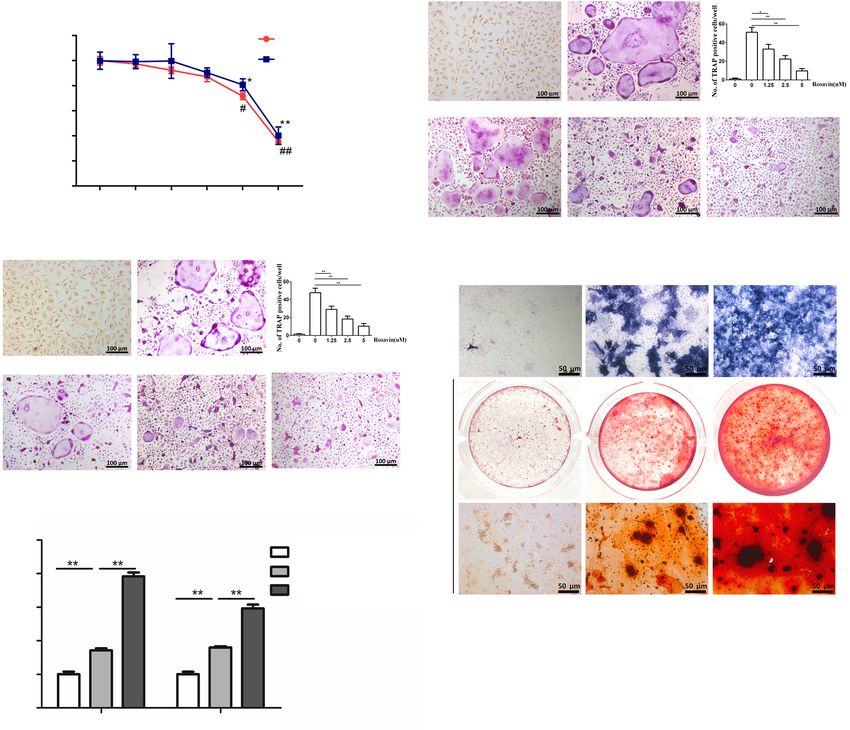
Page 2 of 14                                                                          Zhang et al. Rosavin suppresses osteoclastogenesis

                Keywords: Rosavin; osteoclastogenesis; NF-κB; mitogen-activated protein kinase (MAPK); postmenopausal
                osteoporosis (PMOP)

                Submitted May 26, 2020. Accepted for publication Nov 20, 2020.
                doi: 10.21037/atm-20-4255
                View this article at: http://dx.doi.org/10.21037/atm-20-4255

Introduction                                                               and tartrate-resistant acid phosphatase (TRAP) (15,16).
                                                                           Targeting RANKL-mediated signaling to decrease the
In bone remodeling, the homeostasis between osteoblast-
                                                                           expression of NFATc1 is a potential treatment strategy
mediated bone formation and osteoclast-mediated
                                                                           for osteoclastogenesis-related disorders, including
bone resorption is essential (1). The imbalance of bone
                                                                           osteoporosis (17,18).
homeostasis due to excessive osteoclastogenesis or reduced
                                                                              Natural medicines are important in clinical treatment (19).
osteogenesis can result in various disorders, such as
                                                                           Rhodiola Rosea L is a traditional Chinese herb used for
postmenopausal osteoporosis (PMOP), rheumatoid arthritis,
                                                                           inflammation and pulmonary fibrosis, as well as for its
and Paget’s disease (2-4). Promoting bone formation and
                                                                           antioxidant properties (20,21). Rosavin is one of the most
inhibiting bone resorption are effective treatment strategies
                                                                           important active alkylbenzene diglycoside compound
for bone loss diseases (5,6).
                                                                           ingredients of Rhodiola Rosea L, and has proven efficacy in
    Osteoporosis is the most common bone loss disease, and
is accompanied by an obvious decrease in bone density and                  nootropic, anti-depressant, anti-cancer, and antioxidative
lesions in bone tissue microstructure (7). The incidence                   treatment (22-24). In the present study, we explored the
of osteoporosis is often associated with age and sex (8). In               role of rosavin on ovariectomy-induced osteoporosis
recent years, there has been a dramatic increase in PMOP,                  in mice, which is an effective and widely used model to
with patients over 60 years old experiencing distinctly                    simulate PMOP, as well as the effects of rosavin on the
increased morbidity (9). Estrogen deficiency plays an                      differentiation and function of osteoclasts in vitro. The
important role in PMOP. During the postmenopausal                          molecular mechanisms involved in this process were also
period, various inflammation inhibitors decrease, and                      investigated. We present the following article in accordance
the expression of receptor activator of nuclear factor-                    with the ARRIVE (Animal Research: Reporting of In Vivo
κB ligand (RANKL) increases significantly, resulting                       Experiments) reporting checklist (available at http://dx.doi.
in excessive activation of osteoclasts and the promotion                   org/10.21037/atm-20-4255).
of bone resorption (10). As a result, the suppression
of osteoclastogenesis remains a potential therapy for                      Methods
osteoclast-related bone diseases, such as osteoporosis.
    Osteoclasts differentiate from monocyte-macrophages                    All procedures were approved by and performed in
under the induction of macrophage colony-stimulating                       accordance with the ethical standards of the Ethics
factor (M-CSF) and RANKL (11-13). RANKL binds to                           Committee of Shanghai Ninth People’s Hospital (No.
receptor activator of nuclear factor-κB (RANK), which                      SH9H-2019-T159-2), and were in compliance with the
then recruits the tumor necrosis factor receptor-associated                NIH Guide for the Care and Use of Laboratory Animals
factors (TRAFs), activating a variety of downstream signaling              [2018].
pathways, such as the mitogen-activated protein kinase
(MAPK) and nuclear factor kappa-light-chain-enhancer
                                                                           Reagents
of activated B cells (NF-κB) pathways (14). Nuclear
factor of activated T-cell cytoplasmic 1 (NFATc1) has                      Rosavin was obtained from Nuodande Standard Technical
been reported to be an essential transcriptional factor in                 Services (Shanghai, China). Reagents were diluted in
osteoclast differentiation and is necessary for the expression             phosphate-buffered saline (PBS) solution. Fetal bovine
of osteoclast-associated genes, such as cathepsin K, matrix                serum (FBS), antibodies, streptomycin, and penicillin were
metalloproteinase (MMP)-9, calcitonin receptor (CTR),                      purchased from BioTNT (Shanghai, China).

© Annals of Translational Medicine. All rights reserved.                         Ann Transl Med 2021 | http://dx.doi.org/10.21037/atm-20-4255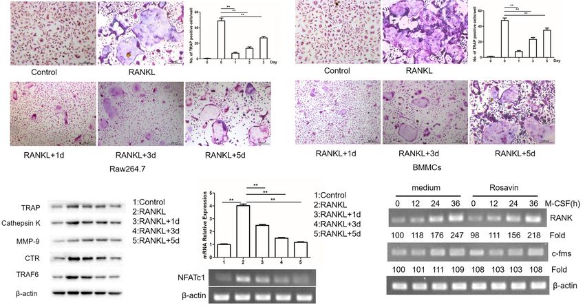
Annals of Translational Medicine, 2021                                                                             Page 3 of 14

Cell proliferation analysis                                    overdose. Bone marrow mesenchymal stem cells
                                                               (BMSCs) were obtained by washing the femurs of the
The MTT assay was conducted according to the
                                                               mice. BMSCs were maintained in a complete medium
manufacturer’s instructions (Sigma-Aldrich, St. Louis, USA).
                                                               supplemented with 1% streptomycin and penicillin,
Cells were placed into 96-well plates (1×10⁴ cells/well).
                                                               20 mM β-glycerophosphate, 10 nM dexamethasone,
The doses of rosavin were as follows: 2.5, 5, 10, 20, and
                                                               and 100 μg/mL ascorbic acid. After 20 days, alkaline
40 μM. Bone marrow monocyte cells (BMMCs), isolated
                                                               phosphatase (ALP) staining and alizarin red staining were
from C57BL/6 mice, and RAW264.7 cells were cultured.
                                                               performed to detect the osteoblasts. The experiments were
After 72 hours, 50 μg/μL of MTT solution was added, and
                                                               independently performed three times.
the absorbance of the plate was detected at 490 nm using a
linked immunosorbent assay plate reader (Olympus, Tokyo,
Japan). The experiments were independently performed           Polymerase chain reaction (PCR) analysis
three times.
                                                               Real-time quantitative reverse transcription polymerase
                                                               chain reaction (qRT-PCR) was used to detect the
In vitro osteoclastogenesis assay                              transcription of osteogenesis and osteoclastogenesis-
                                                               related genes. Briefly, whole-cell ribonucleic acid (RNA)
BMMCs were washed from the femurs of C57 mice. Cells           samples were extracted using TRIzol reagent, as previously
were cultured in minimum essential medium α (α-MEM)            reported, and complementary deoxyribonucleic acid (DNA)
containing 1% penicillin, 1% streptomycin, and 10% FBS.        was generated using a reverse transcriptase kit (25). The TB
Cells were seeded into 96-well plates containing M-CSF         Green premix Ex Taq kit (Takara, CA, USA) was used to
(30 ng/mL) and RANKL (100 ng/mL), and were incubated           detect gene expression. Mouse-specific PCR primers were
with different concentrations of rosavin (0, 1.25, 2.5, and    used. Mouse runt-related transcription factor 2 (Runx2),
5 μM). After 7 days, TRAP staining was conducted to detect     forward: 5'-GACTGTGGTTACCGTCATGGC-3',
the TRAP-positive cells (Sigma-Aldrich, St, Louis, USA).       reverse: 5'-ACTTGGTTTTTCATAACAGCGGA-3';
The experiments were independently performed three             mouse osteocalcin (OCN), forward:
times, and the data were analyzed by one-way analysis of       5'-GGACCATCTTTCTGCTCACTCTGC-3',
variance (ANOVA).                                              reverse: 5'-TGTTCACTACCTTATTGC
                                                               CCTCCTG-3'; mouse NFATc1, forward:
F-actin ring formation and pit formation assay                 5'-ATGACGGGGCTGGAGCAGGA-3', reverse:
                                                               5'-TTAGGAGTGGGGGGATCGTGC-3';
For the F-actin formation assay, cells were initially          m o u s e         R A N K ,            f o r w a r d :
fixed with 4% paraformaldehyde for 1 hour and then             5'-CTGCTCCTCTTCATCTCTGTG-3', reverse:
washed twice with PBS. Next, the cells were conducted          5'- CTTCTGGAACCATCTTCTCCTC-3'; mouse
with fluorescein isothiocyanate (FITC)-phalloidin for          colony-stimulating factor-1 receptor (c-fms), forward:
2 hours and stained with 4’6-diamidino-2-phenylindole for      5'-TTCACTCCGGTGGTGGTGGCCTGT-3', reverse:
20 minutes.                                                    5'-GTTGAGTAGGTCTCCATAGCAGCA-3'. The
   For the pit formation assay, osteoclasts were cultured      experiments were independently performed three times,
on a bone biomimetic synthetic plate (OsteoAssay Surface       and the data were analyzed using the t-test and one-way
plates; Corning, NY, USA), and were induced with               ANOVA.
30 ng/mL of M-CSF and 100 ng/mL of RANKL. The
area resorbed by the osteoclasts was detected using a light
                                                               In vivo experiments
microscope (Lecia CTR400, Germany). The experiments
were independently performed three times, and the data         Eight-week-old female C57BL/6 mice were supplied by
were analyzed by one-way ANOVA.                                the Shanghai SLAC Laboratory Animal Center (SLAC,
                                                               Shanghai, China). All mice were raised in a controlled
                                                               environment at 25 ℃ under a 12-hour light/dark cycle.
In vitro osteogenesis assay
                                                               Sufficient food and water were available.
C57BL/6 mice were sacrificed via pentobarbital sodium             Eighteen mice were randomly separated into three

© Annals of Translational Medicine. All rights reserved.            Ann Transl Med 2021 | http://dx.doi.org/10.21037/atm-20-4255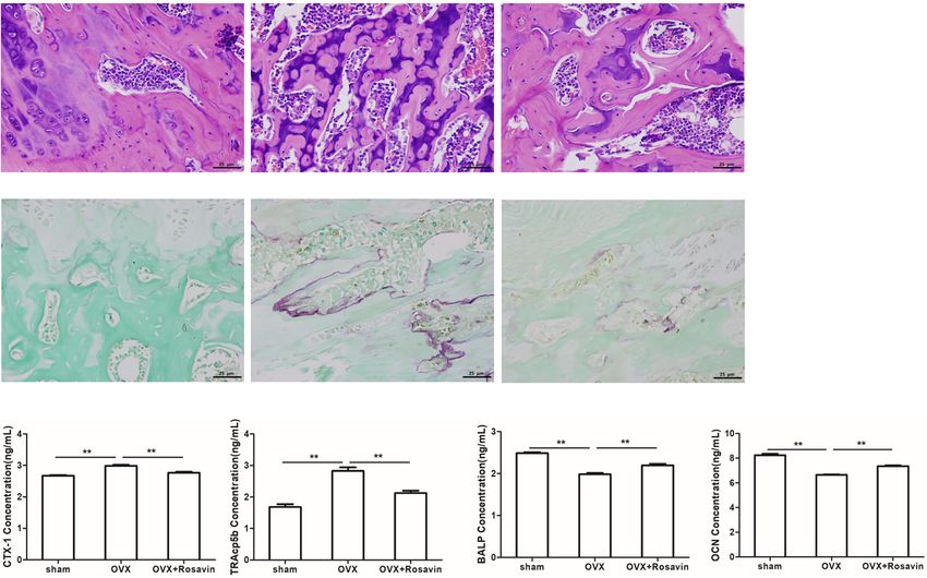
Page 4 of 14                                                                          Zhang et al. Rosavin suppresses osteoclastogenesis

groups; a sham-operated group, an ovariectomized                            (1:1,000, ab201015, abcam, USA), ERK (1:1,000, GB12087,
treatment with vehicle group, and a rosavin treatment                       Servicebio, USA), p-JNK (1:1,000, ab124956, abcam, USA),
group, with six mice in each group. During the operation,                   JNK (1:1,000, ab179461, abcam, USA), p-p38 (1:1,000,
the mice were anesthetized with 1% pentobarbital sodium                     ab195049, abcam, USA), and p38 (1:1,000, ab31828, abcam,
(30 mg/kg). Mice underwent bilateral OVX. Small incisions                   USA). The membranes were subsequently incubated with
were made on the dorsal skin and peritoneum. Two ovaries                    secondary antibodies of anti-rabbit IgG (1:2,000, ab6721,
and part of the oviduct were removed and pressed to stop                    abcam, USA). The experiments were independently
any bleeding. The incision on the skin was closed with                      performed three times, and the data were analyzed using
5-0 nonabsorbable suture lines. After the procedure, mice                   the t-test.
were allowed to recover for 24 h. The mice in the OVX
(ovariectomy) + vehicle group and the OVX + rosavin
                                                                            Bone histomorphometric analysis
group had both ovaries resected, while the mice in the
sham-operated group received surgery to expose the                          The number of osteoclasts and amount of trabecular
ovaries without resection. Rosavin treatment group mice                     bone were evaluated by bone histomorphometric analyses
were intraperitoneally injected with rosavin (10 mg/kg) or                  as previously described. Mouse femurs were fixed and
PBS every day. After 6 weeks, the mice were sacrificed via                  decalcified for at least 4 weeks. The 3-μm-thick bone
pentobarbital sodium overdose (150 mg/kg).                                  sections were then stained with hematoxylin and eosin
                                                                            (H&E) and TRAP.

Immunofluorescence staining
                                                                            Micro-computed tomography (CT) analysis
The effects of rosavin (5 μM) on p65 nuclear translocation were
examined in RAW264.7 cell lines. Immunofluorescence                         We scanned 200 section planes below the growth plate of
was conducted as described previously (26). Cells were                      the mouse femurs (using Skyscan1172, Antwerp, Belgium).
fixed with 4% paraformaldehyde for 20 minutes, and were                     Built-in software was used to detect the trabecular bone and
then washed with Triton X reagents and blocked with 5%                      metaphyseal region.
bovine serum albumin (BSA) for 20 minutes. Next, anti-p65
antibody, anti-mouse immunoglobulin G (IgG) antibody,
                                                                            Enzyme-linked immunosorbent assay
and fluorescein-conjugated streptavidin were added to the
cells. The experiments were independently performed three                   All mice were sacrificed with pentobarbital sodium, and
times.                                                                      the arterial blood of the mice was collected. Next, the
                                                                            serum was centrifuged at 3,000 rpm for 10 minutes. The
                                                                            expression of OCN, Cross Linked C-telopeptide of Type
Western blotting
                                                                            |Collagen (CTX-1), tartrate-resistant acid phosphatase
RAW264.7 cells cultured with RANKL or RANKL &                               5b (TRAcp5b), and bone alkaline phosphatase (BALP)
rosavin (5 μM) for different durations were prepared                        was detected using enzyme-linked immunosorbent assay
for western blotting to determine the expression                            (ELISA) kits according to the manufacturer’s instructions.
of o s t e o c la s t o g e nes i s -rel a ted p ro tei ns , i nc ludin g   The experiments were independently performed three
phosphorylated (p)-p65, p-inhibitory subunit of NF-κB                       times, and the data were analyzed by one-way ANOVA.
(IκB), p-extracellular-signal-regulated kinase (ERK), p-c-
Jun N-terminal kinase (JNK), and p-p38. The samples
                                                                            Statistical analysis
were probed overnight with primary antibodies against
β-actin (1:2,000, GB12001, Servicebio, China), TRAP                         All data were expressed as the mean ± standard deviation
(1:1,000, ab191406, abcam, USA), Cathepsin K (1:1,000,                      (SD). Two independent group comparisons were conducted
ab19027, abcam, USA), MMP-9 (1:1,000, ab219372, abcam,                      using the Student’s t-test. Comparisons among more than
USA), CTR (1:1,000, ab114596, abcam, USA), TRAF6                            three groups were analyzed by one-way analysis ANOVA
(1:500, 13411-1-AP, proteintech, USA), p-p65 (1:10,000,                     via the Statistical Product and Service Solutions (SPSS)
ab76302, abcam, USA), p65 (1:1,000, ab32536, abcam,                         statistical software. A P value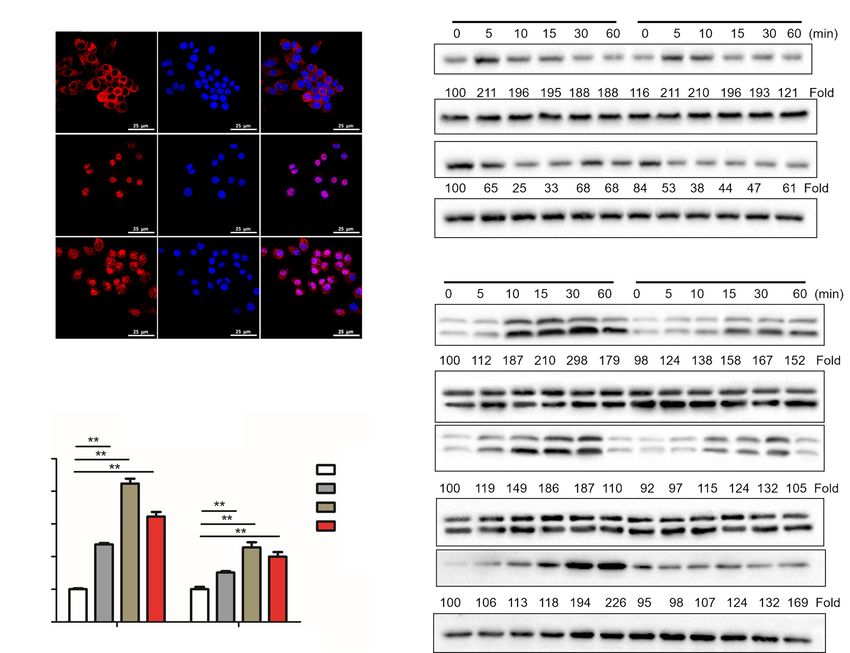
Annals of Translational Medicine, 2021                                                                               Page 5 of 14

Results                                                         Rosavin inhibited osteoclastogenesis at an early stage

Rosavin suppressed osteoclastogenesis                           To investigate the influence of rosavin on the process of
                                                                osteoclastogenesis, we treated BMMCs and RAW264.7
To perform the in vitro experiments, two standard
                                                                cells with rosavin (5 μM) on day 1, 3, or 5 after induction
osteoclastogenesis cellular models, BMMCs and RAW264.7
                                                                with M-CSF and RANKL. TRAP staining results showed
cells, were used for an MTT assay. The results indicated
                                                                that rosavin mainly suppressed osteoclast formation on
that rosavin showed no cytotoxicity below 5 μM (Figure 1A).
                                                                the first day (**, P
Page 6 of 14 Zhang et al. Rosavin suppresses osteoclastogenesis
A B
120 Raw264.7
Proliferation (% of control)
100 BMMCs
80
60 Control RANKL
40
20
0
0 2.5 5 10 20 40
Rosavin μM
RANKL +1.25 μM RANKL +2.5 μM RANKL +5 μM
C
D Control Induction Induction + rosavin
ALP
Control RANKL
Alizarin red
RANKL +1.25 μM RANKL +2.5 μM RANKL +5 μM
E
5
Control
mRNA relative expression
4 Induction
Induction + rosavin
3
2
1
0
Runx2 OCN
Figure 1 Rosavin inhibits osteoclastogenesis in vitro. (A) MTT analysis of the cytotoxic effects in RAW264.7 and BMMCs. (B) Formation
of TRAP-positive cells from BMMCs and quantification of osteoclasts. (C) Formation of TRAP-positive cells from RAW264.7 cells and
quantification of osteoclasts. (D) ALP and alizarin red staining of BMSCs. (E) QRT-PCR of Runx2 and OCN from BMSCs. (*, PAnnals of Translational Medicine, 2021 Page 7 of 14
A
Control RANKL
RANKL +1.25 μM RANKL +2.5 μM RANKL +5 μM
B
Control RANKL
RANKL +1.25 μM RANKL +2.5 μM RANKL +5 μM
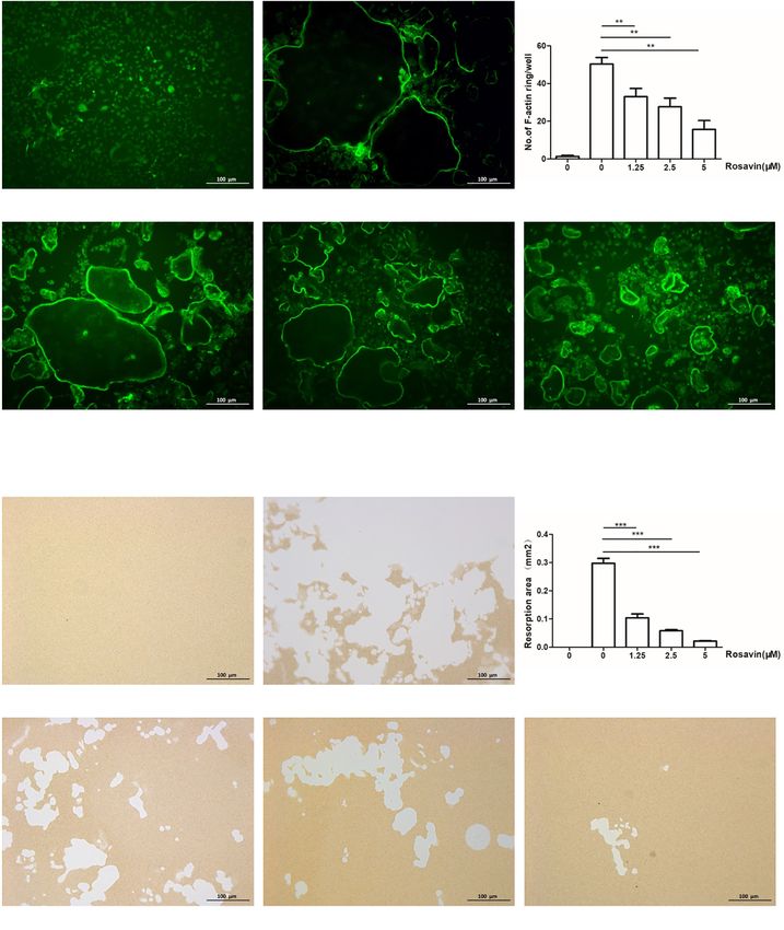
Figure 2 Rosavin inhibits osteoclast function in vitro. (A) Actin ring structures of osteoclasts and quantification of actin rings after rosavin
treatment. (B) Pit formation assay of osteoclasts and quantification of the pits area after rosavin treatment. (**, PA B
Page 8 of 14
© Annals of Translational Medicine. All rights reserved.
C D E
Figure 3 Rosavin inhibits RANKL-induced osteoclast formation at an early stage. (A) Effect of rosavin on RANKL-induced osteoclastogenesis at different stages in
RAW264.7 cells. (B) Effect of rosavin on RANKL-induced osteoclastogenesis at different stages in BMMCs. (C) Western blotting and optical density analysis of the
expression of TRAP, cathepsin K, TRAF6, MMP-9, and CTR with rosavin treatment on day 1, 3, 5. (D) PCR results of NFATc1 in BMMCs at different time points after
rosavin treatment. (E) Q-PCR results of RANK and c-fms in BMMCs at different time points after induction with M-CSF and rosavin treatment. (**, PAnnals of Translational Medicine, 2021 Page 9 of 14
A B RANKL RANKL + rosavin
P65 DAPI Merge
p-p65
Control
P65
IκBα
RANKL
β-actin
RANKL +
D RANKL RANKL + rosavin
rosavin
p-ERK
ERK
C C-FOS
p-JNK
5
0
mRNA relative expression
4 15
30
3 60 JNK
2
p-p38
1
0
RANKL RANKL + rosavin p38
Figure 4 Rosavin inhibits the RANKL-induced NF-κB and MAPK signaling pathways. (A) Rosavin inhibits RANKL-induced p65 nuclear
translocation. (B) Rosavin inhibits the phosphorylation of the p65, p50, and IκB proteins. (C) Results of qRT-PCR of c-Fos in BMMCs. (D)
Rosavin inhibits the phosphorylation of the ERK, JNK, and p38 proteins. (**, PPage 10 of 14 Zhang et al. Rosavin suppresses osteoclastogenesis
A Sham OVX OVX + rosavin
B
Figure 5 Rosavin ameliorates OVX-induced bone loss in vivo. (A) Micro-CT representative graphs of distal femoral sections from each
group 6 weeks after the operation. (B) Tb.Area, BV/TV, Tb.N, and BMD analysis of the distal femur. (**, PAnnals of Translational Medicine, 2021 Page 11 of 14 A Sham OVX OVX + rosavin B Sham OVX OVX + rosavin C Figure 6 Rosavin inhibits the formation of osteoclasts in vivo. (A) H&E staining of distal femoral sections from each group 6 weeks after the operation (B) TRAP staining of distal femoral sections from each group 6 weeks after the operation. (C) Serum levels of CTX-1, TRAcp5b, OCN, and BALP (**, P
Page 12 of 14                                                             Zhang et al. Rosavin suppresses osteoclastogenesis

indicating that rosavin inhibited osteoclastogenesis via        Technology Commission [17411963900, 16411953300]
suppression of the NF-κB and MAPK signaling pathways.           and the Clinical research MDT project of Shanghai
   Osteoporosis, a chronic age-related disease, is typically    Ninth People’s Hospital, Shanghai Jiao Tong University
accompanied by abundant bone loss and a higher incidence        [201701017, 201701018].
of fracture. The main pathogenic factors of this disease
include excessive bone resorption and disruption of bone
                                                                Footnote
formation. Specifically, excessive osteoclastogenesis, which
is caused by abnormal activation of RANKL signaling,            Reporting Checklist: The authors have completed the
has been recognized as being the key aspect in the              ARRIVE (Animal Research: Reporting of In Vivo
development of osteoporosis (37). Therefore, inhibition         Experiments) reporting checklist. Available at http://dx.doi.
of osteoclastogenesis is an effective therapy for osteoclast-   org/10.21037/atm-20-4255
related diseases such as osteoporosis.
   Through the in vivo study, we found that rosavin             Peer Review File: Available at http://dx.doi.org/10.21037/
reduced bone loss in the OVX mouse model. The results           atm-20-4255
of H&E staining and micro-CT showed that rosavin
treatment attenuated trabecular bone loss in the distal         Data Sharing Statement: Available at http://dx.doi.
femur. Furthermore, TRAP staining indicated that rosavin        org/10.21037/atm-20-4255
treatment reduced the number of TRAP-positive cells
around the bone trabecula. The serum levels of CTX-1,           Conflicts of Interest: All authors have completed the
TRAcp5b, OCN, and BALP were determined to                       International Committee of Medical Journal Editors
investigate osteoclast and osteoblast activity. Compared        (ICMJE) uniform disclosure form (available at http://dx.doi.
to the OVX group, the levels of CTX-1 and TRAcp5b               org/10.21037/atm-20-4255). The authors have no conflicts
were reduced after treatment with rosavin, indicating that      of interest to declare.
osteoclastogenesis was inhibited. Furthermore, the levels
of OCN and BALP increased after rosavin treatment,              Ethical Statement: The authors are accountable for all
demonstrating an osteogenesis-promoting effect of               aspects of the work in ensuring that questions related
rosavin. These results demonstrated that rosavin treatment      to the accuracy or integrity of any part of the work are
attenuated bone loss in OVX mice via promotion of               appropriately investigated and resolved. All procedures were
osteogenesis and inhibition of osteoclastogenesis.              approved by and performed in accordance with the ethical
   However, there are still some limitations to this study.     standards of the Ethics Committee of Shanghai Ninth
For instance, it did not investigate the exact drug target of   People’s Hospital (No. SH9H-2019-T159-2), and were in
rosavin, and further studies are needed to explore whether      compliance with the NIH Guide for the Care and Use of
rosavin could be used to treat osteoporosis patients.           Laboratory Animals [2018].

                                                                Open Access Statement: This is an Open Access article
Conclusions
                                                                distributed in accordance with the Creative Commons
Via a series of in vivo and in vitro experiments, the present   Attribution-NonCommercial-NoDerivs 4.0 International
study demonstrated that rosavin promoted osteogenesis and       License (CC BY-NC-ND 4.0), which permits the non-
suppressed RANKL-induced osteoclastogenesis. We found           commercial replication and distribution of the article with
that rosavin inhibits osteoclastogenesis at an early stage by   the strict proviso that no changes or edits are made and the
suppressing the NF-κB and MAPK signaling pathways.              original work is properly cited (including links to both the
Our research indicated that rosavin could be a potentially      formal publication through the relevant DOI and the license).
effective therapy for osteoclast-related disorders.             See: https://creativecommons.org/licenses/by-nc-nd/4.0/.

Acknowledgments                                                 References

Funding: This work was supported by the Key Biological          1.   Tikhonova AN, Dolgalev I, Hu H, et al. The bone marrow
and Pharmaceutical Projects of the Shanghai Science and              microenvironment at single-cell resolution. Nature

© Annals of Translational Medicine. All rights reserved.              Ann Transl Med 2021 | http://dx.doi.org/10.21037/atm-20-4255
Annals of Translational Medicine, 2021 Page 13 of 14
2019;569:222-8. 16. Cui Y, Zhao X, Mei L, et al. Osteon Myospalacem
2. Drake MT, Clarke BL, Oursler MJ, et al. Cathepsin K Baileyi attenuates osteoclast differentiation through
Inhibitors for Osteoporosis: Biology, Potential Clinical RANKL induced NFAT pathways. J Ethnopharmacol
Utility, and Lessons Learned. Endocr Rev 2017;38:325-50. 2018;213:65-71.
3. Singer FR. Paget's disease of bone-genetic and 17. Liu W, Zhou L, Zhou C, et al. GDF11 decreases bone
environmental factors. Nat Rev Endocrinol 2015;11:662-71. mass by stimulating osteoclastogenesis and inhibiting
4. Puchner A, Saferding V, Bonelli M, et al. Non-classical osteoblast differentiation. Nat Commun 2016;7:12794.
monocytes as mediators of tissue destruction in arthritis. 18. Wang X, Wei W, Krzeszinski JY, et al. A Liver-Bone
Ann Rheum Dis 2018;77:1490-7. Endocrine Relay by IGFBP1 Promotes Osteoclastogenesis
5. Cheung LC, Tickner J, Hughes AM, et al. New and Mediates FGF21-Induced Bone Resorption. Cell
therapeutic opportunities from dissecting the pre-B Metab 2015;22:811-24.
leukemia bone marrow microenvironment. Leukemia 19. Xin Z, Jin C, Chao L, et al. A Matrine Derivative M54
2018;32:2326-38. Suppresses Osteoclastogenesis and Prevents Ovariectomy-
6. Farr JN, Xu M, Weivoda MM, et al. Targeting cellular Induced Bone Loss by Targeting Ribosomal Protein S5.
senescence prevents age-related bone loss in mice. Nat Front Pharmacol 2018;9:22.
Med 2017;23:1072-9. 20. Yuan Y, Wu SJ, Liu X, et al. Antioxidant effect of
7. Morris JA, Kemp JP, Youlten SE, et al. An atlas of genetic salidroside and its protective effect against furan-induced
influences on osteoporosis in humans and mice. Nat Genet hepatocyte damage in mice. Food Funct 2013;4:763-9.
2019;51:258-66. 21. Liang SZ, Meng YC, An XZ, et al. Studies on antioxidative
8. Zhang X, Dai Z, Lau EHY, et al. Prevalence of bone activities of different polarity fractions of Rhodiola rosea L.
mineral density loss and potential risk factors for extract in vitro. Med Plant 2010;1:68-70.
osteopenia and osteoporosis in rheumatic patients in 22. Buchwald W, Mordalski R, Kucharski WA, et al. Effect
China: logistic regression and random forest analysis. Ann of fertilization on roseroot (Rhodiola rosea L.) yield and
Transl Med 2020;8:226. content of active compounds. Acta Sci Pol Hortorum
9. Baccaro LF, Conde DM, Costa-Paiva L, et al. The Cultus 2015;14:109-21.
epidemiology and management of postmenopausal 23. Xin X, Yao D, Zhang K, et al. Protective effects of Rosavin
osteoporosis: a viewpoint from Brazil. Clin Interv Aging on bleomycin-induced pulmonary fibrosis via suppressing
2015;10:583-91. fibrotic and inflammatory signaling pathways in mice.
10. Zhu X, Zhang K, Lu K, et al. Inhibition of pyroptosis Biomed Pharmacother 2019;115:108870.
attenuates Staphylococcus aureus-induced bone injury in 24. Panossian A, Nikoyan N, Ohanyan N, et al. Comparative
traumatic osteomyelitis. Ann Transl Med 2019;7:170. study of Rhodiola preparations on behavioral despair of
11. Silbermann R, Bolzoni M, Storti P, et al. Bone marrow rats. Phytomedicine 2008;15:84-91.
monocyte-/macrophage-derived activin A mediates the 25. Chen X, Zhi X, Cao L, et al. Matrine derivate MASM
osteoclastogenic effect of IL-3 in multiple myeloma. uncovers a novel function for ribosomal protein S5 in
Leukemia 2014;28:951-4. osteoclastogenesis and postmenopausal osteoporosis. Cell
12. Zou W, Reeve JL, Liu Y, et al. DAP12 couples c-Fms Death Dis 2017;8:e3037.
activation to the osteoclast cytoskeleton by recruitment of 26. Chen X, Zhi X, Yin Z, et al. 18beta-Glycyrrhetinic Acid
Syk. Mol Cell 2008;31:422-31. Inhibits Osteoclastogenesis In Vivo and In Vitro by
13. Wu M, Chen W, Lu Y, et al. Galpha13 negatively Blocking RANKL-Mediated RANK-TRAF6 Interactions
controls osteoclastogenesis through inhibition of the Akt- and NF-kappaB and MAPK Signaling Pathways. Front
GSK3beta-NFATc1 signalling pathway. Nat Commun Pharmacol 2018;9:647.
2017;8:13700. 27. Zhang Y, Xu S, Li K, et al. mTORC1 Inhibits NF-
14. Ha H, Kwak HB, Le SW, et al. Lipid rafts are important kappaB/NFATc1 Signaling and Prevents Osteoclast
for the association of RANK and TRAF6. Exp Mol Med Precursor Differentiation, In Vitro and In Mice. J Bone
2003;35:279-84. Miner Res 2017;32:1829-40.
15. Zarei A, Morovat A, Javaid K, et al. Vitamin D receptor 28. Choi HK, Kang HR, Jung E, et al. Early estrogen-induced
expression in human bone tissue and dose-dependent gene 1, a novel RANK signaling component, is essential
activation in resorbing osteoclasts. Bone Res 2016;4:16030. for osteoclastogenesis. Cell Res 2013;23:524-36.
© Annals of Translational Medicine. All rights reserved. Ann Transl Med 2021 | http://dx.doi.org/10.21037/atm-20-4255Page 14 of 14 Zhang et al. Rosavin suppresses osteoclastogenesis
29. Xiao Y, Li K, Wang Z, et al. Pectolinarigenin prevents Cell Metab 2014;20:483-98.
bone loss in ovariectomized mice and inhibits RANKL- 34. Klein-Hessling S, Muhammad K, Klein M, et al. NFATc1
induced osteoclastogenesis via blocking activation controls the cytotoxicity of CD8(+) T cells. Nat Commun
of MAPK and NFATc1 signaling. J Cell Physiol 2017;8:511.
2019;234:13959-68. 35. Liu H, Liu Z, Du J, et al. Thymidine phosphorylase exerts
30. Fujita K, Iwasaki M, Ochi H, et al. Vitamin E decreases complex effects on bone resorption and formation in
bone mass by stimulating osteoclast fusion. Nat Med myeloma. Sci Transl Med 2016;8:353ra113.
2012;18:589-94. 36. Ikebuchi Y, Aoki S, Honma M, et al. Coupling of bone
31. Lian JB, Stein GS, van Wijnen AJ, et al. MicroRNA resorption and formation by RANKL reverse signalling.
control of bone formation and homeostasis. Nat Rev Nature 2018;561:195-200.
Endocrinol 2012;8:212-27. 37. Bartelt A, Behler-Janbeck F, Beil FT, et al. Lrp1 in
32. Chen X, Zhi X, Wang J, et al. RANKL signaling in bone osteoblasts controls osteoclast activity and protects against
marrow mesenchymal stem cells negatively regulates osteoporosis by limiting PDGF-RANKL signaling. Bone
osteoblastic bone formation. Bone Res 2018;6:34. Res 2018;6:4.
33. Jin Z, Wei W, Yang M, et al. Mitochondrial complex
I activity suppresses inflammation and enhances bone (English Language Editors: A. Kassem and J. Reynolds)
resorption by shifting macrophage-osteoclast polarization.
Cite this article as: Zhang W, Zhang W, Huo L, Chai Y, Liu
Z, Ren Z, Yu C. Rosavin suppresses osteoclastogenesis in vivo
and in vitro by blocking the nuclear factor kappa-light-chain-
enhancer of activated B cells (NF-κB) and mitogen-activated
protein kinase (MAPK) signaling pathways. Ann Transl Med
2021. doi: 10.21037/atm-20-4255
© Annals of Translational Medicine. All rights reserved. Ann Transl Med 2021 | http://dx.doi.org/10.21037/atm-20-4255You can also read