Alpha Mangostin and Xanthone from Mangosteen (Garcinia mangostana L.) Role on in Preclinical Model Diabetes Mellitus - Lifescience Global
←
→
Page content transcription
If your browser does not render page correctly, please read the page content below

Journal of Pharmacy and Nutrition Sciences, 2018, 8, 83-90                              83

Alpha Mangostin and Xanthone from Mangosteen (Garcinia
mangostana L.) Role on Insulin Tolerance and PPAR-γ in
Preclinical Model Diabetes Mellitus

Welly Ratwita1,*, Elin Yulinah Sukandar2, Neng Fisheri Kurniati2 and I Ketut Adnyana2

1
 Department of Pharmacology and Toxicology, School of Pharmacy, Bandung Institute of Technology-
Jenderal Achmad Yani University, Indonesia
2
 Department of Pharmacology and Toxicology, School of Pharmacy, Bandung Institute of Technology,
Indonesia
            Abstract: Objective: This research elaborated role of alpha mangostin and xanthone on insulin resistance and
            peroxisome proliferator–activated receptor (PPAR)-γ by measuring blood glucose level and PPAR- γ expression on
            adipocyte cell culture.
            Methods: Insulin tolerance test were conducted using male wistar rat divided into 9 groups, which were normal, control
            (D-Glucose induced only), glibenclamide, various doses of α-mangostin and xanthone (5, 10, 20 mg/kgbw). All group
                                                                                                                      th        th
            induced by D-glucose 3 g/kg orally 30 minutes later. Blood glucose levels changes were observed at 90 and 150
            minute. While other study observed PPAR-γ expression on adipocyte cell culture that treated with α-
            mangostin/xanthone/pioglitazone in various concentration.
            Results: KITT in all treatment groups were significantly different (p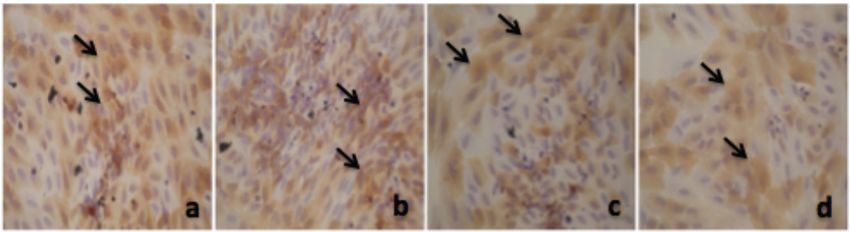
84   Journal of Pharmacy and Nutrition Sciences, 2018, Vol. 8, No. 3                                                   Ratwita et al.

concentration gradient by using sodium co-transport as                 protection with the antioxidant effects. Xanthone most
an energy source [13].                                                 researched are alpha, beta and gamma mangostin,
                                                                       garcinone E, 8-deoksigartanin and gartanin. Xanthone
    The other group of transporters convey glucose by                  could be found on the skin of fruit, fruit, bark and leaves
facilitated  diffusion   down      glucose-concentration               of mangosteen [16-19].
gradients. This group consists of five homologous
transmembrane proteins, GLUT-1, 2, 3, 4, and 5, that                   MATERIAL AND METHODS
are encoded by distinct genes. The GLUT proteins
have distinct substrate specificities, kinetic properties,             Insulin Tolerance Test
and tissue distributions that dictate their functional
                                                                       Preparation of Fat Emulsion
roles. Muscle is the principal site of insulin-stimulated
glucose disposal in vivo; less glucose is transported                     A constant volume of 100 mL fat emulsion
into adipose tissue [14]. Previous studies have                        containing 20 g lamb's fat, 1 g thyreostat, 5 g
indicated that α-mangostin and xanthone increase                       cholesterol, 1 g sodium glutamate, 5 g sucrose and 5 g
GLUT-4 expression on cardiac cell muscle and                           saccharose, 20 mL Tween 80, 30 mL propylene glycol
adipocyte.                                                             was prepared by adding distilled water and stored at
                                                                        0
                                                                       4 C [20].
     Glucose transporter (GLUT)-4 is a high-affinity
glucose transporter predominantly expressed in insulin-                Animal Treatment
sensitive tissues such as muscle and adipocytes.
                                                                          Fourty five Wistar rats were randomly divided into
Increasing GLUT-4 expression and plasma membrane
                                                                       normal group, control group which only got high fat
translocation of GLUT-4 have been found in low blood
                                                                       emulsion, treatment group that got high fat diet and α-
glucose. This transporter enhance glucose transport
                                                                       mangostin/xanthone 5, 10, 20 mg/kgbw respectively, or
and utilization [15]. In normal muscle cells and
                                                                       metformin. Rats in normal group received common
adipocytes, GLUT-4 will be recycled between the
                                                                       water. Rats in control and treatment group got high fat
plasma membrane and intracellular storage pools.                       emulsion group received fat emulsion for 10 d.
GLUT-4 is different from others because 90% of them
will be broken on the intracellular network when there is                  Insulin resistance sensitivity assay by short insulin
no stimulation of insulin, physical activity or other                  tolerance test using capillary blood glucose [21]. Rats
stimuli. The presence of insulin or other stimuli will                 were weighed and placed into mouse cage after fasting
trigger the translocation of GLUT-4 from the plasma                    overnight. Blood sugar in rats was detected six times
membrane to intracellular network. At cardiac muscle,                  after insulin (0.05 U/kgbw) intraperitoneally, every 30
GLUT-4 translocation will lead to the transverse tubules               minutes for 150 minutes. Blood glucose were checked
[7].                                                                   using glucose oxidase method with a glucometer.

     Insulin stimulates translocation of GLUT-4 by                         Absis indicates time and ordinate expresses nature
initiating insulin binding to receptors on the plasma                  logarithm of blood sugar. Regression coefficient (r) or
membrane. This bond will activate tyrosine                             slope was determined by linear regression and KITT
phosphorylation of the receptor intracellular kinase.                  was calculated by multiplying r by 100. K value
Stimulation of glucose transport in muscle cells and                   indicates insulin sensibility with smaller K values for
adipocytes need the phosphoinositide-3 kinase.                         lower sensibilities.
Phosphoinositide-3 kinase will activate the protein
kinase B (serine-threonine kinase). Stimulation of                     PPAR- γ Expression on Adipocytes
glucose transport in diabetic subjects impaired by the
change of insulin levels, while activation of protein                      Preadipocytes were isolated from rat retroperitoneal
kinase remained normal [7].                                            tissue [22]. Fibrous tissue and blood vessels were
                                                                       removed first. Tissue was washed and chopped.
    One alternative way that can be used to overcome                   Tissue suspension incubated with 0.2% collagenase
                                                                                                               0
the disease of diabetes is to utilize native medicinal                 type 1 (Sigma) for 45 minutes, at 37 C on shaking
plants. One of Asia native plant is Mangosteen                         condition. Incubation was stopped by the addition of
(Garcinia mangostana L.). Phytochemical studies show                   culture medium, Dulbecco’s Modified Eagle Media
that mangosteen contains oxygenated and prenylated                     (DMEM/F12 (1:1) supplemented with 15 mmol/l
xanthones. Xanthone is believed to have anti-cancer                    HEPES, 14 mmol/NaHCO3, 33 µmol/l of biotin, 17
effects, anti-inflammatory, anti-viral and cardiovascular              µmol l D-panthotenate and 10% Fetal Bovine Serum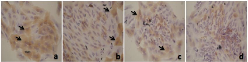
Alpha Mangostin and Xanthone from Mangosteen                   Journal of Pharmacy and Nutrition Sciences, 2018, Vol. 8, No. 3   85

(FBS). The filtration using a nylon mesh (250 µm). Cell     chamber was performed using a light microscope. The
suspension then rotated 1500 rpm for 7 minutes. Fat         data were calculated by the formula k = n x p x 2500,
layers (mature adipocyte and fat droplets) in the           with k = cell density (cells/ml), n = the total number of
supernatant was discarded. Pellet containing fibroblast-    cells in the four counting chamber, and p is the dilution
like preadipocyte then resuspended in cell culture          rate used [24].
media, rotated 1500 rpm for 7 minutes and then
resuspended again using the culture medium.                 Immunocytochemistry Staining

Cell Culture                                                    Adipocyte fixed in 10% formalin (v/v) in PBS (pH
                                                            7.4) for 20 minutes. Cells then washed with PBS (pH
    Preadipocytes were grown in Dulbecco’s Modified
                                                            7.4 ) three times, and treated by 0.02% (w/v) sodium
Eagle Media (DMEM) containing 10% Fetal Bovine
                                                            azide. Cells then washed again with PBS (pH 7.4)
Serum (FBS), 1% penicillin (10.000 U/mL), and 1%
                                                   0        three times for 5 minutes, then treated by H2O2 in PBS
streptomycin (10.000 !g/mL supplemented in 37 C
                                                            for 10 minutes. Reaction then stopped with 0.25%
incubator in a humidified atmosphere of 5% CO2). Cells
                                                            Triton-X blocking serum added in 5% FBS for 1 h, then
were subcultured every 3 to 4 days at approximately
                                                            washed with PBS. Adipocytes were treated with anti-
80% confluence. Mature adipocytes were seeded in
                                                            PPAR- γ antibody diluted in serum 1:500 for 24 h, then
96-well plates and grown until confluence. Alpha                             0
                                                            incubated at 4 C for 24 h. After incubated they were
mangostin/xanthone/pioglitazone was dissolved in
                                                            washed with PBS 3 times for 5 minutes each. Cells
dimethyl sulfoxide (DMSO) and treated for 48 h. Cells
                                                            were incubated in anti-rabbit secondary antibody 1:500
were then washed two times with PBS [23].
                                                            for 1 h at room temperature, then washed with PBS 3
Induction of Adipocytes Differentiation                     times for 5 minutes each. Cells treated by SA-HRP for
                                                            40 minutes, then washed with PBS 3 times for 5
    Before and after incubated in adipogenic media          minutes each. Cells then treated with Diamino
(DMEM/F12) 100 U/ml penicillin and 100 U/ml                 benzidine (DAB) in the DAB buffer. Cells treated with
streptomycin, 66 nM insulin, 100 nM dexamethasone,          courstexin with Mayer hematoxilin for 10 minutes, then
0.5 mM Methyl Iso buthyl Xantine (IBMX) and 10 µg ml        washed with tap water, followed by distilled water for
transferrin were added for adipocytes differentiation.      10 minutes. Oil Red O staining was used to confirm
Suspension cells grew in culture plates, incubated at       that cells that were differentiated were adipocytes.
    0
37 C, 5 % CO2 and 95 % humidity for 24 hours. Cells
were washed once every 3 days.                              Oil Red O Staining

Adipocyte Differentiation                                       Eight days after the differentiation induction, cells
    Cells were seeded into 22-well plates at a density of   were washed three times with PBS and fixed with 10%
      4
2×10 cells/well. Two days after confluence (defined as      formalin for 1 h at room temperature. After fixation,
day-0), cells were stimulated to differentiate with         cells were washed once with PBS and stained with
differentiation medium containing DMEM with 10% FBS         freshly diluted Oil Red O solution (3 parts of 0.6% Oil
and MDI [0.5 mM 3-isobutyl-1-methylxanthine (IBMX),         Red O in isopropanol and 2 parts of water) for 1 h.
0.25 µM dexamethasone, and 1 µg/mL insulin] for 2           Cells were then washed twice with distilled water and
days. In the course of screening adipocyte                  visualised under a microscope [25]. Images were
differentiation inhibitory activity, preadipocytes were     collected on an Olympus microscope.
treated with differentiation medium in the presence of
                                                            RESULT AND DISCUSSION
various concentrations of test compound (0.78 µM,
1.56 µM, 3.125 µM, 6.25 µM, 12.5 µM, 25 and 50 µM of
                                                                Insulin tolerance test was performed on insulin
α-mangostin/xanthone/ pioglitazone) at day-0. At day-
                                                            resistant animal models. Resistant animals were
2, differentiating medium was replaced with 10%
                                                            obtained with a fatty emulsion diet for 10 days. This
FBS/DMEM medium containing 1 µg/mL insulin and
                                                            diet will induce oxidative stress, which increases blood
incubated for another two days (day-4). Thereafter, the
                                                            glucose levels, which are immediately followed by an
cells were maintained in 10% FBS/DMEM medium for
                                                            increase in blood insulin levels, leading to insulin
an additional 4 days (day-8) with medium changes
                                                            resistance. Insulin-resistant animals are then given
every 2 days. Before and after incubated in adipogenic
                                                            intraperitoneal insulin (0.05 U/kgbw), then glucose
media, adipocytes were calculated. Calculating the
                                                            levels of experimental animals are measured every 15
number of cells in Neubauer Improved counting
                                                            minutes.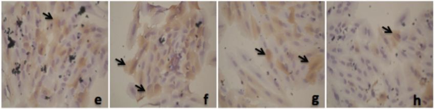
86   Journal of Pharmacy and Nutrition Sciences, 2018, Vol. 8, No. 3                                                      Ratwita et al.

    In order to clarify if the insulin-resistant animal                    superfamily and are ligand-activated transcription
model was established in our study, we detected the                        factors. There are 3 subtypes of PPAR, PPAR-α, δ,
dynamic characteristics of blood sugar after insulin                       and γ. PPAR-α reduces the triglyceride level, but
injection by short insulin tolerance test using capillary                  increase plasma HDL-cholesterol. PPAR-δ is potential
blood glucose. The result demonstrated that the KITT                       therapeutic target for metabolic syndrome, insulin
value decreased markedly after high fat emulsion for                       resistance, and obesity. PPAR-γ agonist are being
10 d compared to normal group, indicating that the rats                    developed to increase insulin sensitivity and to
are insensitive to exogenous insulin, i.e. insulin                         simultaneously     prevent diabetic  cardiovascular
resistance. Insulin resistance refers to the insensitivity                 complication [26].
of tissues (such as sceletal muscle, liver, kidney, and
adipose tissue) to insulin action, i.e. the weaker                            PPAR-γ also having a key role in adipogenesis.
glucose utilization of body after insulin action that                      The PPAR-γ has been identified as the receptor for
results in hyperglycemia. Absis shows time and                             the thiazolidinediones, a new class of oral
ordinate showing blood glucose levels. Lower insulin                       antidiabetic agents which improves glycaemic
tolerance test (KITT) co-efficient indicates low insulin                   control by lowering peripheral insulin resistance. In
sensitivity                                                                patients with Type II (non-insulin-dependent)
                                                                           diabetes    mellitus,    thiazolidinediones    reduce
Table 1: Insulin Tolerance Test                                            peripheral insulin resistance, but also seem to
                                                                           improve the pattern of insulin secretion [27].
                   Group                         Mean KITT ± SD
                                                                  a
                                                                               From Figure 1 we could see that PPAR-γ
                  Normal                           94.82 ± 3.70
                                                                           expression in adipocyte will increase when the
                  Control                          56.62 ± 11.25
                                                                  a
                                                                           concentration bigger. Thiazolidinediones are potent
         Alpha Mangostin 5 mg/kgbw                 90.59 ± 2.40
                                                                  a
                                                                           antidiabetic that lower the hyperglycemia, and
         Alpha Mangostin 10 mg/kgbw                93.39 ± 2.96
                                                                  a
                                                                           hypertriglyceridemia observed in human and animal
         Alpha Mangostin 20 mg/kgbw                93.69 ± 2.92
                                                                  b
                                                                           models of NIDDM [28]. In contrast to sulfonylureas,
            Xanthone 5 mg/kgbw                     72.40 ± 9.50
                                                                  a
                                                                           which improve insulin secretion, thiazolidinediones act
            Xanthone 10 mg/kgbw                    90.23 ± 3.33
                                                                  a
                                                                           by enhancing the peripheral sensitivity to insulin. Some
            Xanthone 20 mg/kgbw                    91.76 ± 2.59
                                                                  a
                                                                           insights into their mode of action has been provided by
                 Metformin                         94.55 ± 2.77
                                                                           the finding that thiazolidinedione compounds are high-
     a                                                     b
Note: Mann-Whitney test, p < 0.05 compared to control group; compared to
normal group.
                                                                           affinity ligands for PPAR-γ, a subtype of the nuclear
                                                                           receptor superfamily of ligand-activated transcription
   The results of the insulin tolerance test showed that                   factors.
the lowest KITT was seen in the positive control group,
which was given only 10 days of fatty emulsion. The                            Almost similar with thiazolinedione, in Figure 2 we
Kruskal Wallis results (p < 0.05), than it was analized                    could see that PPAR-γ expression in adipocyte will
using Mann-Whitney test and showed that there’s                            increase when the consentration bigger. The
significant difference between positive control group                      antihyperglycemic activity of various thiazolidinediones
when compared to the normal group who were not                             is closely linked to their PPAR-γ agonist activity [29].
given a high-fat diet. This suggests that the induction of                 The PPAR-γ gene codes for two PPAR-γ isoforms,
insulin resistance states using a fatty emulsion for 10                    PPAR-γ1 and PPAR-γ2, with PPAR-γ2 being the
days was success.                                                          predominant form expressed in adipose tissue [30].
                                                                           This suggests that adipose tissue might be an
    KITT in all treatment groups were significantly                        important target for the effect of thiazolidinediones and
different when compared to the positive control group,                     α-mangostin on insulin sensitivity, as suggested
except xanthone 5 mg/kgbw. This suggests that alpha                        previously [31-32].
mangostin 5, 10 and 20 mg/bw, xanthone 10 and 20
mg/kgbw, as well as metformin, have the effect of                              In Figure 3 we can see that xanthone also increase
lowering insulin resistance in animal model that given a                   PPAR-γ expression in adipocyte. But its effect not as
10-day fatty emulsion.                                                     good as α-mangostin or thiazolinedione effect. We may
                                                                           suggest that adipose tissue might be an important
   Peroxisome     proliferator–activated     receptors
                                                                           target for the effect of xanthone, like thiazolinedione
(PPARs) belong to a subfamily of the nuclear receptors
                                                                           and α-mangostin.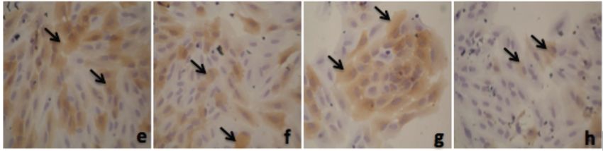
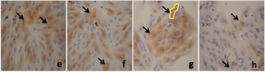
Alpha Mangostin and Xanthone from Mangosteen                          Journal of Pharmacy and Nutrition Sciences, 2018, Vol. 8, No. 3   87

Figure 1: Thiazolinedione effect on PPAR-γ expression in adipocyte.
a. 50 µM; b. 25 µM; c. 12.5 µM; d. 6.25 µM; e. 3.125 µM; f. 1.56 µM; g. 0.78 µM, h. 0 µM.

Figure 2: Alpha mangostin effect on PPAR-γ expression in adipocyte.
a. 50 µM; b. 25 µM; c. 12.5 µM; d. 6.25 µM; e. 3.125 µM; f. 1.56 µM; g. 0.78 µM, h. 0 µM.

Figure 3: Xanthone effect on PPAR-γ expression in adipocyte.
a. 50 µM; b. 25 µM; c. 12.5 µM; d. 6.25 µM; e. 3.125 µM; f. 1.56 µM; g. 0.78 µM, h. 0 µM.
88   Journal of Pharmacy and Nutrition Sciences, 2018, Vol. 8, No. 3                                                 Ratwita et al.

Figure 4: Alpha mangostin and xanthone effect on PPAR-γ expression in adipocyte cell culture (counting by immunoratio-
JPEG2000 virtual slide microscope).
                                     #
*) p < 0.05 compared to control; ) p < 0.05 compared to pioglitazone.

    From this figure we can see that alpha mangostin                   [39], but has also been acknowledged to play an
effect not differ significantly to pioglitazone, except on             important role in the control of energy metabolism [40-
consentration 0.78 µM, on which alpha mangostin                        41]. A number of transcription factors have been
effect bigger than pioglitazone. Xanthone effect on                    documented to be involved in the adipogenesis,
each consentration also not differ significantly if                    glucose uptake, and glycolysis pathway [42]. These
compared to pioglitazone. But it’s effect still below                  transcription factors include PPAR-", glucose
mangostin’s. On the other hand, PPAR-γ is a key factor                 transporter-4 (GLUT-4), and adipokines such as leptin.
for adipocyte differentiation, as shown using cell lines
[33] and thiazolidinediones are efficient promoters of                     PPAK-γ is predominantly expressed in adipose
adipocyte differentiation in vitro [2,14-16, 34-37]. Thus,             tissues and plays a central role in adipose tissue
it could be questioned whether a thiazolidinedione                     functions [43]. PPAK-γ regulates the expression of
therapy aimed at improving insulin sensitivity would                   genes associated with insulin signalling and glucose
promote the recruitment of new adipocytes in vivo, an                  and lipid metabolism in mature adipocytes [11].
effect that could be deleterious, since most of the                    Reduced expression of PPAK-γ has been shown to be
NIDDM patients were obese. Glucose utilization is                      effective in inhibiting the adipogenesis of 3T3-L1 cells
obviously linked to an increased capacity of taking up                 [44-45].
and metabolizing glucose through the activation of
                                                                       CONCLUSION
genes involved in glucose transport (GLUT-4) and
metabolism into lipids (FAS and PEPCK).
                                                                          In conclusion, the present study has shown that α-
                                                                       mangostin and xanthone isolated from mangosteen
    Garcinia mangostana Linn or mangosteen found in
South East Asia. Its pericarps have been used as                       (Garcinia mangostana L) are two substance that
                                                                       showed potential effect on preventing tolerance, and
traditional medicine. Phytochemical studies shown that
                                                                       increasing PPAR-γ expression on adipocyte.
mangosteen contained secondary metabolite as
oxygenated and prenylated xanthones (α, β, γ                           CONFLICT OF INTEREST
mangostin). Previous study has shown that α-
mangostin and xanthone isolated from mangosteen (G.                        The authors declare that there is no conflict of
Mangostana L.) are two substance that showed                           interests.
protective effect to glucose tolerance and also potential
to improve insulin resintency by increasing GLUT-4 on                  ACKNOWLEDGEMENT
muscle and adipocyte [38].
                                                                           This work was supported by Jenderal Achmad Yani
   Adipose is not only known for its capacity to store                 University and School of Pharmacy, Bandung Institute
the excess of dietary energy in the form of triglyceride               of Technology, Indonesia.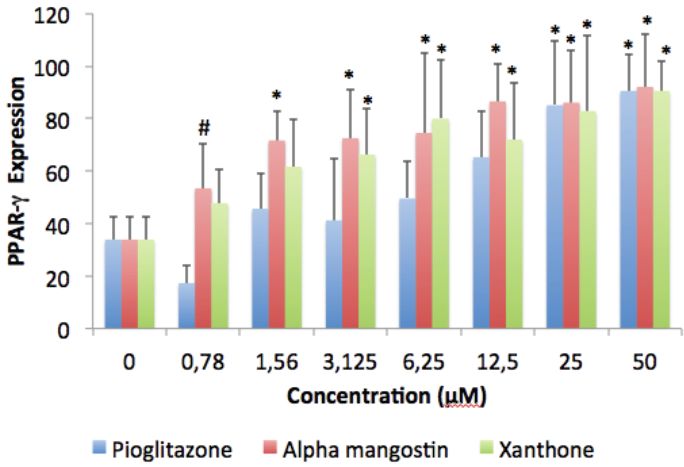
Alpha Mangostin and Xanthone from Mangosteen                                       Journal of Pharmacy and Nutrition Sciences, 2018, Vol. 8, No. 3   89

REFERENCES                                                                  [18]      Chaverri J, Rodrỉguez N, Ibarra M, Rojas J. Medical
                                                                                      properties of mangosteen (Garcinia mangostana). Food and
[1]    Gorelick J, Kitron A, et al. Anti-diabetic activity of Chiliadenus             Chemical Toxicology 2008; 46: 3227-3239.
       iphionoides. Journal of Ethnopharmacology 2011; 137(3):                        https://doi.org/10.1016/j.fct.2008.07.024
       1245-1249.                                                           [19]      Reddy J, Ravikumar N, Gaddamanugu G, Naresh K, Rajan
       https://doi.org/10.1016/j.jep.2011.07.051                                      S, Solomon K. Synthesis, crystal structure, spectral charac-
[2]    Yu Z, Yin Y, Zhao W, Liu J, Chen F. Anti-diabetic activity                     terization and fluorescence studies of salts of α-mangostin
       peptides from albumin against α-glucosidase and α-amylase.                     with APIs. Journal of Molecular Structure 2013: 137-143.
       Journal of Food Chemistry 2012; 135: 2078-2085.                                https://doi.org/10.1016/j.molstruc.2013.01.058
       https://doi.org/10.1016/j.foodchem.2012.06.088                       [20]      Ryu H, Cho J, Long M, Yuk H, Kim Y, Jung S, Lee B, Park K.
[3]    Yu Z, Yin Y, et al. Characterization of ACE-inhibitory peptide                 α-glukosidase inhibition and antihyperglycemic activity of
       associated with antioxidant and anticoagulation properties.                    prenylated xanthones from Garcinia mangostana. Journal of
       Journal of Food Science 2011; 76 (8): 1149-1155.                               Phytochemistry 2011; 72: 2148-2154.
       https://doi.org/10.1111/j.1750-3841.2011.02367.x                               https://doi.org/10.1016/j.phytochem.2011.08.007
[4]    Yu Z, Yin Y, et al. Novel peptides derived from egg white            [21]      Ai J, Wang N, Yang M, Du ZM, Zhang YC, Yang BF.
       protein inhibiting alpha-glucosidase. Food Chemistry 2011;                     Development of Wistar rat model of insulin resistance, World
       129(4): 1376-1382.                                                             Journal of Gastroenterology 2005; 11(24): 3675-3679.
       https://doi.org/10.1016/j.foodchem.2011.05.067                                 https://doi.org/10.3748/wjg.v11.i24.3675

[5]    Orme CM, Bogan JS. Sorting out diabetes. Science 2009;               [22]      Winzell MS, Ahre´n B. The high-fat diet-fed mouse a model
       324: 1155-1156.                                                                for studying mechanisms and treatment of impaired glucose
       https://doi.org/10.1126/science.1174841                                        tolerance and type 2 diabetes. Diabetes 2004; 53(3): 215-19.
                                                                                      https://doi.org/10.2337/diabetes.53.suppl_3.S215
[6]    Yu Z, Yin Y, Zhao W, Liu J, Chen F. Anti-diabetic activity
       peptides from albumin against α-glucosidase and α-amylase.           [23]      Gregoire FM, Smas CM, Sul HS. Understanding adipocyte
       Food Chemistry 2012; 135: 2078-2085.                                           differentiation. American Physiological Society Journal 1998;
       https://doi.org/10.1016/j.foodchem.2012.06.088                                 78(3): 783-809.
                                                                                      https://doi.org/10.1152/physrev.1998.78.3.783
[7]    Shepherd PR, Kahn BB, Glucose transporters and insulin
       action, implication for insulin resistance and diabetes mellitus.    [24]      Mosmann T. Rapid colorimetric assay for cellular growth and
       The New England Journal of Medicine 1999; 341(4): 248-256.                     survival: application to proliferation and cytotoxicity assays.
       https://doi.org/10.1056/NEJM199907223410406                                    Journal of Immunological Methods 1993; 65(2): 55-63.

[8]    Klaus S. Adipose tissue as a regulator of energy balance.            [25]      Susantia D, Amiroudineb MZAM, Rezalic MF, Taherb M.
       Curr Drug Targets 2004; 5: 241-250.                                            Friedelin and lanosterol from Garcinia prainiana stimulated
       https://doi.org/10.2174/1389450043490523                                       glucose uptake and adipocytes differentiation in 3T3-L1
                                                                                      adipocytes. Natural Product Research 2013; 27(4-5): 417-
[9]    Faraj M, Lu HL, Cianflone K. Diabetes, lipids, and adipocyte                   424.
       secretagogues. Biochem Cell Biol 2004; 82: 170-190.                            https://doi.org/10.1080/14786419.2012.725399
       https://doi.org/10.1139/o03-078
                                                                            [26]      Shin E, Choi KM, Yoo HS, Lee CK, Hwang BY, Lee MK.
[10]   Das M, Gabriely I, Barzilai N. Caloric restriction, body fat and               Inhibitory effects of coumarins from the stem barks of
       ageing in experimental models. Obes Rev 2004; 5: 13-19.                        Fraxinus rhynchophylla on adipocyte differentiation in 3T3-L1
       https://doi.org/10.1111/j.1467-789X.2004.00115.x                               cells. Biological and Pharmaceutical Bulletin 2010; 33(9):
[11]   Fluck CE, Slotboom J, Nuoffer JM, Kreis R, Boesch C, Mullis                    1610-1614.
       PE. Normal hepatic glycogen storage after fasting and                          https://doi.org/10.1248/bpb.33.1610
       feeding in children and adolescents with type 1 diabetes.            [27]      Matsuura N, Gamo K, Miyachi H, Iinuma M, Kawada T,
       Pediatr Diabetes 2003; 4: 70-76.                                               Takahashi N, Akao Y, Tosa H. γ-Mangostin from Garcinia
       https://doi.org/10.1034/j.1399-5448.2003.00015.x                               mangostana Pericarps as a dual agonist that activates both
[12]   El-Serag HB, Tran T, Everhart JE. Diabetes increases the                       PPAR-α and PPAR-γ. Biosci Biotechnol Biochem 2013;
       risk of chronic liver disease and hepatocellular carcinoma.                    77(12): 2430-2435.
       Gastroenterology 2004; 126: 460-468.                                           https://doi.org/10.1271/bbb.130541
       https://doi.org/10.1053/j.gastro.2003.10.065                         [28]      Dubois M, Pattou F, Kerr-Conte J, Gmyr V, Vandewalle B,
[13]   Cohen SE, Tseng YH, Michael MD, Kahn CR. Effects of                            Deremaux P, Auwerx J, Schoonjans K, Lefebvre J.
       insulin-sensitizing agents in mice with hepatic insulin                        Expression of peroxisome proliferator-activated receptor γ
       resistance. Diabetologia 2004; 47: 407-411.                                    (PPAR-γ) in normal human pancreatic             islet cells.
       https://doi.org/10.1007/s00125-003-1320-4                                      Diabetologia 2000; 43: 1165-1169.
[14]   Wright EM, Turk E, Zabel B, Mundlos S, Dyer J. Molecular                       https://doi.org/10.1007/s001250051508
       genetics of intestinal glucose transport. J Clin Invest 1991;        [29]      Hoftnann CA, Colca JR. New oral thiazolidinedione
       88: 1435-40.                                                                   antidiabetic agents act as insulin sensitizers. Diabetes Care
       https://doi.org/10.1172/JCI115451                                              1992; 15: 1075-1078.
[15]   DeFronzo RA. Pathogenesis of type 2 diabetes: metabolic                        https://doi.org/10.2337/diacare.15.8.1075
       and molecular implications for identifying diabetes genes.           [30]      Willson TM, Cobb JE, Cowan DJ, Wiethe RW, Correa ID,
       Diabetes Rev 1997; 5: 177-269.                                                 Prakash SR, Beck KD, Moore LB, Kliewer SA, Lehmann JM.
[16]   Taher M, Amiroudine MZAM, Zakaria TMFST, Susanti D,                            The structure-activity relationship between peroxisome
       Ichwan SJA, Kaderi MA, Ahmed OU, Zakaria ZA.                                   proliferator-activated   receptor    γ    agonist   and   the
       #-Mangostin improves glucose uptake and inhibits                               antihyperglycemic activity of thiazolidinediones. J Med Chem
       adipocytes differentiation in 3T3-L1 Cells via PPAR-", GLUT-                   1996; 39: 665-668.
       4, and leptin expressions. Evidence-Based Complementary                        https://doi.org/10.1021/jm950395a
       and Alternative Medicine 2015; 2015: 1-9.                            [31]      Tontonoz P, Hu E, Graves RA, Budavari AI, Spiegelman BM.
[17]   Aisha A, Abu-Salah K, Siddiqui M, Ismail Z, Majid A.                           mPPAR72, tissue specific regulator of an adipocyte
       Quantification of α-, β- dan γ-mangostin in Garcinia                           enhancer. Genes Dev 1994; 8: 1224-1234.
       mangostana fruit rind extracts by a reverse phase high                         https://doi.org/10.1101/gad.8.10.1224
       performance liquid chromatography. Journal of Medicinal              [32]      Hotamisligil GS, Spiegelman BM. Tumor necrosis factor α: a
       Plants Research 2012; 6(29): 4526-4534.                                        key component. Diabetes 1997; 46.
90 Journal of Pharmacy and Nutrition Sciences, 2018, Vol. 8, No. 3 Ratwita et al.
[33] Hofmann C, Lorenz K, Braithwaite SS, Colca JR, Palazuk BJ, [40] Taher M, Amiroudine MZAM, Syafiq TMF, Zakaria T, Susanti
Hotamisligil GS, Spiegelman BM. Altered gene expression D, Ichwan SJA, Kaderi MA, Ahmed QU, Zakaria ZA. α-
for tumor necrosis factor-α and its receptors during drug and Mangostin improves glucose uptake and inhibits adipocytes
dietary modulation of insulin resistance. Endocrinology 1994; differentiation in 3T3-L1 cells via PPAR-γ, GLUT-4, and leptin
134: 264-270. expressions. Evidence-Based Complementary and
https://doi.org/10.1210/endo.134.1.8275942 Alternative Medicine 2015; 1-9.
https://doi.org/10.1155/2015/740238
[34] Zncke YF, Hallakou RS, Doare L, Foufelle F, Kergoat M,
Guerre-Millo M, Berthault MF, Dugail I, Morin J, Auwerx J, [41] Spiegelman BM, Flier JS. Obesity and the regulation of
Ferre P. Pioglitazone induces in vivo adipocyte differentiation energy balance. Cell 2001; 104(4): 531-543.
in the obese. Diabetes 1997; 1393-1399. https://doi.org/10.1016/S0092-8674(01)00240-9
[35] Lehmann JM, Moore LB, Smith-Oliver TA, Wilkinson WO, [42] Yun JW. Possible anti-obesity therapeutics from nature-a
Wilson TM, Kliewer SA. An antidiabetic thiazolidinedione is a review. Phytochemistry 2010; 71(14-15): 1625-1641.
high affinity ligand for peroxisome proliferator-activated https://doi.org/10.1016/j.phytochem.2010.07.011
receptor γ (PPAR-γ). J Biol Chem 1995; 270: 12953-12956. [43] V´azquez-Vela MEF, Torres N, Tovar AR. White adipose
https://doi.org/10.1074/jbc.270.22.12953 tissue as endocrine organ and its role in obesity. Archives of
[36] Kletzien RF, Clarke SD, Ulrich RG. Enhancement of Medical Research 2008; 39(8): 715-728.
adipocyte differentiation by an insulin-sensitizing agent. Mol https://doi.org/10.1016/j.arcmed.2008.09.005
Pharmacol 1992; 41: 393-398. [44] Brun RP, Spiegelman BM. PPAR-gamma and the molecular
[37] Sandouk T, Reda D, Hofmann C. Antidiabetic agent control of adipogenesis. Journal of Endocrinology 1997;
pioglitazone enhances adipocyte differentiation of 3T3-F442A 155(2): 217-218.
cells. Am J Physiol 1993; 264: 1600-1608. https://doi.org/10.1677/joe.0.1550217
https://doi.org/10.1152/ajpcell.1993.264.6.C1600 [45] Choi SS, Cha BY, Iida K. Artepillin C. as a PPAR-γ ligand,
[38] Brun RP, Tontonoz P, Forman BM, Ellis R, Chen J, Evans enhances adipocyte differentiation and glucose uptake in
RM, Spiegelman BM. Differential activation of adipogenesis 3T3-L1 cells. Biochemical Pharmacology 2011; 81(7): 925-
by multiple PPAR isoforms. Genes Dev 1996; 10: 974-984. 933.
https://doi.org/10.1101/gad.10.8.974 https://doi.org/10.1016/j.bcp.2011.01.002
[39] Ratwita W, Sukandar EY, Adnyana IK, Kurniati NF, Alpha [46] Huang C, Zhang Y, Gong Z. Berberine inhibits 3T3-L1
mangostin and xanthone from mangosteen (Garcinia adipocyte differentiation through the PPARγ pathway.
mangostana l.) role on glucose tolerance and glucose Biochemical and Biophysical Research Communications
transporter-4 in diabetes mellitus. International Journal of 2006; 348(2): 571-578.
Pharmacognosy and Phytochemical Research 2017; 9(9): https://doi.org/10.1016/j.bbrc.2006.07.095
1206-1212.
Received on 19-04-2018 Accepted on 04-05-2018 Published on 25-06-2018
DOI: https://doi.org/10.6000/1927-5951.2018.08.03.1
© 2018 Ratwita et al.; Licensee Lifescience Global.
This is an open access article licensed under the terms of the Creative Commons Attribution Non-Commercial License
(http://creativecommons.org/licenses/by-nc/3.0/) which permits unrestricted, non-commercial use, distribution and reproduction in
any medium, provided the work is properly cited.You can also read