Safety study of malapposition of the bio-corrodible nitrided iron stent in vivo
←
→
Page content transcription
If your browser does not render page correctly, please read the page content below

Nanotechnology Reviews 2021; 10: 839–846
Research Article
Xiaoli Shi, Jin Wang*, Gui Zhang, Lu Zhang, Wanqian Zhang, Ping Cao, Geqi Wang,
Deyuan Zhang, and Li Qin
Safety study of malapposition of the
bio-corrodible nitrided iron stent in vivo
https://doi.org/10.1515/ntrev-2021-0062 which works by opening the narrowed arteries thus
received April 13, 2021; accepted July 15, 2021 improving blood flow [1–3]. Coronary stents have under-
Abstract: To evaluate the safety of stent malapposition of gone enormous development since their first introduction
corrodible nitride iron stent as biodegradable cardiovas- in 1987 [4]. Bio-absorbable coronary stents have been
cular implants, a total of 108 stents were implanted into widely acknowledged to be the 4th revolution in vascular
the abdominal aortas, iliac arteries, and iliac artery bifur- intervention, following Percutaneous Transluminal Cor-
cations of 36 New Zealand white rabbits separately. Each onary Angiography, Bare Metal Stent, and Drug-Eluting
rabbit was implanted with three stents. After a follow-up Stent (DES), getting over the adverse event of late stent
period of 3 months, no thrombus and embolism were thrombosis occurred in the third generation DES, pro-
found in local and downstream vessels. And no other viding mechanical support and anti-restenosis properties
adverse events occurred either. Stent strut covered by in short term and then metabolized physiologically, thus
endothelial layer started to show signs of degradation, avoiding the permanent metallic enclosing of the treated
while struts exposed to bifurcated blood flow covered coronary artery. Therefore, bio-absorbable coronary stents
by a layer of tissue and no rust particle was found on have extensive research significance.
the surface. Also, there were no traces of thrombosis Iron-based stents are a promising candidate for bio-
and traces of excess inflammation. The authors conclude absorbable stents, due to their good biocompatibility and
that the risk brought by stent malapposition in less than outstanding mechanical performances like 316L stainless
9 months is acceptable. steel [5–10]. According to reported studies [8–10], it has
been demonstrated that safety of iron stents implantation
Keywords: bio-absorb iron stent, corrosion, iron over- without the significant obstruction of the stented vessel
load, stent malapposition, thrombosis as a result of inflammation, thrombotic events, or neo-
intimal proliferation, but a faster degradation rate is demanded
theoretically; an ideal bio-absorbable iron stent should
provide enough support to ensure that preventing vascular
1 Introduction retraction in the initial stage after implantation, after 3–6
months of action, most of the loss of support caused by the
Coronary stent implantation is the most common and corrosion process is beneficial to tissue regeneration. How-
effective treatment means for coronary artery disease, ever, some studies demonstrated that the corrosion rate of
the pure iron stents was slower than required. For the past
few years, further researches focused on modifying the
* Corresponding author: Jin Wang, Key Lab of Advanced composition, surface, and microstructure of pure iron
Technologies of Materials, Ministry of Education, School of stents to increase their corrosion rate [11–14]. Here vacuum
Materials Science and Engineering, Southwest Jiaotong University,
plasma nitriding was employed to enhance its strength
Chengdu 610031, Sichuan, China, e-mail: wangjin@swjtu.edu.cn
Xiaoli Shi, Lu Zhang: Key Lab of Advanced Technologies of and corrosion rate by incorporating nitrogen into the
Materials, Ministry of Education, School of Materials Science and iron matrix to form dispersive iron nitride precipitation
Engineering, Southwest Jiaotong University, Chengdu 610031, phase [9,15]. But along with the increase of corrosion
Sichuan, China rate of the iron stent, the release of iron ions will also
Gui Zhang, Wanqian Zhang, Geqi Wang, Deyuan Zhang, Li Qin:
increase during the short term after implantation, espe-
Biotyx Medical (Shenzhen) Co., Ltd, Jinxiu Scientific Park,
Wuhe Road, Longhua, Shenzhen 518109, China
cially the stage when the struts were not completely covered
Ping Cao: Shenzhen Testing Center of Medical Device, Nanshan by endothelium cells. And too much iron in the body is
District, Shenzhen 518057, China at risk [16,17]. Some studies show that iron overload has
Open Access. © 2021 Xiaoli Shi et al., published by De Gruyter. This work is licensed under the Creative Commons Attribution 4.0
International License.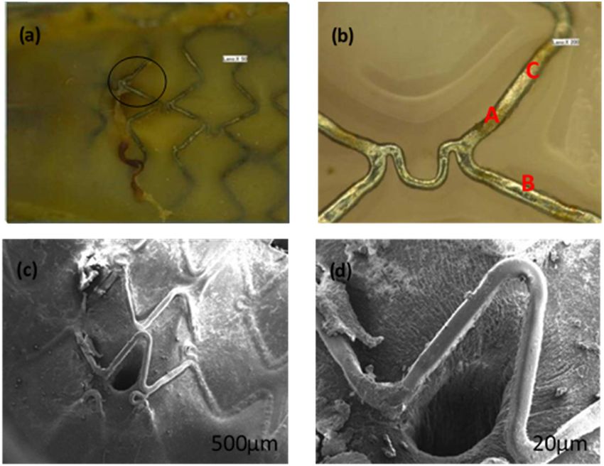
840 Xiaoli Shi et al.
a direct relationship with heart and liver disease, diabetes, hardness, radial strength, and stiffness of the nitrided
and other relevant diseases [18,19]. Conversely, after the iron stent were significantly increased. After the treating
stent strut was completely covered by endothelium cells, process of vacuum nitriding, the nitrided iron stents were
the human body has a series of iron control mechanisms polished to achieve a strut thickness of 70 ± 5 µm. The
(an iron regulation mechanism) to control the systematic stent weighs 10–15 mg. The stents were sterilized with
or local iron overload risk [20]. Hence, it is very necessary ethylene oxide and stored in vacuumed packages before
to evaluate the iron overload risk in the short term after the implantation operation.
implantation of the nitrided iron stent.
Another safety concern about bio-corrodible stent is
late thrombogenicity or embolism of corrosion products
when exposed to blood flow for a long time, probably 2.2 Animals
occurs in stent malapposition, which has not been reported
yet. The stent malapposition is usually caused by inade- The study was conducted with the approval of the local
quate expansion or plaque existed at the vascular wall governmental authorities and adhered to the Guide for
[21]. And this phenomenon will eventually cause delayed the care and use of laboratory animals (NIH publication
endothelialization, stent thrombosis complications, and 85–23, 1985). Thirty six adult New Zealand white rabbits
other diseases [22–25]. In vitro studies cannot fully simu- (mean weight 2.2 kg, range 2.0–2.5 kg) were purchased from
late the real physical and chemical environment in vivo. Guangdong province medical animal test center (China).
Therefore, appropriate animal models are necessary to
further study the implantation of bio-corrosive and
degradable iron nitride stents.
Given these considerations, this study adopted implanting 2.3 Procedure
the bio-absorbable stent in the iliac artery bifurcation posi-
tion of the New Zealand rabbit, to simulate the stent Anesthesia was induced by pentobarbitone (30 mg/kg)
implantation state in a vessel. And through monitoring intravenously. Antibiotic prophylaxis was administered
the thrombus and embolism of supported vessels and intramuscularly. The right carotid artery was surgically
downstream vessels, the topography of nitrided iron stent exposed and a 5 French sheath was introduced over a
after implantation for 1, 3, and 9 months separately, the 0.018-inch guidewire after being immersed in the hepar-
corrosion rate and the late risk brought by the implanted inized saline for 0.5 h. Quantitative angiography of the
stents were evaluated. descending aorta was performed to determine the luminal
diameter of the descending aorta at the site of the implan-
tation (5 French Berman angiography catheter, Arrow,
Reading, Pennsylvania, USA). Balloon catheters with a
diameter of 3 mm (Savy, Cordis, Miami, Florida, USA)
2 Materials and methods were chosen to achieve a balloon to vessel ratio of about
1.2 ± 0.1. The degradable iron stent was manually crimped
2.1 Stents to the balloon catheter. Under fluoroscopic control, the
stent was introduced and positioned at the predetermined
The degradable stents, with a pattern similar to a commer- implantation site. The stents were implanted with 8 atm for
cially available permanent coronary stent of a nominal 10 s. After removal of the catheter and sheath, the carotid
inflated outer diameter of 3.0 mm and a nominal length artery was ligated and the incision of the skin was closed
of 18 mm, were laser cut from iron tubes (>99.8% iron, with sutures. The animals were returned to the recovery
made by Biotyx Medical (Shenzhen) Co., Ltd). Vacuum area. Figure 1 shows the implantation position of a testing
plasma nitriding technique is added into the manufac- rabbit sacrificed after 3 months of implantation.
turing process after laser cutting using a self-designed
vacuum nitriding furnace. Vacuum plasma nitriding in
an engineering field is originally a surface modification
technology. However, when applied to small coronary 2.4 Follow-up study
stents, it becomes a bulk alloying method since the ori-
ginal strut thickness is 100 µm or less. As a result of dis- All animals were followed up and results were docu-
persion strengthening and solution strengthening, the mented throughout the study. Vessel morphology was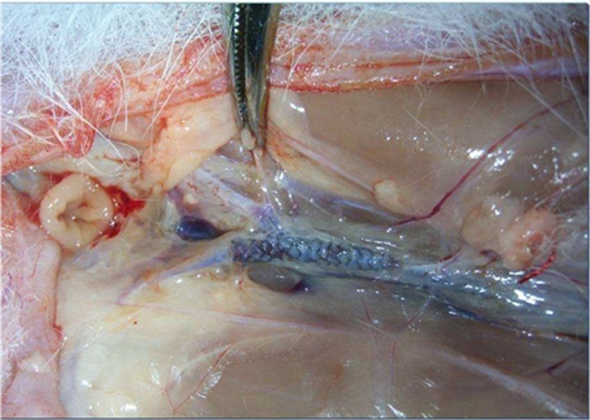
Malapposition of bio-corrodible nitrided iron stent in vivo 841
of heparin were injected into the rabbits, and then the
animals were sacrificed by an intravenous injection of
a lethal dose of pentobarbital. Immediately after death,
the infra diaphragmatic descending aorta was exposed
and the stented segment with stents was identified and
harvested. The segments were placed in 3.5% neutral
buffered formalin and kept for histopathological investi-
gation. The stents were cut longitudinally and the lumen
of the stented vessel was evaluated for signs of adherent
thrombi or overt neointimal obstruction of the vessel. One
specimen of each study group was viewed under a scan-
ning electron microscope after critical point drying and
gold sputtering to assess the endothelialization of the
stent struts.
Figure 1: The implantation position of nitrided iron stent in a rabbit.
observed after 1 month (n = 15), 3 months (n = 15), and
9 months (n = 6). 3 Results
The SEM and EDS of the nitride iron material surface
were shown in Figure S1 (Supporting Information). After
2.5 Morphological investigation nitride, the atom ratio of Nitrogen is about 5.27%, with a
weight percent of 1.37%. The degradable iron stents were
After angiography for the downstream vessels (using an implanted in the predetermined segment of the iliac
x-ray machine with the assistant of diatrizoate), 1,000 U arteries in 36 rabbits without major complications. No
Figure 2: Rabbit iliac aorta with degradable iron stent after 1-month implantation. (b) is the scale-up diagram in (a); (d) is the scale-up
diagram in (c).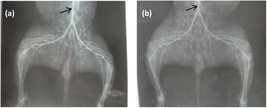
842 Xiaoli Shi et al. abnormalities on macroscopic inspection of the implan- by a layer of uniform complete fibrous tissue membrane, tation site were observed. During the 3 months of the which effectively prevented the direct contact between follow-up period, no cases of animal death or other the strut and the blood flow. In short, it increased safety obvious symptoms of pathological changes occurred. when there was a stent malapposition state occurred. After the animals were put to dissection, no thrombosis Figure 3 shows rabbit iliac artery with degradable or angiemphraxis is observed in the downstream vessels. iron stent after 3 months implantation. As seen from The segment of the iliac arteries was separated and Figure 3, the corrosion level increased compared with cut down for the next study of degradation. There was the case of 1 month. Iron transportation was observed also no blockage or thrombosis observed at the implant from the implantation site towards the reticuloendothe- position. Figure 2 shows the rabbit iliac arteries with lial system after 1 month, and the level became more degradable iron stent after 1 month of implantation. As serious after 3 months. There are obvious black corrosion seen from Figure 2a, the strut in the orifice of vascular products stacked on the surface of the struts which are branch position was not covered by endothelial cells, and located at the transitional position. However, the strut the other struts contacted with the vascular wall were which is located at the vascular branch position is still covered by endothelial cells completely. In addition, shining. No corrosion products could be detected on the the nitrided iron stent was corroded and there was no strut surface. Figure 3c and d show the SEM images of obvious corrosion product formed on the surface of the rabbit iliac artery with degradable iron stent after 3 strut. Compared with the strut contacted with the vas- months of implantation. We can see that the fiber tissue cular wall (position C in Figure 2b) and the transitional membranes covered on the strut located at the branch strut (incompletely covered by endothelial cells, position position became thicker than the ones after 1 month of A in Figure 2b), the strut in the branch position is rela- implantation. The thickness of the fiber tissue membrane tively smooth. In other words, the corrosion level in posi- is about several microns. The fiber tissue membrane tion B is relatively slight. SEM image gives information shown in Figure 3d is deliberately broken for the aim of that the strut in the vascular branch position is covered evaluating the thickness change. Figure 3: Rabbit iliac artery with degradable iron stent after 3 months implantation. (b) is the scale-up diagram in (a); (d) is the scale-up diagram in (c).
Malapposition of bio-corrodible nitrided iron stent in vivo 843
Figure 4: Rabbit iliac artery with degradable iron stent after 9 months implantation. (b) is the scale-up diagram in (a).
Figure 4 shows the strut located at the vascular malapposition is caused by the positive reconstruction
branch position with a degradable iron stent after 9 of vessels or by the chronic rebound of the stent. Research
months of implantation. As shown, the surface of the has shown that late stent malapposition may lead to the
strut is relatively smooth, and also no obvious corrosion formation of stent thrombosis [29,30]. Once the iron stent
product was found. To evaluate the risk of thrombosis was implanted, it corrodes gradually; when there the
brought by the stent malposition, the rabbits’ lower limbs stent malapposition happens, the struts’ endothelializa-
vessels’ X-ray morphology was performed before the rabbit tion process would be delayed. At the same time, the cor-
was sacrificed at 3 and 9 months separately (Figure 5). As roded strut may produce degradation product particles.
seen, there were no obstacles found and there was no And the particles may flow away and even enter into the
abnormal blood flow of the downstream vessels. downstream small vessels. This hazard could cause com-
plications like thrombosis and obstruction. However, the
follow-up animal study of 1 and 3 months found that there
are no attached degradation products found on the surface
4 Discussions of the strut which is located at the vascular branch posi-
tion. In addition, there was a layer of fiber tissue mem-
Stent malapposition refers to improper contact between branes covered on the strut. This kind of fiber layer tissue
the struts and vascular walls [26]. And it includes two membrane isolated the stent strut and the flowing blood
kinds: early acute and late chronic malapposition [27]. [31]. The strut at the branch position remained shining for
Early acute stent malapposition cases are mainly caused 3 months. Corrosion mechanism is a major difference
by the inexperienced operation, partially caused by some between the degradation iron metal stent and the perma-
vascular diseases like calcified lesions [28]. Late stent nent stent, thus the extensive research attention in the
Figure 5: Rabbits’ downstream vessels’ X-ray morphology with a degradable iron stent at the iliac artery position: (a) 3 months after
implantation; (b) 9 months after implantation, arrow position stand for the position of the implanted stent.844 Xiaoli Shi et al.
Figure 6: Surface topography of the strut after cleaning the corrosion products ((a) at the position adjacent to the vascular wall and (b) at
the vascular branch position).
degradation vascular stent area [32–35]. The degradation branch position, the membrane covered on the strut is
process of the nitrided iron stent in vivo could be explained thinner; the iron ion released from the strut is easy to
as follows: First, the iron was oxidized to metal ions fol- pass through the membrane and taken away by the blood
lowing equation (1). The electrons from the anodic reaction flow. So, there was no corrosion product stacked on the
were consumed by a corresponding cathodic reaction and surface of strut. Conversely, as for the strut at the position
the reduction of oxygen dissolved in water, following adjacent to vascular wall, 3 months after implantation it
equation (2). These reactions occurred randomly over the was revealed that it was covered by a relatively thicker
entire surface where a difference existed, at grain bound- and denser layer mainly composed of endothelium. Hence,
aries or the interface between different phases [36]. the iron ion released from the strut is hard to pass through
Anodic reaction: and chemical reactions (3) and (4) occurred. To some
(1) extent, this could explain the result we see from Figure 3.
Fe → Fe 2 + + 2e.
To further distinguish the difference of degradation condi-
Cathodic reaction: tions, SEM analysis was performed to the surface topo-
1/ 2O2 + H2 O+ 2 e→ 2OH− . (2) graphy of strut at different implant positions after cleaning
the corrosion product. As seen in Figure 6, the corrosion
Then, the released metal ions reacted with the hydroxyl depth of the strut adjacent to the vascular wall is slightly
ion (OH−) released from the cathodic reaction to form in- deeper than the one at the branch position (6 months
soluble hydroxides (hydrous metal oxides) according to after implantation). The corrosion extent of the strut at
equations (3) and (4). the transitional position looks more serious (Figure 3b).
2Fe 2 + + 4OH− → Fe(OH)2 or 2FeO ⋅ 2H2 O, (3) This phenomenon may cause by the difference in oxygen
diffusion rate. In another word, the oxygen concentration
Fe4Fe(OH)2 + O2 → 4FeOOH + 2H2 O. (4)
of the tissue fluid around the strut at the two positions
However, the corrosion conditions are differences is different. In addition, the difference in biochemical
between the strut at the branch position and the position environment between the two positions also (galvanic cor-
adjacent to the vascular wall. As for the strut at the rosion) accelerated the corrosion rate at the transitional
position [37].
Figure 7 shows the corrosion speed between different
existed states of an iron element. In a real degradation
situation in vivo environment, corrosion products are
always produced on the surface of an iron stent. The
amount of rust is determined by the difference between
V1 and V2, while V3 is not so important as V1 and V2 since
rust will stay much longer than intact iron during the
lifespan of the iron stent. And the final degradation
time of the stent is mainly determined by V2 since V2 is
Figure 7: The sketch map of corrosion speed between the different much lower than V1. V2 becomes a bottleneck procedure
states of an iron element. in all degradation activities of the iron stent. TheMalapposition of bio-corrodible nitrided iron stent in vivo 845
exception occurred before the iron stent is covered by [2] Naghshtabrizi B, Monfared AM, Emami F, Poorolajal J. Impact
tissue when the bloodstream flows on the surface of of stent type on incidence of major adverse cardiac events in
the iron stent, which will expedite the V2 extremely to large coronary arteries with tubular and diffuse lesions.
Minerva Cardioangiol. 2015;64(5):517–24.
the level faster than V1. So the struts of the stent at the
[3] Liu HF, Wang M, Xu YS, Kumar SM, Lu XR, Qiang LJ. Diagnostic
branch position keep free rust on. accuracy of dual-source and 320-row computed tomography
Currently adopted in vivo experiments are using bare angiography in detecting coronary in-stent restenosis: a sys-
nitride iron stents, without being coated with drug-eluting tematic review and meta-analysis. Acta Radiol.
coatings. After being covered with coatings, the corrosion 2018;60(2):149–59.
[4] McKavanagh P, Zawadowski G, Ahmed N, Kutryk M. The evo-
rate should be slower, especially at the initial contacting
lution of coronary stents. Expert Rev Cardiovasc Ther.
stage, which ultimately causes less corrosion products 2018;16(3):219–28.
accumulation and thus lead to a safe performance at the [5] Zheng Q, Dong P, Li Z, Lv Y, An M, Gu L. Braided composite
implantable site. However, the endothelialization process stent for peripheral vascular applications. Nanotechnol Rev.
might be affected by the released anti-hyperplasia drugs, 2020;9(1):1137–46.
[6] Shlofmitz E, Iantorno M, Waksman R. Restenosis of Drug-
as drug-releasing would possibly cause delayed healing of
eluting stents: A new classification system based on disease
endothelium cell layer [38–41]. Future study will focus on
mechanism to guide treatment and state-of-the-art review.
this consideration. Overall, the data presented in this work Circ Cardiovasc Interv. 2019;12(8):e007023.
properly supported the safety of the malapposition of the [7] Shi Y, Zhang L, Chen J, Zhang J, Yuan F, Shen L, et al. In vitro
bio-corrodible nitrided iron stent. and in vivo degradation of rapamycin-eluting Mg–Nd–Zn–Zr
alloy stents in porcine coronary arteries. Mater Sci Eng C Mater
Biol Appl. 2017;80:1–6.
[8] Omar WA, Kumbhani DJ. The current literature on bioabsorb-
5 Conclusion able stents: a review. Curr Atherosc Rep. 2019;21(12):54.
[9] Nogic J, McCormick LM, Francis R, Nerlekar N, Jaworski C,
West NEJ, et al. Novel bioabsorbable polymer and polymer-free
After 3 months of implantation of the nitrided iron stents, metallic drug-eluting stents. J Cardiol. 2018;71(5):435–43.
no thromboembolic complications and no adverse events [10] Li X, Zhang W, Lin W, Qiu H, Qi Y, Ma X, et al. Long-term efficacy
occurred; iron stent strut covered by endothelial layer of biodegradable metal-polymer composite stents after the
started to show signs of degradation without evidence of first and the second implantations into porcine coronary
stent particle embolization, thrombosis traces, and traces of arteries. ACS Appl Mater Interfaces. 2020;12(13):15703–15.
[11] Boland EL, Shine R, Kelly N, Sweeney CA, McHugh PE. A review
excess inflammation. This animal study together with corro-
of material degradation modelling for the analysis and design
sion mechanism calculation concludes that the risk brought of bioabsorbable stents. Ann Biomed Eng. 2016;44(2):341–56.
by stent malapposition within 9 months is acceptable. [12] Lin W, Qin L, Qi H, Zhang D, Zhang G, Gao R, et al. Long-term
in vivo corrosion behavior, biocompatibility and bioresorption
Funding information: This program was financially sup- mechanism of a bio-absorbable nitrided iron scaffold. Acta
Biomater. 2017;54:454–68.
ported by the National 863 Program (Grant No. 2011AA030103).
[13] Fagali NS, Grillo CA, Puntarulo S, Fernandez Lorenzo de
Mele MA. Is there any difference in the biological impact of
Author contributions: All authors have accepted respon- soluble and insoluble degradation products of iron-containing
sibility for the entire content of this manuscript and biomaterials. Colloids Surf B Biointerfaces. 2017;160:238–46.
approved its submission. [14] Peuster M, Hesse C, Schloo T, Fink C, Beerbaum P, von
Schnakenburg C. Long-term biocompatibility of a corrodible
peripheral iron stent in the porcine descending aorta.
Conflict of interest: The authors state no conflict of interest.
Biomaterials. 2006;27(28):4955–62.
[15] Moravej M, Purnama A, Fiset M, Couet J, Mantovani D.
Ethical approval: The research related to animals’ use has Electroformed pure iron as a new biomaterial for degradable
been complied with all the relevant national regulations stents: in vitro degradation and preliminary cell viability stu-
and institutional policies for the care and use of animals. dies. Acta Biomater. 2010;6(5):1843–51.
[16] Mariot P, Leeflang MA, Schaeffer L, Zhou J. An investigation on
the properties of injection-molded pure iron potentially for
biodegradable stent application. Powder Technol.
2016;294:226–35.
References [17] Gallart F, Prat N, Garcia-Roger EM, Latron J, Rieradevall M,
Llorens P, et al. A novel approach to analysing the regimes of
[1] Suryawan D. Design and modeling balloon-expandable cor- temporary streams in relation to their controls on the com-
onary stent for manufacturability. IOP Conf Ser Mater Sci Eng. position and structure of aquatic biota. Hydrol Earth Syst Sc.
2017;172:012014. 2012;16(9):3165–82.846 Xiaoli Shi et al.
[18] Kong LB, Zhou YJ, Song KX, Hui D, Hu H, Guo BJ, et al. Effect of coherence tomography after drug‐eluting stent implantation.
aging on properties and nanoscale precipitates of Cu–Ag–Cr J Am Heart Assoc. 2019;8(7):e011817.
alloy. Nanotechnol Rev. 2020;9(1):70–8. [31] Guo N, Mintz GS. Drug-eluting stent malapposition and its
[19] Xie JL, Jiang HN, Li JY, Huang F, Zaman A, Chen XX, et al. relationship to drug-eluting stent thrombosis. Interv Cardiol.
Improved impedance matching by multi-componential metal- 2012;4(5):521–5.
hybridized rGO toward high performance of microwave [32] Naganuma T. Acute stent malapposition: harmful or harmless?
absorption. Nanotechnol Rev. 2021;10(1):1–9. Int J Cardiol. 2020;299:106–7.
[20] Dejen KD, Zereffa EA, Murthy HCA, Merga A. Synthesis of ZnO [33] Lee SY, Ahn JM, Mintz GS, Hong SJ, Ahn CM, Park DW, et al.
and ZnO/PVA nanocomposite using aqueous Moringa Oleifeira Ten-year clinical outcomes of late-acquired stent malapposi-
leaf extract template: antibacterial and electrochemical activ- tion after coronary stent implantation. Arterioscl Throm Vas.
ities. Rev Adv Mater Sci. 2020;59(1):464–76. 2020;40(1):288–95.
[21] Lin W, Zhang G, Cao P. Cytotoxicity and its test methodology [34] Takano T, Ozaki K, Hoyano M, Yanagawa T, Minamino T. Stent
for a bioabsorbable nitrided iron stent. J Biomed Mater Res malapposition occurred 17 days following percutaneous cor-
Part B Appl Biomater. 2014;103(4):764–76. onary intervention for a severe calcified lesion in acute myo-
[22] Fagali NS, Madrid MA, Maceda B, Fernández M, Puerto R, cardial infarction. J Cardiol Cases. 2019;20(1):4–7.
Mele M. Effect of degradation products of iron-bio-absorbable [35] Jiao ZY, Zhang DP, Xia K, Wang LF, Yang XC. Clinical analysis of
implants on the physiological behavior of macrophages acute myocardial infarction caused by coronary embolism.
in vitro. Metallomics. 2020;12(11):1841–50. J Thorac Dis. 2017;9(9):2898–903.
[23] Lin W, Zhang H, Zhang W, Qi H, Ding J. In vivo degradation and [36] Roth C, Gangl C, Dalos D, Delle‐Karth G, Neunteufl T, Berger R.
endothelialization of an iron bio-absorbable scaffold. Bioact Incidence of late-acquired stent malapposition of drug eluting
Mater. 2021;6(4):1028–39. stents with second generation permanent and biodegradable
[24] Cocato ML, Lobo AR, Azevedo-Martins AK, Filho JM, Rose M, polymer coatings – a prospective, randomized comparison
Colli C. Effects of a moderate iron overload and its interaction using optical coherence tomography. J Interv Cardiol.
with yacon flour, and/or phytate, in the diet on liver anti- 2018;31(6):780–91.
oxidant enzymes and hepatocyte apoptosis in rats. Food [37] Guan GP, Yu CL, Xing MY, Wu YF, Hu XY, Wang HJ, et al.
Chem. 2019;285(Jul 1):171–9. Hydrogel small-diameter vascular graft reinforced with a
[25] Zhang P, Chen L, Zhao Q, Du X, Jiang H. Ferroptosis was more braided fiber strut with improved mechanical properties.
initial in cell death caused by iron overload and its underlying Polymers (Basel). 2019;11(5):810.
mechanism in Parkinson’s disease. Free Radic Biol Med. [38] Zhu WQ, Shao SY, Xu LN, Chen WQ, Yu XY, Tang KM, et al.
2020;152(5):227–34. Enhanced corrosion resistance of zinc-containing nanowires-
[26] Gkouvatsos K, Papanikolaou G, Pantopoulos K. Regulation of modified titanium surface under exposure to oxidizing micro-
iron transport and the role of transferrin. Biochim Biophys Acta environment. J Nanobiotechnol. 2019;17:55.
Gen Subj. 2012;1820(3):188–202. [39] Zhang B, Yao R, Li L, Wang Y, Luo R, Yang L, et al. Green
[27] Im E, Lee SY, Hong SJ, Ahn CM, Kim JS, Kim BK, et al. Impact of tea polyphenol induced Mg2+ -rich multilayer conversion
late stent malapposition after drug-eluting stent implantation coating: toward enhanced corrosion resistance and promoted
on long-term clinical outcomes. Atherosclerosis. in situ endothelialization of AZ31 for potential cardiovascular
2019;288:118–23. applications. ACS Appl Mater Interfaces. 2019;11(44):
[28] O’Brien CC, Lopes AC, Kolandaivelu K, Kunio M, Brown J, 41165–77.
Kolachalama VB, et al. Vascular response to experimental [40] Zhang H, Xie L, Shen X, Shang T, Luo R, Li X, et al. Catechol/
stent malapposition and under-expansion. Ann Biomed Eng. polyethyleneimine conversion coating with enhanced
2016;44(7):2251–60. corrosion protection of magnesium alloys: potential applica-
[29] Poon EKW, Thondapu V, Hayat U, Barlis P, Yap CY, Kuo PH, tions for vascular implants. J Mater Chem B.
et al. Elevated blood viscosity and microrecirculation resulting 2018;6(43):6936–49.
from coronary stent malapposition. J Biomech Eng. [41] Zhang F, Hu C, Yang L, Liu K, Ge Y, Wei Y, et al. A conformally
2018;140(5):051006. adapted all-in-one hydrogel coating: towards robust hemo-
[30] Im E, Hong S, Ahn C, Kim J, Kim B, Ko Y, et al. Long‐term clinical compatibility and bactericidal activity. J Mater Chem B.
outcomes of late stent malapposition detected by optical 2021;9(11):2697–708.You can also read