Unveiling the morphology of the Oriental rare monotypic ant genus Opamyrma Yamane, Bui & Eguchi, 2008 (Hymeno ptera: Formicidae: Leptanillinae) ...
←
→
Page content transcription
If your browser does not render page correctly, please read the page content below
Myrmecological News
ISSN 1997-3500
myrmecologicalnews.org
Myrmecol. News 30: 27-52 doi: 10.25849/myrmecol.news_030:027 16 January 2020
Original Article
Unveiling the morphology of the Oriental rare monotypic ant genus Opamyrma
Yamane, Bui & Eguchi, 2008 (Hymenoptera: Formicidae: Leptanillinae)
and its evolutionary implications, with first descriptions of the male, larva,
tentorium, and sting apparatus
Aiki Yamada, Dai D. Nguyen, & Katsuyuki Eguchi
Abstract
The monotypic genus Opamyrma Yamane, Bui & Eguchi, 2008 (Hymenoptera, Formicidae, Leptanillinae) is an ex-
tremely rare relictual lineage of apparently subterranean ants, so far known only from a few specimens of the worker
and queen from Ha Tinh in Vietnam and Hainan in China. The phylogenetic position of the genus had been uncertain
until recent molecular phylogenetic studies strongly supported the genus to be the most basal lineage in the cryptic
subterranean subfamily Leptanillinae. In the present study, we examine the morphology of the worker, queen, male, and
larva of the only species in the genus, Opamyrma hungvuong Yamane, Bui & Eguchi, 2008, based on colonies newly
collected from Guangxi in China and Son La in Vietnam, and provide descriptions and illustrations of the male, larva,
and some body parts of the worker and queen (including mouthparts, tentorium, and sting apparatus) for the first time.
The novel morphological data, particularly from the male, larva, and sting apparatus, support the current phylogenetic
position of the genus as the most basal leptanilline lineage. Moreover, we suggest that the loss of lancet valves in the
fully functional sting apparatus with accompanying shift of the venom ejecting mechanism may be a non-homoplastic
synapomorphy for the Leptanillinae within the Formicidae.
Key words: Relictual lineage, China, male genitalia, subterranean ant, venom ejecting mechanism, Vietnam.
Received 30 August 2019; revision received 21 November 2019; accepted 27 November 2019
Subject Editor: Herbert Zettel
Aiki Yamada (contact author) & Katsuyuki Eguchi, Systematic Zoology Laboratory, Department of Biological
Sciences, Graduate School of Science, Tokyo Metropolitan University, 1-1 Minami-Osawa, Hachioji-shi, Tokyo,
192-0397, Japan. E-mail: aiki.ymd@gmail.com
Dai D. Nguyen, Systematic Zoology Laboratory, Department of Biological Sciences, Graduate School of Science,
Tokyo Metropolitan University, 1-1 Minami-Osawa, Hachioji-shi, Tokyo, 192-0397, Japan; Institute of Ecology and
Biological Resources, Vietnam Academy of Science and Technology, 18 Hoang Quoc Viet Road, Cau Giay District,
Hanoi, Vietnam.
Introduction
The monotypic genus Opamyrma Yamane, Bui & Eguchi, incipient colony) of O. hungvuong from Hainan in China.
2008 (Hymenoptera, Formicidae, Leptanillinae) is an ex- The form of the male and larva, and morphology of some
tremely rare relictual lineage of apparently subterranean body parts of the worker and queen (e.g., palpi, sting ap-
ants, so far known only by a few specimens of the worker paratus), and most of biological features of this species
and queen from Ha Tinh in Vietnam and Hainan in China remain unknown.
(Yamane & al. 2008, Chen & al. 2017). This genus was The phylogenetic position of the genus Opamyrma
established for a unique species Opamyrma hungvuong had been uncertain until recently. Opamyrma was first
Yamane, Bui & Eguchi, 2008, which was described based considered by Yamane & al. (2008) to be a relative of
on only two worker specimens discovered from Ha Tinh in the Afrotropical monotypic genus Apomyrma Brown,
Vietnam. The type series was the only known specimens of Gotwald & Levieux, 1971 (but at least one undescribed
the genus until a recent rediscovery by Chen & al. (2017) species is known, see Boudinot 2015) based on some
who recovered a single worker and a dealate queen (likely shared morphological characteristics of the worker (the
© 2020 The Author(s). Open access, licensed under CC BY 4.0
https://creativecommons.org/licenses/by/4.0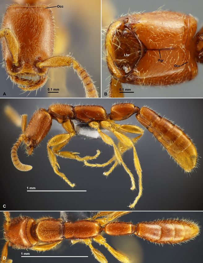
generic name is the anagram of the name of Apomyrma).         specimens used for descriptions, see the species account
Yamane & al. (2008) tentatively assigned Opamyrma to          in “Results”. Voucher specimens are or will be deposited
the subfamily Amblyoponinae together with Apomyrma            in the following collections:
by following definition of the subfamily by Saux & al.        AKYC        Ant Collection of A. Yamada (currently depos-
(2004). However, recent molecular phylogenetic analysis                   ited in the TMUZ).
(Ward & Fisher 2016) indicated a large phylogenetic           ACEG        Ant Collection of K. Eguchi (currently deposited
distance between these two genera, and the phylogenetic                   in TMUZ).
position of the two genera, that is, Apomyrma as a sister     GXNU        Insect Collection, Guangxi Normal University,
lineage of the Amblyoponinae, and Opamyrma as a sis-                      Guilin, Guangxi, China.
ter lineage of the subfamily Leptanillinae. Consequently,     IEBR        Institute of Ecology and Biological Resources,
Opamyrma was transferred to the Leptanillinae by Ward                     Vietnam Academy of Science and Technology,
& Fisher (2016), whereas the Apomyrma is now assigned                     Hanoi, Vietnam.
to own distinct subfamily Apomyrminae by Fisher &             MCZC        Museum of Comparative Zoology, Cambridge,
Bolton (2016).                                                            Massachusetts, USA.
    The subfamily Leptanillinae is a little-known group of    MHNG Muséum d’Histoire Naturelle, Geneva, Switzer-
cryptic subterranean ants which are apparently special-                   land.
ized predators. The subfamily has been recovered as one       TMUZ        Systematic Zoology Laboratory, Tokyo Metro-
of the most basal extant lineages of ants together with                   politan University, Tokyo, Japan.
the Amazonian monotypic subfamily Martialinae which               Morphological examination and imaging: Ex-
is also represented by a single known species, Martialis      ternal morphology of the adult body was observed using
heureka Rabeling & Verhaagh, 2008. However, the               Nikon SMZ1270 stereomicroscope and an LED epi-illu-
position of the root has been controversial (Rabeling &       mination device. For transmitted light observations of
al. 2008, Kück & al. 2011, Branstetter & al. 2017). The       mouthparts, tentorium and sting apparatus of the worker
most recent molecular phylogenetic analysis by Borowiec       and queen, male genitalia, and larval head, whole body or
& al. (2019) supported Martialinae and Leptanillinae (in-     focal parts were dissected and slide-mounted with Euparal
cluding Opamyrma) together as a clade that is sister to       after cleaning by using the Chelex-TE protocol (for detail
all other extant ants. Therefore, knowledge of Opamyrma       see Yamada & Eguchi 2016); and then slide-mounted
has great importance for understanding evolutionary           specimens were observed using Nikon Eclipse E600 mi-
history of not only Leptanillinae but also the whole of the   croscope. The worker, queen, and larva were also observed
Formicidae.                                                   and photographed using scanning electron microscope
    In the last three years, we discovered two Opamyrma       (SEM), JEOL JSM-6510. Ethanol-preserved specimens
hungvuong colonies in the course of our field explorations    were dissected, dried and then mounted on specimen
in Guangxi in China and Son La in Vietnam, and had an         stubs and coated with platinum before SEM observation;
opportunity to examine the morphology of the worker,          the t-butyl alcohol freeze-dry method (Inoue & Osatake
queen, male, and larva using an optical microscope and        1988) was used for preparation of larval specimens. Source
scanning electron microscope (SEM). Herein we provide         images for focus stacking and measurements were taken by
descriptions and illustrations of the male, larva, and body   a Canon EOS Kiss X9 digital camera attached to a Nikon
parts of the worker and queen (including mouthparts,          AZ100 stereomicroscope (for dry-mounted specimens) or
tentorium, and sting apparatus) for the first time. We also   Nikon Eclipse E600 microscope (for slide-mounted spec-
highlight morphological characteristics of Opamyrma by        imens). Multi-focused images were produced by Helicon
contrasting it with other ant lineages, particularly other    Focus Pro 7.5.0 (Helicon Soft Ltd., Ukraine), and then
leptanilline genera, Apomyrma, and Martialis, to provide      improved using the retouching function of Helicon Focus.
supporting morphological evidence for the phylogenetic        The color balance, contrast, and sharpness of the images
position and novel evolutionary implications.                 were adjusted using Adobe Lightroom Classic CC 8.1 and
                                                              GIMP 2.10 (The GIMP Development Team, available at
Material and methods
                                                              http://www.gimp.org/).
Material examined: As source materials for morpho-                The morphological terminology follows Gotwald
logical examination, the present study used two colony        (1969), Keller (2011), Richter & al. (2019), and Liu &
series AKY05vii17-06 (from Guangxi, China containing a        al. (2019) for general morphology of the worker, Kugler
total of 57 workers and 12 larvae) and Dai19iii2019-029       (1978) for sting apparatus, Kubota & al. (2019) for tento-
(from Son La, Vietnam, containing a total of 18 workers,      rium, and Boudinot (2013, 2015) for general morphology
18 alate queens, 11 dealate queens, 2 males) that were        of the male and queen. For wing terminology, the style in
identified as Opamyrma hungvuong Yamane, Bui &                Boudinot (2015) is followed: Brown & Nutting (1949) for
Eguchi, 2008 by referring the original description and        wing venation, Mason (1986) for wing vein development,
images of the holotype on AntWeb (CASENT0178347). As          and Yoshimura & Fisher (2011) for cellular terminology
many detached wings were included in the ethanol pre-         with the modifications proposed in Boudinot & al. (2013);
served stock from the Son La locale, many of the dealate      the wing venation typification system presented by Ogata
queens were probably alate when collected. For a details of   (1991) is also used.

28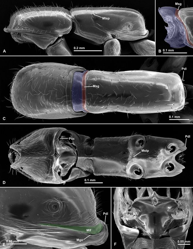
Fig. 1: General habitus of Opamyrma hungvuong worker, nontype (AKY05vii17-06, China, Guangxi). (A) head in full-face view;
(B) head in anteroventral view; (C) body in lateral view; (D) body in dorsal view. Abbreviations: Occ = occipital carina; Lbr =
labrum; Pgr = postgenal ridge.

                                                                                                                            29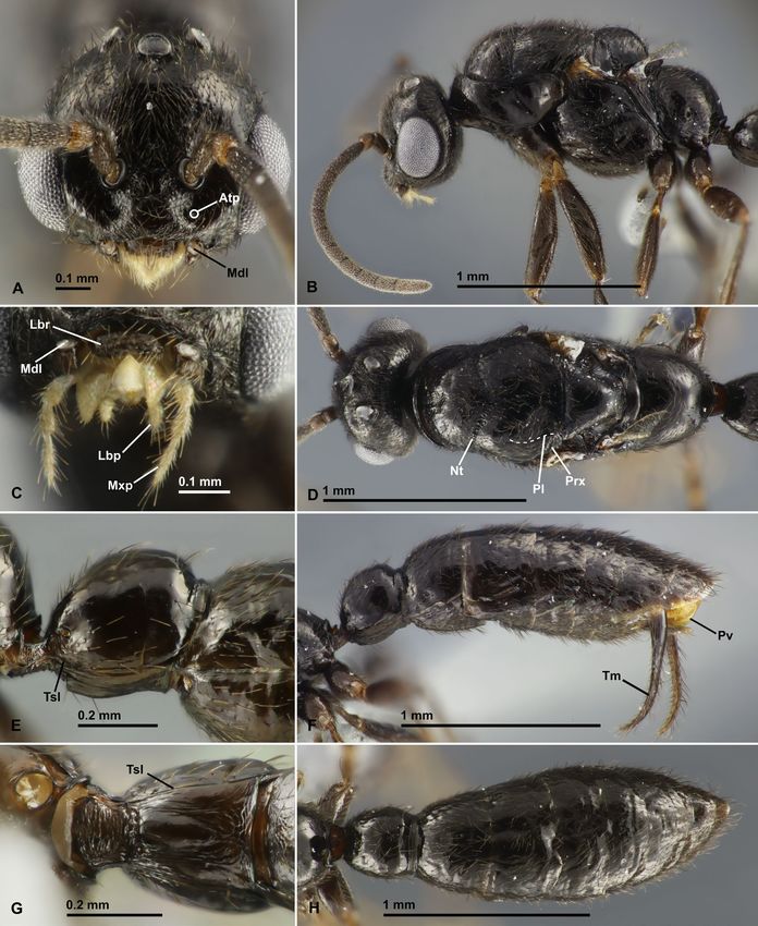
The following parts of bodies were measured using            combination of characteristics: 1) occipital carina vir-
ImageJ 1.52a (National Institute of Mental Health, USA,         tually uninterrupted and anteriorly located before the
available at http://imagej.nih.gov/ij/), and then indices       posterior margin of cranium; 2) outer face of labrum bears
were calculated.                                                numerous peg-like setae; 3) waist 1-segmented; 4) petiole
CI         Cephalic index: HW / HL × 100.                       without distinct anterior peduncle; 5) tergosternal fusion
EI         Eye index: EL / HW × 100.                            of petiole present only anteriorly; 6) gaster elongated and
EL         Eye length: maximum length of major axis of          flattened laterally, with distinct presclerites in abdominal
           eye in lateral view (for queen and male only).       segment IV. The male may be recognizable by the following
HL         Head length: minimal length of cranium in            combination of characteristics: 1) mandible reduced and
           full-face view, measured from the anteromedian       nub-like; 2) wing venation reduced (Ogata’s venation type
           margin of clypeus to the posterior margin of         IVb) with only three closed cells, that is, basal, subbasal,
           cranium.                                             and discal cells; 3) propodeal lobes inconspicuous; 4)
HW         Head width: maximum width of cranium in              waist 1-segmented; 5) petiole not tergosternally fused;
           full-face view (excluding eyes).                     6) pygostyli absent; 7) abdominal sternite IX without
MFI        Metafemur index: MFL / HW × 100.                     prongs or teeth, and with posteromedian lobe; 8) genitalia
MFL        Metafemur length: the maximum length of the          conspicuous with extremely elongate telomere directed
           metafemur, measured in dorsal view.                  ventrad in repose.
OI         Ocellus index: OL / HW × 100.                            Measurements and indices: Worker: CI 76 - 82;
OL         Ocellus length: maximum length of major axis         HL 0.62 - 0.71 mm; HW 0.48 - 0.54 mm; MFI 91 - 96; MFL
           of median ocellus (for queen and male only).         0.44 - 0.50 mm; PTH 0.28 - 0.38 mm; PTI 46 - 50; PTL
PTH        Petiolar height: maximum height of petiole in        0.42 - 0.55 mm; PTW 0.21 - 0.27 mm; PW 0.35 - 0.43 mm;
           lateral view.                                        SI 64 - 71; SL 0.33 - 0.38 mm; WL 1.01 - 1.18 mm (n = 7).
PTI        Petiolar index: PTW / PTL × 100.                         Queen: CI 78 - 79; EI 24 - 25; EL 0.17 mm; HL 0.86 -
PTL        Petiolar length: minimal length of petiole in lat-   0.87 mm; HW 0.68 mm; MFI 89 - 92; MFL 0.61 - 0.63 mm;
           eral view, measured from posterodorsal corner        OI 8 - 9; OL 0.05 - 0.06 mm; PTH 0.48 - 0.49 mm; PTI
           of anterior articulation to posterior margin of      50 - 56; PTL 0.69 - 0.70 mm; PTW 0.35 - 0.38 mm; PW
           posterior peduncle, inside which the helcium         0.58 - 0.60 mm; SI 67 - 69; SL 0.45 - 0.47 mm; WL 1.58 -
           articulates.                                         1.63 mm (n = 3).
PTW        Petiolar width: maximum width of petiole in              Male: CI 99 - 106; EI 53 - 56; EL 0.33 mm; HL 0.59 mm;
           dorsal view.                                         HW 0.58 - 0.62 mm; MFI 104 - 109; MFL 0.64 - 0.65 mm;
PW         Pronotal width: the maximum width of the             OI 15; OL 0.09 - 0.10 mm; PTH 0.36 - 0.37 mm; PTI 92 - 99;
           pronotum in dorsal view.                             PTL 0.34 - 0.36 mm; PTW 0.31 - 0.35 mm; PW 0.53 mm;
SI         Scape index: SL / HW × 100.                          SI 19; SL 0.11 - 0.12 mm; WL 1.48 - 1.49 mm (n = 2).
SL         Scape length: maximum length of antennal                 Redescription: Worker (Figs. 1 - 9): C r a n i u m.
           scape excluding basal condylar bulbus.               In full-face view subrectangular, longer than wide, with
WL         Weber’s length of mesosoma: maximum di-              slightly convex lateral margin and slightly concave pos-
           agonal distance of mesosoma in lateral view,         terior margin, in lateral view, flattened dorsoventrally.
           measured from the point at which the pronotum        Median longitudinal cephalic carina absent. Frontal carina
           meets the cervical shield to the posteroventral      and lobes absent. Occiput extended anteriorly to have dis-
           corner of propodeum.                                 tinct dorsal, lateral, and ventral face, delimited anteriorly
                                                                by distinct occipital carina (“Occ” in Figs. 1A - B, 2B, 4C;
Results
                                                                = “preoccipital carina” in the original description); the
Opamyrma hungvuong                                              carina virtually uninterrupted, forming a V-shaped angle
Yamane, Bui & Eguchi, 2008                                      at the middle of the venter (arrow in Fig. 2B). Postgenal
(Figs. 1 - 15)                                                  ridge (“Pgr” in Figs. 1B, 4C) externally visible as a dark
Non-type material examined: China: 25 workers,                  line running on the ventral midline, ending a little before
6 larvae (colony ID: AKY05vii17-06), Guangxi, Guilin,           the level of occipital carina (the dark line was mentioned
Huaping National Nature Reserve, 25.57° N, 109.94° E,           as “median furrow” in the original description, but the
ca. 1000 - 1500 m above sea level (a.s.l.), collected from      ventral midline is not furrowed as seen in Fig. 2B). Hy-
soil under stone on forest floor, coll. A. Yamada, 5 July       postomal process (“Hysp” in Fig. 2B, D) conspicuous,
2017 (AKYC, ACEG, GXNU, MCZC, MHNG). Vietnam: 3                 in lateral view broad with rounded apex. Eye and ocelli
workers, 2 alate queens, 5 dealate queens, 2 males (colony      completely absent. Antennal socket completely exposed
ID: Dai19iii2019-029), Son La, Ta Xua Nature Reserve, Bac       in full-face view, directing almost dorsad, located in a
Yen, Hang Dong, 21.3158° N, 104.5213° E, 1533 m a.s.l.,         large, roundly excavated area of which anterior wall is
collected from soil on forest floor, coll. D. D. Nguyen, 14     steep a little behind the anterior margin of clypeus; the
March 2019 (AKYC, ACEG, IEBR, MCZC, MHNG).                      area not clearly defined posteriorly. Antennal torulus
    Diagnosis: The female (worker and queen) of the             distinct, simple annular, located distant from anterior
unique species is easily recognizable by the following          clypeal margin, posterolaterally surrounded by deep tear-

30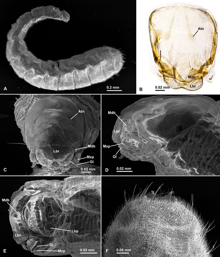
Fig. 2: Scanning electron microscope images of cephalic parts of Opamyrma hungvuong worker, nontype (AKY05vii17-06,
China, Guangxi). (A) anterior part of head in frontal view; (B) cranium in ventral view; (C) right mandible in dorsal view; (D)
right hypostomal process in lateral view; (E) left mandible in ventral view; (F) labrum in outer view. Abbreviations: Ctl = can-
thellus; Hysp = hypostomal process; Occ = occipital carina; Lbr = labrum; Lps = labral peg-like seta; Mdl = mandalus; Mps =
mandibular peg-like seta; Pat = preapical tooth; Ptg = peritorular groove; Ptp = posterior tentorial pit; Sca = supraclypeal area.

drop-shaped peritorular groove (“Ptg” in Figs. 2A, 4A - B;         in Figs. 2B, 4C) located laterally to postocciput. Median
the term “peritorular groove” is borrowed from Richter &           portion of clypeus rather clearly divided into anterior steep
al. 2019). Anterior tentorial pit not externally visible (see      slope and posterior horizontal area that is raised dorsad
tentorium description below). Posterior tentorial pit (“Ptp”       and posteriorly roundly delimited by a continuous steep

                                                                                                                               31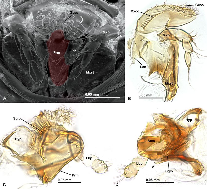
Fig. 3: Maxillolabial complex of Opamyrma hungvuong worker, nontype (AKY05vii17-06, China, Guangxi). (A) Scanning elec-
tron microscope image of maxillolabial complex in ventral view, labrum removed; (B) right maxilla in outer view; (C) labium
in lateral view; (D) labium in dorsal view. Abbreviations: Ams = anteromedian sclerite; Gcss = galeal crown’s stout seta; Hyp =
hypopharynx; Lbp = labial palp; Lcn = lacinia; Mxco = maxillary comb; Mxp = maxillary palp; Mxst = maxillary stipes; Prm =
prementum; Sglb = subglossal brush.

declivity; posterior area broadly inserted between anten-        long but somewhat bluntly tapering apical tooth followed
nal sockets, nearly reaching the level of posterior margin       by a broad trapezoidal preapical tooth (“Pat” in Fig. 2C,
of antennal torulus; supraclypeal area (“Sca” in Fig. 2A)        E; in the original description, the preapical tooth was
small, subtriangular; median longitudinal clypeal carina         mentioned as “trapezoidal lobe” that was interpreted as
absent; lateral portion of clypeus in front of antennal          fusion of two preapical teeth, but it could well be a single
socket very narrow anteroposteriorly: posterior limit of         preapical tooth corresponding in location to, for example,
lateral portion of clypeus externally not obvious, but inter-    Apomyrma, †Gerontoformica Nel & Perrault, 2004, and
nal line that might be paroculoclypeal sulcus (dashed line       Prionopelta Mayr, 1866) and three or four inconspicuous
in Fig. 4A) tracing the anterior outline of excavated area       teeth; ventral face with a single peg-like seta (“Mps” in
around antennal socket is recognized under transmitted           Fig. 2A, C, E; the seta morphology is similar to that of
light microscope; anterior clypeal margin broadly concave        labrum) which is located near the base of the second and
without any peg-like setae or cuticular denticles.               third inconspicuous teeth of the masticatory margin, and
    M o u t h p a r t s. Mandible short and sublinear,           two long apically spatulate setae (arrows in Fig. 2C, E);
strongly curved near the distal end of mandalus, with            trulleum apparently absent; canthellus (“Ctl” in Fig. 2C, E)

32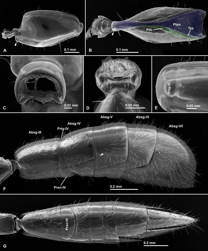
Fig. 4: Tentorium of Opamyrma hungvuong worker, nontype (AKY05vii17-06, China, Guangxi). (A) anterior part of dorsal sclerite
of cranium in dorsal view; (B) part of dorsal sclerite of cranium around right antennal socket with anterior part of tentorium, in
inner ventral view; (C) part of ventral sclerite of cranium with posterior part of tentorium in inner dorsal view; (D) right half of
tentorium in dorsal view (lacking posterior tentorial arm). Abbreviations: Ata = anterior tentorial arm; Atp = anterior tentorial
pit; Ct = corpotendon; Dta = dorsal tentorial arm; Ep = external plate; Ip = internal plate; Lclp = lateral portion of clypeus; Mdb
= mandible; Occ = occipital carina; Pgr = postgenal ridge; Pta = posterior tentorial arm; Ptg = peritorular groove; Ptp = posterior
tentorial pit; Tb = tentorial bridge.

less-defined, not differentiated from the basal margin of           of short thin setae; maxillary palp (“Mxp” in Fig. 3A - B)
mandible; mandalus (“Mdl” marked by red color in Fig. 2C)           4-segmented, becoming shorter and narrower apically;
elongate and narrow club-shaped (it is visible as whitish           apical segment with bluntly tapering apex. Premental
membranous area in dry specimen under an optical micro-             shield (ventral surface of prementum, marked by red
scope, and it was misinterpreted as trulleum in the orig-           color in Fig. 3A) convex oblong in ventral view, without
inal description). Labrum (Fig. 2F, “Lbr” in Fig. 1B) large,        transverse premental groove. Labium in dorsal view with
entirely concealing prementum (“Prm” in Fig. 3A, C) and             conspicuous anteromedian sclerite (“Ams” in Fig. 3D)
maxillary stipes (“Mxst” in Fig. 3A - B) when mouthparts            that is apparently dorsal extension of prementum; base
retracted, almost as long as wide, with rounded distal mar-         of subglossal brush (“Sglb” in Fig. 3C - D) forming strong
gin (median cleft absent); labral tuberculi absent; basal           anterolateral projection in dorsal view (arrow in Fig. 3D);
third of the outer face bearing numerous peg-like setae             paraglossa unrecognizable in our observation; labial palp
that are arranged regularly but not in strict transverse            (“Lbp” in Fig. 3A, C - D) 2-segmented; second segment
rows (“Lps” in Fig. 2A, F); distal area of the outer face with      about as long as first segment, with rounded apex.
at least five pairs of long apically spatulate setae (halves            T e n t o r i u m. Anterior tentorial arm (“Ata” in Fig. 4B
of the pairs are indicated by arrows in Fig. 2F) that are           - D) originated from endoskeletal structure of antennal
regularly arranged. Maxilla with conspicuous maxillary              socket; anterior tentorial pit apparently located on medi-
comb (“Mxco” in Fig. 3B); transverse stipital groove absent;        oventral wall of antennal socket (“Atp” in Fig. 4A - B). But-
galeal comb absent; galeal crown flattened with a series of         tress-like extension absent. Internal plate (“Ip” in Fig. 4B,
thick apically rounded (not spatulate) setae, one of which          D) relatively narrow but much broader than external plate
is particularly stout (“Gcss” in Fig. 3A - B), without ventral      (“Ep” in Fig. 4D), with rounded anterodistal corner. Dorsal
comb; lacinia (“Lcn” in Fig. 3B) small subtriangular, with          tentorial arm (“Dta” in Fig. 4D) distinct, with long branch-
relatively acute apex; lacinial comb present, composed              like apical part. Tentorial bridge (“Tb” in Fig. 4C - D) thin

                                                                                                                                33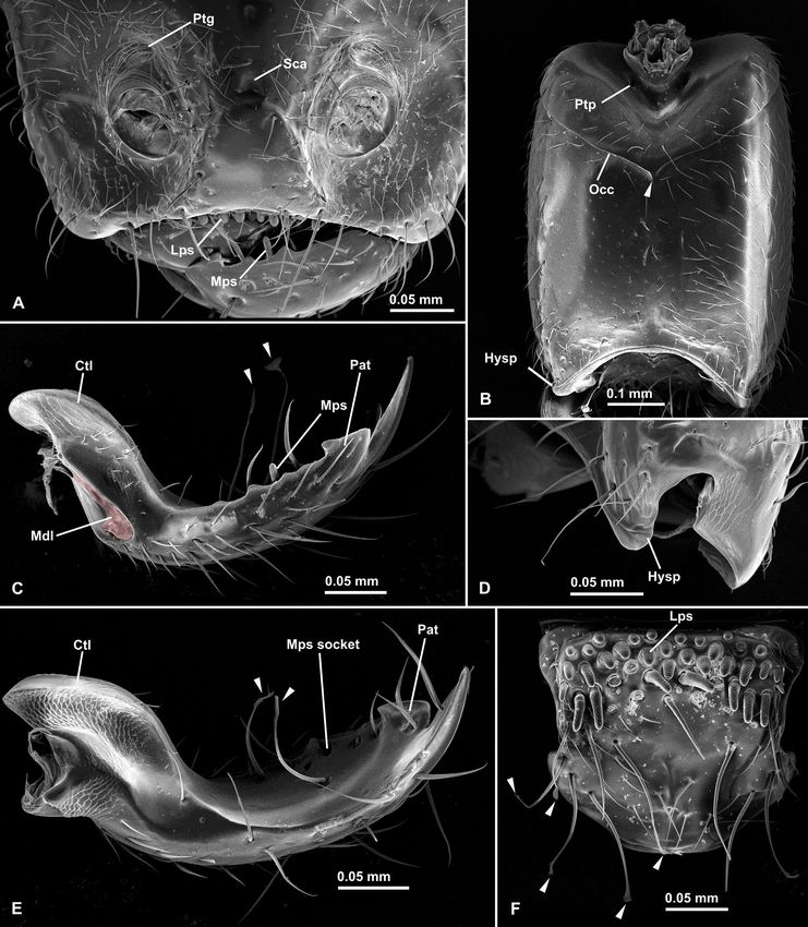
Fig. 5: Scanning electron microscope images of antenna of Opamyrma hungvuong worker, nontype (AKY05vii17-06, China,
Guangxi). (A) entire right antenna in dorsal view; (B) apical antennomere in dorsal view.

tubular. Corpotendon (“Ct” in Fig. 4C - D) long. Posterior    spiracle unrecognizable in our observation. Notopleural
tentorial arm (“Pta” in Fig. 4C) thin tubular.                suture of mesothorax absent. Longitudinal mesopleural
    A n t e n n a. Antenna 12-merous, gradually incrassate    sulcus absent. Metanotal groove absent. Mesometapleu-
from antennomeres II to XII (Fig. 5A). Antennal scape,        ral suture present as weak groove. Metanotal spiracle
when laid backward, extending past midlength of cranium,      (“Mtsp” in Fig. 6A) small and apparently closed, located
flattened dorsoventrally, narrowed toward base, without       high on lateral face. Propodeum with rather flat dorsum
distinct basal flange distal to bulbus; antennomere II        and steep posterior face; posterior face roundly continues
subconical bead-like, in dorsal view strongly narrowed at     to dorsal and lateral faces without any delimiting carina.
base, slightly longer than wide; antennomore III slightly     Propodeal spiracle located relatively low on the lateral
longer than wide and narrowed basally; antennomeres           face of propodeum. Outline of metapleural gland bulla
IV and V almost as long as wide; antennomeres VI - XI         conspicuously recognized through cuticle under natural
wider than long; apical antennomere longer than wide          lighting, subcircular, occupying posterior two-fifths of
and bluntly pointed at apex. Apical antennomere, with at      ventrolateral part of the pleuron; metapleural gland orifice
least two types of sensilla recognizable: basiconic (black    (“Mgo” in Fig. 6E) narrow slit-like, located in the lower
arrows in Fig. 5B), and trichodic ones; trichodic sensilla    posterior corner of the metapleuron; metapleural longitu-
become small on basal marginal area; pit-like structures      dinal flange (“Mlf”, marked by green color in Fig. 6E) anter-
that might be coeloconic / ampullaceous sensilla or socket    oposteriorly long, projecting laterad and overhanging (but
of broken basiconic / trichodic sensilla also recognizable    not concealing) metapleural gland orifice. Ventral part of
(white arrows in Fig. 5B). Scape bulbus hemispherical with    metapleuron below the orifice also laterally produced to
short tubular neck; anterior basal margin of the bulbus       form longitudinal flange. Propodeal lobes (“Pdl” in Fig. 6C
apparently without a distinct notch.                          - E) weakly present, low and round. Anteroventral face of
    M e s o s o m a. Mesosoma slender and consisting of       mesopectus with distinct median carina and submedial
two distinct portions, prothorax and meso-metathoraci-        deep subrectangular depressions that accommodate the
co-propodeal complex, which is oblong and slightly longer     forecoxae; posterior remaining face just medially weakly
and narrower than pronotum and almost parallel-sided          raised without forming distinct median carina. Mesos-
in dorsal view: articulation between prothorax and mes-       ternal pit present (“Mstp” in Fig. 6D). Metasternal pit
othorax unfused and fully flexible in fresh condition.        apparently absent. Meso- and metacoxal cavities small,
Pronotum longer than wide in dorsal view, with slightly       fully closed with a complete cuticular annuli surrounding
convex dorsal face that roundly continues to lateral face;    the cavities; metacoxal cavity separated from propodeal
anterior slope short and steep. Propleurae unfused relative   foramen by a cuticular band.
to one another, but are strongly attached along the ventral       L e g s. Relatively broad gap present between pro-
midline. Procoxal cavity (“Pcc” in Fig. 6D, F) as small as    and mesocoxae. Metacoxal dorsum unarmed. Profemur
meso- and metacoxal cavities, in ventral view bounded         broader than meso- and metafemur. Protibia broader than
anteriorly by propleuron and laterally and posteriorly by     meso- and metatibia. Anterior face of protibia without
prosternum (virtually not bounded by pronotum). Dor-          “protibial anterior sulcus” sensu Keller (2011). The calcar
sum of anterior articulatory area of mesonotum (inserted      of the strigil (“Ca” in Fig. 7A - C) fully pectinated; basal
under the pronotum, marked by blue color in Fig. 6B - C)      one third of the calcar bearing narrow unnotched lamina
posteriorly delimited by deep narrow transverse groove        (arrow in Fig. 7B); anterior surface with brush that is com-
(“Msg”, marked by red color in Fig. 6B - C) which contin-     posed of dense seta-like cuticular projections; posterior
ues along the ventral margin of mesopleuron. Mesonotal        surface with sparse seta-like cuticular projections. Pos-

34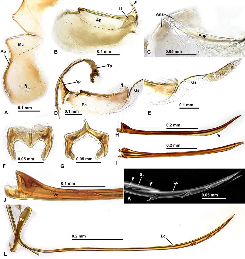
Fig. 6: Scanning electron microscope images of mesosoma of Opamyrma hungvuong worker, nontype (AKY05vii17-06, China,
Guangxi). (A) whole mesosoma in lateral view; (B) anterior articulation of mesonotum in lateral view, prothorax removed; (C)
whole mesosoma in dorsal view; (D) whole mesosoma in ventral view, all legs removed; (E) metapleuron and propodeum in lat-
eral view; (F) prosternite in ventral view, propleurae and prolegs removed. Abbreviations: Mgo = metapleural gland orifice; Mlf
= metapleural longitudinal flange; Msg = mesonotal groove; Mstp = mesosternal pit; Mtsp = metanotal spiracle; Pcc = procoxal
cavity; Pdl = propodeal lobe.

                                                                                                                            35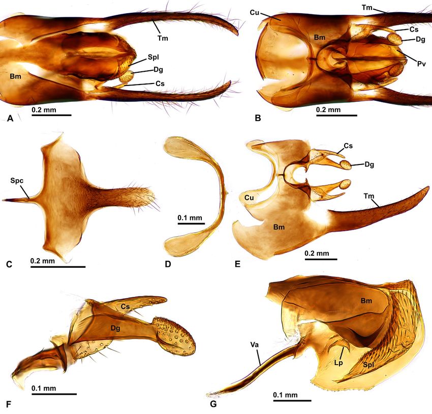
Fig. 7: Scanning electron microscope images of legs of Opamyrma hungvuong worker, nontype (AKY05vii17-06, China, Guangxi).
(A) strigil of right proleg in anterior view; (B) calcar of strigil of right proleg in anterior view; (C) strigil of right proleg in posterior
view; (D) distitarsus of right proleg in posterior view; (E) tibial spurs and basitarsus of right mesoleg in anterior view; (F) tibial spurs
and basitarsus of right metaleg in posterior view; (G) pretarsal claws of right metaleg in posteroventral view; (H) tibial spurs of left
metaleg in anterior view. Abbreviations: Ats = anterior spur; Ca = calcar; Pts = posterior spur; Ptb = protibia; Pbts = probasitarsus;
Ptss = protibial stout seta; Mnb = manubrium; Mstb = mesotibia; Msbts = mesobasitarsus; Mttb = metatibia; Mtbts = metabasitarsus.

terodistal apex of protibia with a single stout seta (“Ptss”             late anterior spur (“Ats” in Fig. 7E, H) and a well-developed
in Fig. 7C), located close to the insertion of the calcar of the         pectinate posterior spur (“Pts” in Fig. E - F, H); posterior
strigil. Meso- and meta- tibiae each with a reduced barbu-               spurs with dense seta-like cuticular projections except

36
Fig. 8: Scanning electron microscope images of metasoma of Opamyrma hungvuong worker, nontype (AKY05vii17-06, China,
Guangxi). (A) petiole in lateral view; (B) petiole in ventral view; (C) helcium in anterior view; (D) helcium in ventral view; (E) pretergite
of abdominal segment IV in dorsal view; (F) gaster in lateral view; (G) gaster in ventral view. Abbreviations: Absg = abdominal
segment; Prsn = presternite; Prtg = pretergite; Ptlt = petiolar laterotergite; Ptsn = petiolar sternite; Tss = tergosternal suture.

for basiposterior surface of the metatibial spur. Apically              similar setae also present posterior face of metabasitarsus
truncated and somewhat flattened setae present on poste-                along its inner margin (arrows in Fig. 7F). Anterior surface
rior face of metatibia near insertion of the posterior spur;            of probasitarsal notch with numerous acute scale-like

                                                                                                                                         37
Fig. 9: Sting apparatus of Opamyrma hungvuong worker, nontype (AKY05vii17-06, China, Guangxi). (A) spiracular plate in lat- eral view; (B) quadrate plate in lateral view; (C) anal arcs and anal plate in flattened dorsal view; (D) oblong plate and triangular plate in lateral view; (E) gonostylus in lateral view; (F) furcula in anterodorsal view; (G) furcula in posterior view; (H) sting in lateral view; (I) sting in dorsal view; (J) basal part of sting in lateral view; (K) Scanning electron microscope image of apical parts of sting and lancet in lateral view; (L) lancet and fulcral arm in lateral view. Abbreviations: Ana = anal arc; Anp = anal plate; Ap = anterior apodeme; Fa = fulcral arm; Gs = gonostylus; Lc = lancet; Ll = lateral lobe; Mc = medial connection; Pa = posterior arm; St = sting; Tp = triangular plate; Vc = valve chamber. cuticular projections (Fig. 7A - B); posterior surface of I - III respectively with some conspicuous stout spiniform the probasitarsal notch without any stout spiniform seta setae (arrows in Fig. 7D). Stout spiniform setae absent on (Fig. 7C). Anterior surfaces of distal portion of protibia and mesotibia, mesobasitarsus, and metabasitarsus (except for probasitarsus bearing numerous spatulate setae (Fig. 7A - stout setae near distal margin of mesobasitarsus). Pretar- B). Basiventral margin of probasitarsus just rounded, not sal manubrium (“Mnb” in Fig. 7G) relatively large, flat and strongly produced. Posteroventral corner of protarsomeres longitudinal elliptical, with a pair of stout long setae (no 38
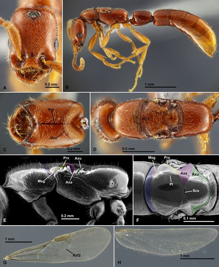
Fig. 10: Opamyrma hungvuong queen, nontype (Dai19iii2019-029, Son La, Vietnam). (A) head in full-face view; (B) body in lateral
view; (C) head in ventral view; (D) head and mesosoma in dorsal view; (E) Scanning electron microscope image of mesosoma
in lateral view; (F) Scanning electron microscope image (SEM) of meso- and metanotum in dorsal view; (G) forewing in dorsal
view; (H) hindwing in dorsal view. Abbreviations: Axa = axilla; Axu = axillula; Msg = mesonotal groove; Occ = occipital carina;
Pgr = postgenal ridge; Pl = parapsidal line; Prx = preaxilla; Rs = radial sector; Scs = scutoscutellar sulcus.

                                                                                                                            39
significant differences between those of pro-, meso- and (abdominal tergite IX) much smaller than its main disc,
meta legs). Pretarsal claws simple, without teeth (Fig. 7G). delimited by distinct midplate line (partially indicated
M e t a s o m a. Waist 1-segmented, that is, consist- by dashed line in Fig. 9B), with large lateral lobe (“Ll” in
ing of only petiole (abdominal segment II). Petiole sub- Fig. 9B); anterodorsal corner (arrow in Fig. 9B) long and
rectangular to oblong in lateral view, virtually sessile sharp. Anal arcs and plate present (“Ana” and “Anp” in
without distinct anterior peduncle, longer than wide in Fig. 9C), weakly sclerotized apparently without anal sen-
dorsal view, with slightly convex dorsal face that roundly silla. Anterior apodeme (“Ap” in Fig. 9D) of oblong plate
continues to lateral face; petiolar sternite in ventral view (gonocoxa IX; = second valvifer) forming small subtrian-
(“Ptsn”, marked by blue color in Fig. 8B) disproportionate gular sclerite in lateral view, that is posteriorly margined
dumbbell-shaped, with narrow elongated anterior part, by diverging thickened ridge that is connected with dor-
only anteriorly fused with the tergite; posterior part of the sal ridge of the posterior arm (“Pa” in Fig. 9D); posterior
sternite delimited from the tergite by distinct tergoster- arm relatively large, more than twice as long as high in
nal suture (“Tss” in Fig. 8B); petiolar laterotergite (“Ptlt”, lateral view: dorsal subterminal part of the arm (arrow
marked by green color in Fig. 8B) present as narrow area in Fig. 9D) forming weakly sclerotized flange protruding
along the tergosternal suture; petiolar spiracle located from the dorsal ridge; most of ventral arm inconspicuous
anteriorly on the lateral face of the tergite at its mid- and apparently membranous; fulcral arm (“Fa” in Fig. 9L)
height; anteroventral corner of petiole with flange-like large and linear. Basal part of triangular plate (gonangu-
structure (arrow in Fig. 8A - B); petiolar levator process lum; = first valvifer; “Tp” in Fig. 9D) long and thin, weakly
complete, without lacuna; very short tubular posterior curved; lateral tubercle apparently absent; dorsoapical
peduncle present inside which the helcium articulates. and ventroapical processes short and stout. Gonostylus
Gaster very elongate and laterally compressed especially (gonoplac; = third valvula; “Gs” in Fig. 9D, E) long and
in posterior segments, in lateral view highest at the pos- slender, composed of 2 distinct segments; first segment
terior end of abdominal segment V. Helcium axial (sensu long and feebly sclerotized except for well-sclerotized
Keller 2011), tergosternally fused; helcium sternite lat- dorsal margin, with sparse short erect setae along dorsal
erally enclosed in the tergite. Postsclerites of abdominal margin and on posterior part of its outer face; second
segment III tergosternally unfused, having a free ante- segment short, relatively well-sclerotized, with denser and
rior face above the helcium, longer than high, narrowed longer erect setae on its outer face. Furcula (Fig. 9F - G)
basally in dorsal view, longer than segments IV, V and thick, Y-shaped in posterior view, with short dorsal arm,
VI. Prora of abdominal sternite III present as a strong unfused with sting base. Sting (gonapophysis IX; = stylet,
corner that is produced anteriad to reaching the level of second valvula; Fig. 9H - K) very narrow elongate and
the anteriormost point of tergite III. Abdominal spiracle blade-like; sting bulb conspicuously wider and higher than
III located on lower lateral face of the tergite. Abdominal base of the shaft; valve chamber (“Vc” in Fig. 9J) present
segment IV with externally visible presclerites; pretergite but narrow; sting shaft more than twice as long as valve
(“Prtg-IV” in Fig. 8E - F) short and inconspicuous, just chamber, upcurved, with two pairs of small barbs on the
weakly constricted; presternite (“Prsn-IV” in Fig. 8F - G) apex (arrows at “St” in Fig. 9K); distiventral edge of the
long and conspicuous, posteriorly delimited by strong con- shaft produced to form broad lamina (arrow in Fig. 9H).
striction. Abdominal spiracles IV - V visible, but VI - VII Lancet (gonapophysis VIII; = first valvula; “Lc” in Fig. 9L)
concealed by preceding tergites (the original description completely lacks valves, with 5 apical barbs of which basal
stated that spiracles V is concealed, but it is clearly visible three are conspicuously large and directed ventrad, and
as indicated by arrow in Fig. 8F). Abdominal segment VII apical two are small and inconspicuous.
longest among the segments III - VII. Pygidium (abdom- C o l o r , s c u l p t u r e , a n d p i l o s i t y. Body
inal tergite VII) very large and simple, unarmed, convex entirely light orangish brown, with slightly yellowish
and downcurved posteriorly in lateral view. Hypopygium antennae and legs. Body largely smooth and shining.
(abdominal sternite VII) unarmed, in ventral view long Body largely covered with sparse to dense decum-
subtriangular. bent / standing hairs as shown in figures: hairs most
S t i n g a p p a r a t u s. Lateral hemitergites of spi- dense in dorsum of cranium, and most sparse in pos-
racular plate (abdominal tergite VIII) narrowly attached terolateral face of mesonotum and lateral face of pet-
each other by large medial connection (“Mc” in Fig. 9A iole; a series of particularly thicker and longer hairs
); the attachment present as a suture-like, strongly scle- present along anterior margin of cranium in full-face
rotized midline; median connection distinctly delimited view.
from the main disc (= “body” sensu Kugler, 1978) by a Queen (Fig. 10): Fully winged, largely similar to the
weak carina (indicated by dashed line in Fig. 9A); main worker except for optic- and flight-related characters de-
disc subrectangular with broadly concave posterodorsal scribed below. Eyes and ocelli large and conspicuous; eyes
margin, without distinct dorsal notch; spiracle (arrow in circular with about 15 ommatidia at maximum diameter in
Fig. 9A) relatively large, located lower (ventrad) center lateral view, located a little lower of mid-length of cranium
of the disc; posteroventral corner without posterodorsal in full-face view; ocelli located high close to occipital ca-
lobe and tubercle; anterior apodeme (“Ap” in Fig. 9A) nar- rina. Occipital carina (“Occ” in Fig. 10C) nearly complete,
row. Anterior apodeme (“Ap” in Fig. 9B) of quadrate plate but interrupted at midline of the venter. Postgenal ridge
40Fig. 11: Opamyrma hungvuong male, nontype (Dai19iii2019-029, Son La, Vietnam). (A) head in full-face view; (B) head and
mesosoma in lateral view; (C) mouthparts in anteroventral view; (D) head and mesosoma in dorsal view; (E) petiole in lateral view;
(F) metasoma in lateral view; (G) petiole in ventral view; (H) metasoma in dorsal view. Abbreviations: Atp = anterior tentorial
pit; Lbp = labial palp; Lbr = labrum; Mdl = mandalus; Mxp = maxillary palp; Nt = notauli; Pl = parapsidal line; Pv = penisvalva;
Tm = telomere; Tsl = tergosternal line.
41(“Pgr” in Fig. 10C) extending more posteriorly, beyond the nub-like, forming a broad gap when fully closed (Fig. 11A,
ventral interruption of occipital carina. Mesosoma having C); masticatory margin edentate; mandalus (“Mdl” in
full complement of flight sclerites but still slender, with Fig. 11A, C) large but still ringed by sclerite in dorsal view.
almost linear dorsal outline in lateral view; mesonotum Labrum (“Lbr” in Fig. 11C) subrectangular, more than
not raised dorsally. Pronotum having large dorsal face as twice as wide as long, with almost straight distal margin
that of the worker. Dorsum of anterior articulatory area (median cleft absent); labral tuberculi absent; peg-like
of mesonotum that inserted under the pronotum (marked dentiform setae absent. Maxillary palp (“Mxp” in Fig. 11C)
by blue color in Fig. 10E - F) posteriorly delimited by a 4-segmented. Labial palp (“Lbp” in Fig. 11C) 2-segmented.
faint line and submedial weak narrow transverse grooves M e s o s o m a. Pronotum with conspicuously nar-
(“Msg”, marked by red color in Fig. 10E - F) that is pos- rowed cervical shield; median pronotal area behind
sibly homologous with that of the worker. Mesoscutum cervical shield convex and short in lateral view; max-
in dorsal view oval, much wider than long (excluding the imum height of pronotum almost as long as mesoscu-
anterior articulatory area); notauli absent; parapsidal lines tum height in lateral view. Mesoscutum large in dorsal
(“Pl” in Fig. 10F) faintly present. Parascutal carinae weak. view, much longer than wide; lateral margin concave
Preaxilla (“Prx”, marked by yellow color in Fig. 10E - F) around the anterior terminus of notauli. Notauli (“Nt” in
distinctly visible as narrow area in dorsal view. Axillae Fig. 11D) distinct and weakly scrobiculate, meeting each
(“Axa”, marked by purple color in Fig. 10E - F) in dorsal other at the midline, but not extending to transscutal line
view large, strongly extending medially between mesoscu- (the posterior terminus located far from the transscutal
tum and mesoscutellum but not meeting at midline. Ax- line). Parapsidal line (“Pl”, indicated by dashed line in
illulae (“Axu”, marked by green color in Fig. 10E - F) large Fig. 11D) present, weakly undulate. Parascutal carinae
and conspicuous in dorsal view, virtually meeting each weak. Preaxilla distinctly visible as small area in dorsal
other behind mesoscutellum. Scutoscutellar sulcus (“Scs” view (“Prx” in Fig. 11D). Axillae small and conspicuous in
in Fig. 10F) weak, very narrow and only faintly scrobicu- dorsal view, moderately inserted between mesoscutum
late. Mesoscutellum in dorsal view circular, a little wider and mesoscutellum, not meeting each other at midline.
than long. Metascutellum large and conspicuous in dorsal Axillulae large and conspicuous in dorsal view, virtually
view, not strongly produced in lateral view. Wing venation meeting each other behind mesoscutellum. Scutoscutellar
largely same as that of male (Fig. 10G - H, see also male sulcus present as narrow and faintly scrobiculate groove,
description below), but Rsf2 faintly recognized as a ves- without crossribbing. Mesoscutellum in lateral view as
tigial spectral line in the queen forewing. Sting apparatus high as mesoscutum, with convex dorsal margin, in dor-
largely same as that of the worker. sal view rounded subtrapezoidal, almost as long as wide.
Male (Figs. 11 - 13): C r a n i u m. In full-face view Metascutellum broad and conspicuously visible in dorsal
circular, almost as long as wide excluding eyes, with view, in lateral view strongly produced. Mesopectus with
strongly convex posterior margin. Frontal carinae and oblique and weakly sinuate sulcus; anterior terminus of
lobes absent. Occipital carina absent. Eye and ocelli large the sulcus located well ventral to pronotal corner. Meta-
and conspicuous; ocelli distantly located from eyes: me- pleural spiracular plate absent. Anterior metapleural area
dian ocellus located just posterior to two-thirds of poste- weakly separated from posterior metapleural area by an
rior part of cranium above eyes in full-face view. Antennal inconspicuous transverse sulcus, and from propodeum
socket located in a large, roundly excavated area of which by a deep conspicuous groove. Metapleural gland orifice
anterior wall is steep just behind the posterior margin of occluded; internal structure of metapleural gland un-
clypeus; the area not clearly defined posteriorly. Antennal recognizable through metapleural sclerite. Propodeum in
torulus distinct, simple annular, distantly located from lateral view with roundly convex dorsal margin; posterior
posterior clypeal margin (the distance slightly less than face roundly meeting dorsal and lateral faces without any
one torulus diameter). Anterior tentorial pit (“Atp” in delimiting carina; propodeal spiracle circular, large, low
Fig. 11A) situated anterior to antennal torulus. Median on lateral propodeal surface; propodeal lobe inconspicu-
portion of the clypeus roundly raised dorsad; posterior ous, just faintly developed. Metacoxal cavities fully closed
area not inserted between antennal toruli, not quite reach- with complete cuticular annuli surrounding the cavities,
ing the level of anterior margin of the torulus; supraclypeal separated from propodeal foramen by a cuticular band.
area distinct but indistinctly margined; lateral portion of M e t a s o m a. Waist 1-segmented, that is, consisting
clypeus in front of antennal socket narrow anteroposte- of only petiole (abdominal segment II). Petiole roundly
riorly; median anterior clypeal margin weakly broadly swollen, virtually without distinct anterior peduncle, al-
concave, without any peg-like dentiform setae. Antenna most as long as wide and high, in lateral view with strongly
13-merous, filiform without becoming incrassate apically. convex dorsal margin; petiolar sternite unfused with the
Antennal scape short cylindrical, when laid backward, not tergite, delimited by distinct tergosternal line (“Tsl” in
reaching the level of posterior margin of eye in full-face Fig. 11E, G) even in anterior articulatory area; the sternite
view; antennomere II bead-like and shortest among anten- broad in ventral view, in lateral view with weakly convex
nomeres; antennomeres III–XII longer than wide, almost ventral margin; subpetiolar process absent; petiolar lat-
same length; antennomere XIII longest, with bluntly ta- erotergite absent. Helcium axial (sensu Keller 2011), with
pering apex. Mandibles strongly reduced, subtriangular sternite visible in lateral view, not enclosed by pretergite.
42Fig. 12: Male wings of Opamyrma hungvuong, nontype (Dai19iii2019-029, Son La, Vietnam). (A) forewing in dorsal view; (B)
hind wing in dorsal view. Abbreviations: 1A = first anal vein; Bc = basal cell; C = costal vein; Cc = costal cell; Cu = cubital vein;
Mc1 = marginal cell 1; R = radial vein; Rs = radial sector; Sbc = subbasal cell; Sc = subcostal vein; Sdc1 = subdiscal cell 1; Smc
= submarginal cell.
Abdominal postsclerites III tergosternally unfused. Prora 2rs-m absent (submarginal cell 2 absent); 1 m-cu nebulous
of abdominal sternum III present just as weak carina de- (discal cell 1 closed); M + Cu tubular (basal cell closed);
limiting poststernite from helcium; anteromedian area of Cuf2 - 3 nebulous; 1A tubular, disappearing distally after
helcium sternite concave. Abdominal tergites IV - VIII and the connection with cu-a (subdiscal cell 1 open; subbasal
abdominal sternites IV - IX well developed, not reduced or cell closed). Hindwing venation reduced, only with tubular
obscured. Abdominal segment IV without distinct pres- R + Rs and 1A, with six hamuli; R not reaching anterior
clerites. Abdominal spiracles III - V visible but VI - VIII wing margin; 1A short; claval region relatively developed,
concealed by preceding tergites. Abdominal segment IV with rounded margin; jugal lobe absent.
longest among the segments III - VIII, a little longer than G e n i t a l i a ( F i g. 1 3 ) . Genitalia large and ex-
segments III and V. Abdominal tergite VIII unarmed. tremely specialized; most part of telomere and apical part
W i n g s ( F i g. 1 2 ) . Wings hyaline, completely of penisvalvae visible in external lateral view, without
covered by fine setose layer. Forewing venation reduced distension or dissection (see Fig. 11F). Pygostyles absent.
with only three closed cells (basal, subbasal, and discal), Basal disc of abdominal sternite IX (Fig. 13C) distinctly
categorized as Ogata’s venation type IVb, although discal more than twice wider than long when excluding spiculum,
cell enclosed by nebulous Rs + M and 1 m-cu; pterostigma with anterolateral corner just weakly produced; posterior
large and conspicuous; free R distal to pterostigma absent; lobe very narrow, about one sixth as wide as basal disc,
costal vein (C) tubular only in short basal part and soon distinctly longer than basal disc when excluding spiculum,
disappearing distally (costal cell open); Rsf1 tubular, very with strongly convex posterior apex; spiculum (“Spc” in
short and nearly lost; Mf1 tubular, completely closing basal Fig. 13C) long and acute, nearly as long as basal disc. Cu-
cell; Rs + M nebulous; Rsf2 absent; Rsf3 only partially pula (Fig. 13D, “Cu” in Fig. 13B, E) reduced, non-annular,
weakly present as a short diverging branch from Rsf4 only present as short half arc-shaped ventral sclerite.
(submarginal cell 1 unclosed); Rsf4 + tubular, continu- Parameres highly fused with each other both dorsally
ous with 2r-rs which is directed posteroapically, ending and ventrally (therefore a complete annulus formed), and
before wing apex (marginal cell 1 open); Mf3 + absent and also with penisvalvae dorsally, that is, parameres and pe-
43Fig. 13: Male genitalia of Opamyrma hungvuong, nontype (Dai19iii2019-029, Son La, Vietnam). (A) genital capsule in dorsal view; (B) genital capsule in ventral view; (C) abdominal sternite IX in ventral view; (D) cupula in ventral view; (E) left paramere with basiventral part of right paramere and cupula, in unfolded outer view; (F) left volsella in lateral view; (G) left penisvalva in lateral view. Abbreviations: Bm = basimere; Cu = cupula; Cs = cuspis; Dg = digitus; Lp = lateral apodeme; Pv = penisvalva; Spl = spinescent lobe; Spc = spiculum; Tm = telomere; Va = valvura. nisvalvae inseparable without destruction (Fig. 13A - B); standing hairs on basal face. Digitus (“Dg” in Fig. 13A - basivolsellae also strongly fused with each other ventrally B, E - F) club-shaped, with strongly swollen apical part and with basimeres; basimere (“Bm” in Fig. 13A - B, E, directed laterad with numerous short modified setae on G) well-developed, without oblique carina on lower face ventrolateral face. Penisvalvae (Fig. 13G, “Pv” in Fig. 13B) (“BmC” sensu Yamada & Eguchi 2016); telomere (“Tm” in not fused with each other directly, but connected to each Fig. 13A, B, E) extremely elongate, distinctly longer than other via apical extension of basimere (dorsal sclerite basimere, weakly recurved anteroventrad, clearly visible in apparently seem not be part of penisvalvae, but apical external lateral view (see Fig. 11F), gently tapering apicad; extension of basimere that extends lateroventrad and is articulation of basimere to telomere apparently fused, but fused partly with lateral face of valviceps; see Fig. 13G). differentiated by ventral membranous notch. Cuspis (“Cs” Valvura (“Va” in Fig. 13G) elongate liner, directed anter- in Fig. 13A - B, E - F) distinct, elongate digitiform, with oventrad. Valviceps with a modified lateral apodeme (“Lp” several short-modified setae on apical face and normal in Fig. 13G) that is visible as small semielliptic sclerite on 44
Fig. 14: Larva of Opamyrma hungvuong, nontype (AKY05vii17-06, China, Guangxi). (A) Scanning electron microscope (SEM)
image of body in lateral view; (B) transmitted light microscopy image of head in dorsal view; (C) SEM image of head in frontal
view; (D) SEM image of head in lateral view; (E) SEM image of head in ventral view; (F) SEM image of abdominal terminus in
lateral view. Abbreviations: Atn = antenna; Gl = galea; Lbp = labial palp; Lbr = labrum; Mdb = mandible; Mxp = maxillary palp.
mid-height of basal valviceps in lateral view. Valviceps bearing numerous spines (therefore, it seems to be de-
also have a uniquely specialized structure termed here rived by extreme modification and sclerotization of penis
as “spinescent lobe” (“Spl” in Fig. 13A, G); spinescent valvar membrane that commonly has numerous spines).
lobe originated from dorsoapical corner of the valviceps Anteroventral corner of valviceps strongly produced with
and extended ventrolaterally to form arc-shaped sclerite acute angle. Anterior part of ventral margin of valviceps
45Fig. 15: Scanning electron microscope images of larval mouthparts and prothoracic surface of Opamyrma hungvuong, nontype
(AKY05vii17-06, China, Guangxi). (A) left maxilla in dorsal view; (B) labrum in ventral view; (C) labium in ventral view; (D) cu-
ticular spinules on prothorax. Abbreviations: Gl = galea; Lbp = labial palp; Lbr = labrum; Mdb = mandible; Mxp = maxillary palp.
with about 25 small teeth. Apical margin of valviceps two basiconic sensilla (white arrows on “Mxp” in Fig. 15A);
rounded. galea (“Gl” in Fig. 14C - E) slender and digitiform with two
C o l o r , s c u l p t u r e , a n d p i l o s i t y. Body basiconic sensilla (white arrows on “Gl” in Fig. 15A) on the
entirely black, with faintly paler antennae and legs; maxilla apex. Labrum (“Lbr” in Fig. 14B - E) broad with anterior
and labium whitish; telomere black; penisvalvae yellowish. margin strongly and narrowly concave medially; surface
Body largely smooth and shining. Body largely covered near the anteroventral border with several basiconic sen-
with sparse to dense decumbent / standing hairs as shown silla (arrows in Fig. 15B). Labium with dense cuticular
in figures; hairs most dense in dorsum of cranium, and spinules on anteroventral surface; anterior margin in
sparser in mesosoma and metasoma. ventral view weakly broadly concave medially; labial palp
Larva (Figs. 14 - 15): Following description is based stout, with two basiconic sensilla (arrows in Fig. 15C).
on relatively developed larvae whose instar is unknown Anteroventral surface of prothorax with dense transverse
(with head width around 0.11 mm). Body elongate and series of tiny cuticular spinules (Figs. 14E, 15D). Body
slender with proportionally small head. Cranium longi- hairs unbranched, with two types: 1) short thin standing
tudinally oval in full-face view, with smooth and hairless hairs that very densely cover abdominal segments and
surface; posteromedian part just behind the level of an- sparsely present in thoracic segments; 2) stout standing
tennae strongly depressed. Antenna (“Atn” in Fig. 14B - C) hairs that very sparsely present in thoracic and abdominal
consisting of three sensilla located at the anterior end of a segments, and especially numerous in around abdominal
sulcus which extends from posterior end of cranium (the terminus (see Fig. 14F). Specialized structures such as
“sulcus” well-recognized as internal ridge in Fig. 14B). “prothoracic projection” and “hemolymph tap” known in
Mandible (“Mdb” in Fig. 14B - E) well-sclerotized, in dorsal the Leptanilla larva absent. Spiracles unrecognizable in
view mostly concealed under labrum in the closed posi- our observation (probably tiny and inconspicuous, as in
tion, in dorsal view subtriangular with acute apex curved the genus Leptanilla).
medially; masticatory margin linear and edentate. Maxilla Distribution: This species has been recorded from
with some cuticular spinules (black arrows in Fig. 15A); southern area of China (Hainan, Guangxi) and northern
maxillary palp (“Mxp” in Fig. 14C - E, Fig. 15A) stout with central to northern Vietnam (Ha Tinh and Son La). Re-
46Fig. 16: Distribution map of Opamyrma hungvuong. Type locality, record by Chen & al. (2017), and new records by the preset
study are indicated by circle, square, and stars, respectively.
corded elevations ranges from ca. 640 m a.s.l (in Hainan) full-face view, and is complete and uninterrupted (in the
to ca. 1500 m a.s.l (in Son La); the elevation of the type worker) or is almost complete with a short medioventral in-
locality was not recorded. The confirmed distributional terruption (in the queen). To our knowledge, such anterior
records are mapped in Fig. 16. location of occipital carina (and accompanying anterior
Bionomics: There are little data on the biology. Both extension of occiput) is unknown from any other extant
of the two colonies examined in the present study were col- or extinct ant lineage, and thus likely be an autapomorphy
lected from soil on forest floor. The workers run agilely but of Opamyrma. The location of the anterior invagination
didn’t climb up smooth plastic walls. All 12 of the larvae of tentorium in Opamyrma is also unusual. The anterior
from colony AKY05vii17-06 were approximately the same tentorial arm is invaginating from the anterior tentorial
size, suggestive of brood cycles. pit that is usually externally visible. The pits are located
anteriorly on the dorsal surface of the cranium, at or very
Discussion
close to the posterior clypeal margin, and usually close to
We here highlight the distinctive morphological char- the antennal socket (from glossary in Fisher & Bolton
acteristics of Opamyrma by comparison with other ant 2016). However, in Opamyrma, the anterior tentorial pits
lineages, particularly other leptanilline genera, the apo- are not externally visible, and apparently located on medi-
myrmine genus Apomyrma, and the martialine genus oventral wall of antennal socket. Because ant tentoria have
Martialis. been very poorly studied (Kubota & al. 2019, Richter &
Female morphology: The head of female Opa- al. 2019), future comprehensive studies on ant tentoria
myrma is characterized by an occipital carina which is are needed to understand the morphological significance
located well before the posterior margin of cranium in of the state observed in Opamyrma.
47You can also read