Adverse drug reaction with silodosin: a case report of an unusual skin rash - Lynton Lasers
←
→
Page content transcription
If your browser does not render page correctly, please read the page content below
European Journal of Plastic Surgery
https://doi.org/10.1007/s00238-018-1443-y
CASE REPORT
Adverse drug reaction with silodosin: a case report of an unusual
skin rash
Stefania Tenna 1,2 & Marco Morelli Coppola 1 & Beniamino Brunetti 1 & Paolo Persichetti 1
Received: 23 May 2018 / Accepted: 25 July 2018
# Springer-Verlag GmbH Germany, part of Springer Nature 2018
Abstract
Silodosin is a selective alpha-1 adrenergic receptor antagonist approved for the treatment of lower urinary tract symptoms
associated with benign prostatic hypertrophy. Adverse events include retrograde ejaculation, dizziness, diarrhea, vasodilator
symptoms, and postural hypotension. Skin adverse reactions to silodosin such as eruption, purpura, and jaundice are uncommon.
This article reports a case of unusual silodosin-related skin rash, Naranjo score 5, which regressed after applying combined
intense pulsed light (IPL)/fractional CO2 laser/chemical treatments and drug interruption.
Level of evidence: Level V, risk/prognostic.
Keywords Post-inflammatory hyperpigmentation . Silodosin . Intense pulsed light . CO2 laser
Introduction The authors herein report a case of an unusual skin rash,
which occurred after silodosin assumption, introducing a
Benign prostatic hypertrophy (BPH) is a common progressive combined approach to help hasten its resolution.
disease among men with age-dependent incidence. The man-
agement of patients with BPH includes pharmacological and
surgical options, with the choice of therapy typically depend-
Case report
ing on the presence and severity of the symptoms.
Pharmacological treatments include α1-adrenergic receptor
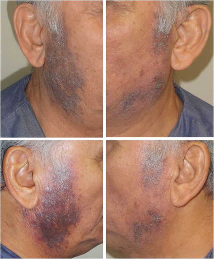
A 77-year-old Caucasian man presented bilateral mandibular
antagonists (or blockers) and 5α-reductase inhibitors [1].
skin rash, which occurred after 6 months of assumption of
In the early 1990s, Kissei Pharmaceutical Co. Ltd. began
silodosin (8 mg a day). The skin lesions presented purple/
the development of α1-adrenergic receptor antagonists that
bluish discolored and confluent spots that did not blanch after
were highly selective for the lower urinary tract without af-
applying pressure (Fig. 1). There were no associated itch,
fecting blood pressure. This led to the discovery of silodosin, a
sting, or any other symptoms and clinical manifestations.
novel indoline derivative approved by the US Food and Drug
The physical examination of the patient was normal. The pa-
Administration (FDA) in 2008. Phase II/III studies reported
tient did not assume any other drugs. At first, systemic lupus
several silodosin-related adverse events with a variable inci-
erythematosus was suspected, but the diagnostic hypothesis
dence. Retrograde ejaculation was the most frequent (28%),
was rejected after some investigations, as the patient did not
followed by dizziness (3.2%), diarrhea (2.6%), orthostatic hy-
meet any other clinical or immunologic (ANA, anti-DNA,
potension (2.6%), headache (2.4%), and nasal congestion
anti-Sm, antiphospholipid Ab, low complement, direct
(2.1%). Toxic skin eruption, purpura, and jaundice were un-
Coombs test) criteria. Histopathological examination revealed
common [2].
chronic inflammation of the epidermis with melanin pigment
deposits in the reticular dermis, as for a post-inflammatory
* Stefania Tenna hyperpigmentation (PIH).
s.tenna@unicampus.it The drug was discontinued 11 months after the appearance
of the skin rash; another alpha1-blocker, doxazosin, was pre-
1
Unit of Plastic Surgery and Dermatology, BCampus Bio Medico^ scribed, with no adverse reactions reported in the following
University, Rome, Italy 12 months.
2
Plastic and Reconstructive Surgery Unit, Via Alvaro del Portillo, According to the Naranjo questionnaire [3], our patient was
200-00128 Rome, Italy evaluated using the questions reported in Table 1, and his final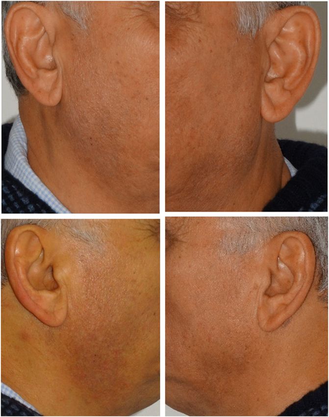
Eur J Plast Surg

                                                                             treatments and patient was advised to avoid direct sun expo-
                                                                             sure for 3 weeks after treatment. Then the patient underwent
                                                                             three fractional CO2 (SmartXide Punto, DEKA M.E.L.A.,
                                                                             Calenzano, Italy) laser treatments, 3 months interval, with
                                                                             these parameters: H-pulse, a DOT spacing of 500 μm at
                                                                             9 W. Following each fractional CO2 laser session the patient
                                                                             applied for 4 weeks a cream containing kojic acid in 4% con-
                                                                             centration, formulated with glycolic acid (10%) and hydroqui-
                                                                             none (1%), to increase efficacy [4].
                                                                                A good cosmetic result was achieved with a complete res-
                                                                             olution of the eruption that remained at an improved state
                                                                             1 year after final treatment (Fig. 2).

                                                                             Discussion

                                                                             Post-inflammatory hyperpigmentation (PIH) is an acquired
                                                                             hypermelanosis occurring after cutaneous inflammation that
                                                                             may arise in all skin types, but more frequently affects darker
                                                                             skin types (Fitzpatrick types IV through VI). These pigmen-
                                                                             tary changes may alter the quality of life of the patients, as
                                                                             they often occur in exposed areas such as the face, the neck, or
                                                                             the décolleté, and when their treatment tends to be difficult
Fig. 1 Pretreatment view of bilateral mandibular skin rash. Skin lesions     and long [5].
are purple/bluish and do not blanch with pressure                                A wide range of etiologies exists for PIH: infections such as
                                                                             dermatophytoses or viral exanthems, allergic reactions from
score was 5, meaning that the adverse reaction can be proba-                 insect bites, or a contact dermatitis, cutaneous injuries from
bly ascribed to the drug.                                                    irritants, burns, or cosmetic procedures, skin inflammatory
   The patient started our combined treatment 1 month after                  (papulosquamous) diseases like atopic dermatitis, lichen
silodosin discontinuation as hyperpigmentation did not clear,                planus, psoriasis, or lupus erythematosus as well as hypersen-
and it was refractory to topical agents. We performed two                    sitivity reactions to medications [6].
treatment sessions, 2 months apart, with intense pulsed light                    PIH results from the overproduction of melanin or an irreg-
(IPL), (Synchro HP, DEKA M.E.L.A., Calenzano, Italy), at                     ular dispersion of pigment after cutaneous inflammation. The
500 nm wavelength, single pulse, at 8 J/cm2. Topical applica-                epidermal inflammatory response, through mediators such as
tion of gentamicin sulfate cream 0.1% twice a day and zinc                   prostaglandins and leukotrienes, stimulates epidermal mela-
oxide cream was recommended on the first few days after                      nocytes to increase the synthesis of melanin and transfer the

Table 1 Naranjo questionnaire
for adverse drug reaction (ADR)       Question                                                                               Answer         Score

                                      Are there previous conclusive reports on this reaction?                                No             0
                                      Did the adverse events appear after the suspected drug was given?                      Yes            2
                                      Did the adverse reaction improve when the drug was discontinued or a specific          Yes            1
                                        antagonist was given?
                                      Did the adverse reaction appear when the drug was readministered?                      Not done       0
                                      Are there alternative causes that could have caused the reaction?                      No             2
                                      Did the reaction reappear when a placebo was given?                                    Not done       0
                                      Was the drug detected in any body fluid in toxic concentrations?                       Not done       0
                                      Was the reaction more severe when the dose was increased or less severe when the       Not done       0
                                        dose was decreased?
                                      Did the patient have a similar reaction to the same or similar drugs in any previous   No             0
                                        exposure?
                                      Did any objective evidence confirm the adverse event?                                  No             0

                                     The final score indicates: ≥ 9, definite ADR; 5–8, probable ADR; 1–4, possible ADR; 0, doubtful ADR
Eur J Plast Surg
published a successful use of fractional CO2 laser in these
kinds of problems [10–15]. Our case first reports a post-
inflammatory hyperpigmentation related to silodosin assump-
tion, showing that recalcitrant PIH should benefit from a com-
bined approach and that pico laser is not the only possibility.
Fractional CO2 treatment in fact is less aggressive on the
tissue as the photocoagulation effect is limited to a few points
and the heat is scattered. Spacing enough the columns within
the spot decreases the thermal effect reducing collateral effects
such as erythema, edema, or crusts. Finally, thanks to the very
low CO2 emission time and the high-peak power (H-Pulse), a
sort of Bcold^ ablation inhibits the immediate inflammatory
response allowing the chemical agent to penetrate easily.
Conclusions
Silodosin cutaneous adverse reactions are rare but may occur.
Early treatment of post-inflammatory hyperpigmentation is
advisable and should be started to help hasten its resolution.
Fractional CO2 lasers combined with chemical agents should
be definitively considered an adjunctive tool in the treatment
of deep hyperpigmentation.
Fig. 2 Post-treatment view with a complete resolution of the eruption
Compliance with ethical standards
melanin granules to keratinocytes, resulting in epidermal Conflict of interest and funding Stefania Tenna, Marco Morelli
melanosis. On the contrary, dermal melanosis, characterized Coppola, Beniamino Brunetti and Paolo Persichetti declare that they have
by a gray or bluish color, occurs when inflammatory processes no conflict of interest. They do not have any commercial associations that
might pose or create a conflict of interest with the information presented
disrupt the basal cell layer and the melanin pigment is trapped in this article. No intramural or extramural funding supported any aspect
by the macrophages in the papillary or reticular dermis [7]. of this work.
Treatments include topical depigmenting agents, chemical
peels, and laser and light therapy. Topical therapy is typically Ethical approval All authors have contributed equally to the scientific
effective for epidermal hyperpigmentation but usually less work.
The study has been performed in accordance with the ethical standards
effective for dermis localization. as laid down in the 1964 Declaration of Helsinki and its later amendments
Due to the wide absorption spectrum of melanin (250– or comparable ethical standards.
1200 nm), many lights and lasers are helpful in recalcitrant
hyperpigmentation, but caution has to be paid to prevent wors- Informed consent Informed consent was obtained from the patient to
ening of PIH. Typically, short wavelength lasers are for epi- use photographs for scientific purposes.
dermal melanin while longer wavelengths penetrate deeper
with selective absorption by dermal targets.
Traditional CO2 resurfacing and intense pulsed light (IPL) References
must be used very carefully as their long pulse duration may
induce hyperpigmentation as well. They should not be con- 1. Masaki Y, Junzo K, Yukio H, Kazuki K (2011) Safety and efficacy
sidered in darker skinned individuals and cooling devices of silodosin for the treatment of benign prostatic hyperplasia. Clin
Interv Aging 6:161–172 (Dovepress)
should be recommended for safety. In case of high melanin
2. National Drug Monograph Silodosin (Rapaflo®) (2012) VA phar-
density, laser energy intended for deeper dermal targets could macy benefits management services, medical advisory panel, and
be absorbed within the pigmented epidermis, leading to com- VISN pharmacist executives, www.pbm.va.gov
plications such as dyschromias, blistering, and scars [8]. 3. Naranjo CA, Busto U, Sellers EM, Sandor P, Ruiz I, Roberts EA,
Nd:Yag Q-switched laser with shorter pulse duration but Janecek E, Domecq C, Greenblatt DJ (1981) A method for estimat-
ing the probability of adverse drug reactions. Clin Pharmacol Ther
long wavelength may also be absorbed in the superficial layers 30(2):239–245
leading to complication, so to date pico laser is the most ap- 4. Halder RM, Richards GM (2004) Topical agents used in the man-
propriate to treat deep marks [9]. Many papers already agement of hyperpigmentation. Skin Therapy Lett 9:1–3Eur J Plast Surg
5. Davis EC, Callender VD (2010) Postinflammatory hyperpigmenta- alone, or a combination of the two: a comparative study. J Drugs
tion: a review of the epidemiology, clinical features, and treatment Dermatol 9(4):315–322
options in skin of color. J Clin Aesthet Dermatol 3(7):20–31 11. Jalaly NY, Valizadeh N, Barikbin B, Yousefi M (2014) Low-power
6. Chang MW (2009) Disorders of hyperpigmentation. In: Bolognia fractional CO2 laser versus low-fluence Q-switch 1, 064 nm Nd:
JL, Jorizzo JL, Rapini RP (eds) Dermatology. 2nd ed. Elsevier, YAG laser for treatment of melasma: a randomized, controlled,
Mosby, pp 333–389 split-face study. Am J Clin Dermatol 15(4):357–363
7. Taylor SC, Grimes PE, Lim J, Im S, Lui H (2009) Postinflammatory 12. Hsiao CY, Sung HC, Hu S, Huang CH (2015) Fractional CO2 laser
hyperpigmentation. J Cutan Med Surg 13:183–191 treatment to enhance skin permeation of tranexamic acid with min-
8. Tan KL, Kurniawati C, Gold MH (2008) Low risk of imal skin disruption. Dermatology 230(3):269–275
postinflammatory hyperpigmentation in skin types 4 and 5 after 13. Katz TM, Goldberg LH, Firoz BF et al (2009) Fractional
treatment with fractional CO2 laser device. J Drugs Dermatol. photothermolysis for the treatment of postinflammatory hyperpig-
7(8):774–777 mentation. Dermatol Surg 35:1844–1848
9. Cho SB, Park SJ, Kim JS, Kim MJ, Bu TS (2009) Treatment of 14. Oram Y, Deniz Akkaya A (2014) Refractory postinflammatory hy-
post-inflammatory hyperpigmentation using 1064-nm Q-switched perpigmentation treated fractional CO2 laser. J Clin Aesthet
Nd: YAG laser with low fluence: report of three cases. J Eur Acad Dermatol 7(3):42–44
Dermatol Venereol 23:1206–1207 15. Tierney EP, Kouba DJ, Hanke CW (2009) Review of fractional
10. Trelles MA, Velez M, Gold MH (2010) The treatment of melasma photothermolysis: treatment indications and efficacy. Dermatol
with topical creams alone, CO2 fractional ablative resurfacing Surg 35:1445–1461You can also read