Refractory chronic osteomyelitis of femur: A case report - Open ...
←
→
Page content transcription
If your browser does not render page correctly, please read the page content below
Medical Case Reports and Reviews

  Case Report                                                                                                                                ISSN: 2517-7214

Refractory chronic osteomyelitis of femur: A case report
Li Chen*, Huibao Hou, Liren Zhang and Qi Pan
Department of orthopedics, Suzhou Hospital of Anhui Medical University (Suzhou Municipal Hospital of Anhui Province), 299 Bianhe Middle Road, Yongqiao District,
Suzhou City, Anhui Province, China

Abstract
Purpose: The purpose of this paper is to introduce a case of refractory chronic osteomyelitis of the femoral shaft.
Case Report: A 51 year old male suffered from chronic osteomyelitis of the femoral shaft for about 40 years. In the first stage, thorough debridement, extensive
lesion resection, antibiotic bone cement spacer and external fixator were applied; in the second stage, the external fixator and bone cement column were changed to
femoral bone marrow internal nail combined with femoral locking plate and autologous bone implantation at the broken end under the premise of complete control
of inflammation and good health. Finally, the patient was cured and very satisfied with the current state.
Conclusion: We report a case of refractory chronic osteomyelitis, which is very rare to have a long history of disease, a large number of fractures, and deformity degree
of lower limbs.

Introduction                                                                           improved after anti-inflammatory treatment and wound cleaning and
                                                                                       dressing change, but the skin intermittent exudation was not cured.
     It is difficult to treat chronic osteomyelitis of femoral shaft, which            Three years later, the symptoms of swelling, ulceration and purulent
often leads to deformity and seriously affects limb function, which                    fluid exudation of the lateral skin of the middle and lower femur were
brings serious burden to patients’ life and economy. The history of                    obvious, and he went to the local hospital for treatment, diagnosis:
chronic osteomyelitis is generally long and it is easy to have repeated                osteomyelitis of femoral shaft. The first operation was performed, soft
attacks. If there is dead cavity, dead bone or scar tissue without blood               tissue debridement, femoral shaft slotting and gentamicin was added to
supply in the lesion, antibiotics can not achieve its efficacy. At the same            the femoral medullary cavity flushing solution for continuous flushing.
time, due to the influence of purulent secretion of wound surface,                     The patient was stable for the next 10 years, no swelling, heat, pain and
antibiotics are easy to be diluted, and even some drugs are difficult                  ulceration were found in the skin of the affected limb. Ten years after
to pass through the pus to act on tissues, long-term use of antibiotics                surgery, when the patient was about 20 years old, pain symptoms of the
will lead to drug resistance. If there is no thorough debridement of                   affected limb recurred, the patient could not bear the pain and went
osteomyelitis lesions, only simple debridement and anti-inflammatory                   to the local hospital for treatment, Physical examination showed that
treatment often can not achieve the goal of cure, and miss the best                    the skin was intact and no ulcerative fluid was found, recurrence of
opportunity for early complete cure of osteomyelitis, eventually,                      osteomyelitis was considered according to imaging findings, The patient
the early infection will progress to chronic osteomyelitis [1-2] .                     was treated again with irrigation and drainage of intramedullary tube.
Therefore, reasonable and correct treatment plan is very important                     During the operation, a large amount of purulent fluid flowed out of the
for patients with osteomyelitis. Surgery is the mainstay of treatment                  medullary cavity, and the symptoms of swelling and pain of the affected
for osteomyelitis patients, including local irrigation and debridement,                limbs were obviously improved after the operation, however, the skin
extensive lesion resection or amputation [1,3-5]. The purpose of this                  sinus of the middle and lower femur was formed, which was not cured.
paper is to introduce a case of refractory chronic osteomyelitis of                    When the patient was about 30 years old, he suffered an accidental
the femoral shaft. In the first stage, thorough debridement, extensive                 fracture of the right femoral shaft. At that time, the patient’s history
lesion resection, antibiotic bone cement spacer and external fixator                   of chronic osteomyelitis and limited medical level were considered,
were applied; in the second stage, the external fixator and bone cement                the patient received plaster external fixation. Unfortunately, over the
column were changed to femoral bone marrow internal nail combined                      next 20 years, the patient experienced six fractures in the same area, the
with femoral locking plate and autologous bone implantation at the                     interval between each fracture was 4-5 years, because plaster external
broken end under the premise of complete control of inflammation                       fixation can not completely fix both ends of fracture, in the process
and good health.

Case report
                                                                                       *Correspondence to: Li Chen, Department of orthopedics, Suzhou Hospital of
    A 51 year old male patient had no history of hypertension and
                                                                                       Anhui Medical University (Suzhou Municipal Hospital of Anhui Province), 299
diabetes mellitus, no smoking and drinking habits. He complained of                    Bianhe Middle Road, Yongqiao District, Suzhou City, Anhui Province, China,
soft tissue swelling around the right knee joint due to sprain when he                 E-mail: 524985750@qq. com
was 8 years old, there was no fracture at that time, after a month of
rest and massage, the lateral skin of the knee joint was broken with                   Key words: chronic osteomyelitis, debridement, intramedullary fixation, external
                                                                                       fixation
fluid exudation, during this period, the body temperature was on
the high side (about39℃). At that time, the fever symptoms were                        Received: August 04, 2020; Accepted: August 11, 2020; Published: August 14, 2020

Med Case Rep Rev, 2020        doi: 10.15761/MCRR.1000153                                                                                                Volume 3: 1-4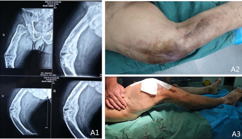
Chen L (2020) Refractory chronic osteomyelitis of femur: A case report
of fracture healing, the angle of femoral shaft forward angulation is
gradually increasing, as a result, the patient’s right lower limb could
not walk upright, in addition, plaster external fixation after multiple
fractures resulted in complete stiffness of the affected knee joint and
loss of function (Figure 1 A2-3).
On April 21, 2020, The patient developed right thigh pain after
sprain three days ago, He went to our hospital for further treatment
for the first time. The X-ray examination showed that: the right femoral
shaft is curved, angulation in front, the outline of the middle femur
became thicker, irregular shape, hyperplasia, thickening and sclerosis of
bone cortex, irregular medullary cavity, a lot of sclerotic bone and dead
bone were formed, the continuous interruption of the bone cortex in
front of the femoral shaft showed the sign of pathological fracture, the
medullary cavity was completely occluded, a large number of envelope
was formed around the dead bone, there were sinus holes formed by
pus erosion on the surface of envelope (Figure 1 A1). Pyogenic fluid
exudates from the skin sinus formed in the middle and lower femur. The
results of laboratory tests showed that erythrocyte sedimentation rate
(ESR): 53. 0 mm\h, high sensitivity C-reactive protein (hs-CRP): 62.
04 mg\L, serum procalcitonin (pct-q): 0. 23 ug\L. The bacterial culture
and drug sensitivity of secretion suggested that Staphylococcus aureus
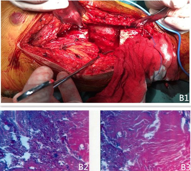
infection was sensitive to glycopeptide antibiotics and aminoglycoside Figure 2. B1. photos of femoral defect after resection of intraoperative lesions; B2-3. after
resection of the femoral lesions, the lesions were observed under a microscope
antibiotics.
On April 28, 2020, the patient was brought into the operating
room for thorough debridement, microbial culture and pathological
examination. During the operation, the lesions were completely
removed and the middle femur was removed about 7 cm (Figure
2 B1) and then sent for pathological examination (Figure 2 B2-3).
After thorough removal of dead bone in medullary cavity, 40g acrylic
resin bone cement (1* 40 g, blomet France SARL) mixed with 2. 0g
vancomycin (0. 5 g, Zhejiang Haizheng) was implanted into the femoral
defect, and negative pressure irrigation tube was implanted in the side
of femur, the combined external fixator (WGJIV, alder Technology)
was used to stabilize the distal and proximal femur, the postoperative
X-ray film of the affected limb is shown in the figure (Figure 3 C1). Figure 3. C1. the X-ray films were reexamined after debridement; C2-3. the X-ray film of
Intraoperative microbial culture indicated Staphylococcus aureus the right femur was reexamined 8 weeks after debridement
infection, after 6 weeks of intravenous infusion of vancomycin,
rifampicin (150mg Shanghai Hengshan) was given orally for 4 weeks. in the operating room again. The X-ray film of the right femur was
Gentamicin was added to the washing solution for 2 weeks, during this reexamined 8 weeks after operation as shown in the figure (Figure 3
period, several times of bacterial culture indicated that no bacterial C2-3), The results of laboratory tests showed that ESR: 30 mm\h, hs-
growth was observed, and then the flushing tube was pulled out. CRP: 14. 18 mg\l, pct-q: 0. 11 ug\l.
During the treatment, the wound around the original skin sinus was On July 6, 2020, the patient returned to the operating room again
not well healed, and it was completely healed after local debridement to remove all external fixators and bone cement mixed with antibiotics.
The blood fluid in the medullary cavity was cultured, the subsequent
results indicated that no bacterial growth was observed. Then, 11*380
mm metal interlocking intramedullary nail (A-UFN-02, Xiamen
Dabo Technology) was implanted after reaming the proximal and
distal femur, the ipsilateral iliac bone was cut into 10*5*5 mm bone
strips, mixed with 2. 0g vancomycin, and implanted into the femoral
defect, considering the large defect of femur, the metal locking plate
(LCLP01-14hole, Xiamen Dabo Technology) was implanted in the
lateral femur to play the role of eccentric fixation (Figure 4 D1-2). The
patient was given vancomycin intravenously, and rifampicin was given
orally one week later, laboratory tests showed that ESR: 64 mm\h, hs-
CRP: 27. 61 mg\l, pct-q: 0. 09 ug\l. During the whole treatment, the
body temperature fluctuated within the normal range. One month
after operation, the X-ray film of femur was reexamined as shown
Figure 1. A1. the X-ray film of right femoral shaft before admission; A2-3. appearance of in Figure (Figure 4 D3-4) and the wound healed well (Figure 5 E1-
right lower limb before operation 2). Reexamination of inflammatory indicators showed that: ESR: 50
Med Case Rep Rev, 2020 doi: 10.15761/MCRR.1000153 Volume 3: 2-4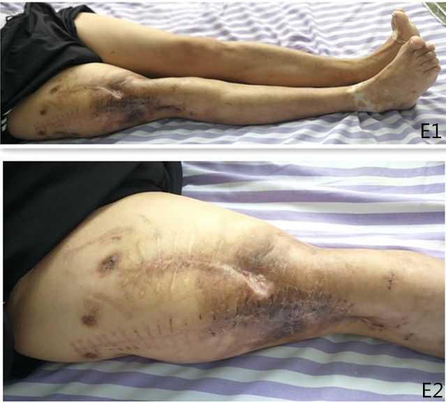
Chen L (2020) Refractory chronic osteomyelitis of femur: A case report

                                                                                         In the process of osteomyelitis treatment, dead cavity, dead bone
                                                                                         or scar tissue with lack of blood supply must be eliminated and the
                                                                                         internal fixation materials were applied until there was evidence that
                                                                                         the inflammation was completely controlled [10]. This also shows that
                                                                                         the cure of chronic osteomyelitis needs a long process of diagnosis and
                                                                                         treatment, can not rush for a while. Kanakaris, et al. [11,12] suggested
                                                                                         that patients with chronic osteomyelitis should be treated in stages,
                                                                                         because there is a risk of reinfection caused by residual pathogenic
                                                                                         bacteria in soft tissue after debridement, early internal fixation is
                                                                                         not allowed in patients with osteomyelitis. In our case, thorough
                                                                                         debridement, catheter irrigation and external fixator will be carried out
                                                                                         in the early stage of treatment, the next stage of treatment will be carried
Figure 4. D1-2. the X-ray films of femur were reexamined after replacement of internal
fixation materials; D3-4. the X-ray films of femur were reexamined after 4 weeks of      out when there is evidence that the infection is completely controlled.
replacement of internal fixation materials                                               At present, an antibiotic impregnated cement interlocking nail [13] or
                                                                                         rod [14,15] has been used to treat patients with infection after femoral
                                                                                         or tibial fractures, however, the service life of interlocking nail or rod
                                                                                         and cement must be considered. Capanna, et al. [16] during a 4. 5-year
                                                                                         follow-up of 76 patients with distal femoral or proximal tibial tumors
                                                                                         treated with interlocking nail and bone cement spacer, they found
                                                                                         that 12 cases (16%) failed (i. e. , broken, bent or displaced rods) and
                                                                                         bone tissue could not grow in polymethylmethacrylate (PMMA), they
                                                                                         suggested that the cement interlocking nail or rod should be removed
                                                                                         2 months after operation. According to the patient’s condition, we
                                                                                         removed the bone cement filled with the broken end and replaced it
                                                                                         with autologous bone 9 weeks after the first-stage debridement, at the
                                                                                         same time, we replaced the external fixator with femoral interlocking
                                                                                         nail combined with metal locking plate to stabilize the broken end of
                                                                                         femur. We hope that in the future, under the premise of good bone
                                                                                         healing, knee orthopedic surgery will restore knee joint function.

                                                                                         Conclusion
                                                                                              We report a case of refractory chronic osteomyelitis, which is very
                                                                                         rare to have a long history of disease, a large number of fractures, and
                                                                                         deformity degree of lower limbs. We adhere to the most basic treatment
                                                                                         principles of osteomyelitis, and constantly adjust and improve the
                                                                                         treatment plan, and finally achieve the goal of cure. The patient is very
             Figure 5. E1-2. appearance of right lower limb after operation
                                                                                         satisfied with the current state.

mm\h, hs-CRP: 16. 55 mg\L, pct-q: 0. 07 ug\ L. The patients were very                    Acknowledgments
satisfied with the current situation.                                                        The authors did not receive any outside funding or grants in support
                                                                                         of their research for or preparation of this work. No commercial entity
Discussion                                                                               paid or directed, or agreed to pay or direct, any benefits to any research
     Osteomyelitis is a well-known disease, most common in adults,                       fund, foundation, division, center, clinical practice or other charitable
staphylococcus aureus is the most common pathogen of osteomyelitis                       or non profit organization with which the authors, or a member of their
[3,4]. Usually associated with open fractures or soft tissue infection                   immediate families, are affiliated or associated.
after a long period of infection through blood transmission [6,7]. To
analyze the patient with osteomyelitis, the main pathogenic factors                      Conflicts of interest statement
are improper treatment of early soft tissue injury, long-term failure to                      None of the authors have relevant conflicts of interest to declare.
recover after infection, and blood borne transmission of pathogenic
bacteria, postoperative pathology showed that a large number of                          References
inflammatory cells infiltrated into the nutrient canal of bone tissue, the               1. Forsberg JA, Potter BK, Cierny G 3rd, Webb L (2011) Diagnosis and management of
vascular channels around bone tissue were infiltrated and occluded by                       chronic infection. J Am Acad Orthop Surg 19: S8-S19.
inflammatory cells [3,7], the resulting weak bone and diseased bone                      2. Beck-Broichsitter BE, Smeets R, Heiland M (2015) Current concepts in pathogenesis
hinder the healing ability of bone tissue and increase the risk of fixation                 of acute and chronic osteomyelitis. Curr Opin Infect Dis 28: 240-245.
failure. Thein, et al. [6] pointed out that two patients with pathological               3. Parsons B, Strauss E (2004) Surgical management of chronic osteomyelitis. Am J Surg
fractures underwent five surgical interventions before they were                            188: 57-66.
cured in their cases. Debridement is the most important method for                       4. Jiang N, Ma YF, Jiang Y, Zhao XQ, Xie GP, et al. (2015) Clinical characteristics
the successful treatment of chronic osteomyelitis. Complete removal                         and treatment of extremity chronic osteomyelitis in Southern China: a retrospective
of the lesion is the main means to prevent the recurrence of chronic                        analysis of 394 consecutive patients. Medicine 94: e1874.
osteomyelitis [8], the long-term recurrence rate of osteomyelitis                        5. Fodor L, Ullmann Y, Soudry M, Calif E, Lerner A (2006) Prophylactic external fixation
patients can be reduced to 20% after thorough debridement [1,4,5,9].                        and extensive bone debridement for chronic osteomyelitis. Acta Orthop Belg 72: 448-453.

Med Case Rep Rev, 2020          doi: 10.15761/MCRR.1000153                                                                                                      Volume 3: 3-4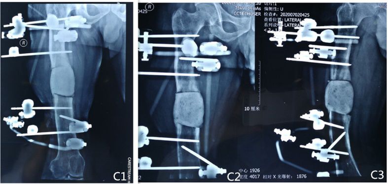
Chen L (2020) Refractory chronic osteomyelitis of femur: A case report

6. Thein R, Tenenbaum S, Chechick O, Leshem E, Chechik A, et al. (2013) Delay in            12. Kanakaris NK, Tosounidis TH, Giannoudis PV (2015) Surgical management of infected
   diagnosis of femoral hematogenous osteomyelitis in adults: an elusive disease with           non-unions: An update. Injury 46: S25-S32.
   poor outcome. Isr Med Assoc 15: 85-88.
                                                                                            13. Thonse R, Conway J (2007) Antibiotic cement-coated interlocking nail for the treatment
7. Lew DP, Waldvogel FA (2004) Osteomyelitis. Lancet 364: 369-379.                              of infected nonunions and segmental bone defects. J Orthop Trauma 21: 258-268.
8. Simpson AH, Deakin M, Latham JM (2001) Chronic osteomyelitis. The effect of the extent   14. Paley D, Herzenberg JE (2002) Intramedullary infections treated with antibiotic cement
   of surgical resection on infection-free survival. J Bone Joint Surg Br 83: 403-407.          rods: preliminary results in nine cases. J Orthop Trauma 16: 723-729.
9. Conterno LO, Turchi MD (2013) Antibiotics for treating chronic osteomyelitis in          15. Qiang Z, Jun PZ, Jie XJ, Hang L, Bing LJ, et al. (2007) Use of antibiotic cement rod
   adults. Cochrane Database Syst Rev 9: CD004439.                                              to treat intramedullary infection after nailing: preliminary study in 19 patients. Arch
                                                                                                Orthop Trauma Surg 127: 945-951.
10. Lew DP, Waldvogel FA (1997) Osteomyelitis. N Engl J Med 336: 999-1007.
                                                                                            16. Capanna R, Biagini R, Ruggieri P, Bettelli G, Casadei R, et al. (1989) Temporary
11. Ryan S, Eward W, Brigman B, Zura R (2017) Chronic Osteomyelitis of the Distal
                                                                                                resection-arthrodesis of the knee using an intramedullary rod and bone cement. Int
    Femur Treated with Resection and Delayed Endoprosthetic Reconstruction: A Report
    of Three Cases. Case Rep Orthop 2017: 5141032.                                              Orthop 13: 253-258.

Copyright: ©2020 Chen L. This is an open-access article distributed under the terms of the Creative Commons Attribution License, which permits unrestricted use,
distribution, and reproduction in any medium, provided the original author and source are credited.

Med Case Rep Rev, 2020           doi: 10.15761/MCRR.1000153                                                                                                         Volume 3: 4-4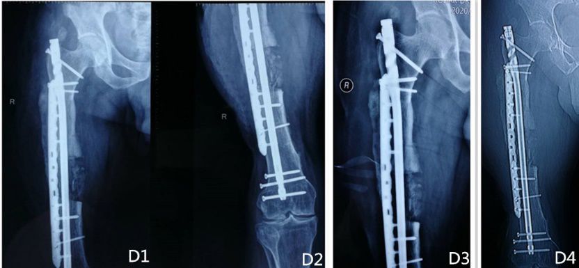
You can also read