Allopurinol-Induced Oral Lichenoid Drug Reaction with Complete Regression after Drug Withdrawal - MDPI
←
→
Page content transcription
If your browser does not render page correctly, please read the page content below
Case Report
Allopurinol-Induced Oral Lichenoid Drug Reaction
with Complete Regression after Drug Withdrawal
Alexandre Perez † , Benjamin Lazzarotto † , Jean-Pierre Carrel and Tommaso Lombardi *
 Unit of Oral Medicine and Pathology, Division of Oral Maxillofacial Surgery, Department of Surgery,
 University Hospitals of Geneva, 1205 Geneva, Switzerland; alexandre.perez@hcuge.ch (A.P.);
 benjamin.lazzarotto@hcuge.ch (B.L.); jean-pierre.carrel@unige.ch (J.-P.C.)
 * Correspondence: tommaso.lombardi@hcuge.ch; Tel.: +41-79-553-32-06
 † These authors are considered equally as co-First authors.

 Received: 15 June 2020; Accepted: 10 August 2020; Published: 12 August 2020

 Abstract: Background: Lichen planus is a chronic mucocutaneous inflammatory disease.
 Oral manifestations are common, and may remain exclusive to the oral mucosa without involvement of
 the skin or other mucosae. A differential diagnosis includes oral lichenoid drug reactions. Allopurinol,
 which is the first line hypo-uricemic treatment, is often quoted as being a possible offending drug,
 though oral reactions have rarely been reported. Case presentation: We describe a 59-year-old male
 gout patient, successfully treated with allopurinol, who developed acute onset of oral lichenoid
 lesions, involving bilaterally the buccal mucosa, the tongue and the labial mucosa. Histopathology
 was consistent with a lichen planus or a drug-induced lichenoid reaction. Improvement of the
 patient’s condition after withdrawal of allopurinol confirmed the lichenoid nature of the lesion.
 Remission was complete after a few weeks. Discussion: Although unusual, allopurinol may induce
 a lichenoid drug reaction. These reactions may mimic clinically and histopathologically idiopathic
 lichen planus. Improvement or complete regression of the lesions may be attempted to confirm
 the diagnosis. According to the latest WHO recommendations, these lesions have a potential for
 malignant transformation.

 Keywords: oral mucosa; lichen planus; lichenoid reaction; adverse drug reaction; allopurinol

1. Introduction
     Lichen planus is a common chronic inflammatory disorder, affecting the skin, oral and genital
mucosa, scalp and nails. Oral manifestations are frequent, and may remain exclusive to the oral cavity
without involvement of other organs [1].
     To the contrary of cutaneous lichen planus, oral lichen planus is a long-term chronic disease
with a dynamic evolution, as a result of successive waves of variably destructive activity at the
epithelium–chorion interface. Thus, progressive changes of the clinical and histopathological aspects
occur over time, increasing the number of possible clinical presentations, going from white keratotic
dots to mucosal atrophy and hyperkeratosis in the late stage. The most characteristic and easily
recognized clinical aspect being the reticular form [2].
     Oral lichenoid drug reactions are a connected entity, the term referring to a lichen planus-like
rash triggered by systemic drug exposure. Allopurinol stands beside many classes of drugs involved,
such as nonsteroidal anti-inflammatory drugs (NSAIDs), B-blockers, ACE inhibitors, thiazide diuretics
as well as some antibiotics [3]. This anti-gout drug is often quoted but only a few reports are described
in the literature.
     This article reports a clinically and histopathologically detailed case of oral lichenoid lesions
associated with allopurinol therapy, that showed complete regression after the withdrawal of the drug.

Dermatopathology 2020, 7, 18–25; doi:10.3390/dermatopathology7020004   www.mdpi.com/journal/dermatopathology
Dermatopathology 2020, 7                                                                             19

Dermatopathology 2020, 7, x                                                                              2

   2. Case Presentation
2. Case Presentation
        A 59-year-old man was referred due to a 4-week history of severe pain within the oral cavity.
      A 59-year-old man was referred due to a 4-week history of severe pain within the oral cavity. The
  The patient give written consent for publication. Medical history revealed skin psoriasis diagnosed
patient give written consent for publication. Medical history revealed skin psoriasis diagnosed with a
  with a biopsy more than 30 years ago, which has since been treated by calcipotriol betamethasone gel
biopsy more®than 30 years ago, which has since been treated by calcipotriol betamethasone gel (Daivo-
  (Daivobet , LEO Pharmaceutical Products Ltd., Regensdorf, Switzerland) intermittently depending
bet®, LEO Pharmaceutical Products Ltd., Regensdorf, Switzerland) intermittently depending on the evo-
   on the evolution of the lesions. Otherwise, the patient suffered episodic gout attacks for which he was
lution of the lesions. Otherwise, the patient suffered episodic gout attacks for which he was under allo-
   under allopurinol for one month prior to consultation. On examination, he had multiple keratinized
purinol for one month prior to consultation. On examination, he had multiple keratinized lesions in
   lesions in plaque form, involving the bilateral buccal and lingual mucosae, with some erythematous
plaque form, involving the bilateral buccal and lingual mucosae, with some erythematous streaks and
   streaks and ulcerated areas (Figure 1).
ulcerated areas (Figure 1).

                                                   (a)

                                                   (b)

                                              Figure 1. Cont.
Dermatopathology 2020, 7 20
Dermatopathology 2020, 7, x 3
Dermatopathology 2020, 7, x 3
(c)
(c)
Figure 1. 1.
(a)(a)
Keratotic
Keratotic lesions
lesionsinvolving
involvingthe theright
rightbuccal
buccalmucosa and the lower lip, with focal erythema
erythema
Figure
Figure 1. (a) Keratotic lesions involving the right buccal mucosa
mucosa and andthe
thelower
lowerlip,
lip,with
withfocal
focal erythema
and anan
and ulcerated
ulcerated area.
area.(b)(b)Keratotic
Keratoticlesions
lesionsinvolving
involvingthe lateral border of the tongue, and the
the median
and an ulcerated area. (b) Keratotic lesions involving the
the lateral
lateral border
borderofofthe
thetongue,
tongue,and
and themedian
median
anterior area.
anteriorarea. (c)
area.(c)Keratotic
(c)Keratotic lesions
Keratoticlesions of the
lesionsofoftheleft
theleftbuccal
leftbuccalmucosa
buccalmucosa and
mucosaand lip.
andlip.
lip.
anterior
AAAbiopsy
biopsy
biopsy ofofthe
oftheright
the
right buccal
right mucosa
buccal
buccal mucosa
mucosa was performed.
waswas Histopathological
performed.
performed. examination
Histopathological
Histopathological revealed
examination
examination aastrat-
revealed
revealed strat-
ified
a squamous
stratified epithelium
squamous with
epithelium focal
withatrophy
focal and
atrophy discreet
and parakeratosis.
discreet The
parakeratosis. superficial
The
ified squamous epithelium with focal atrophy and discreet parakeratosis. The superficial chorion con- chorion
superficial con-
chorion
tained
tained aaband-like
contained band-like dense
a band-like dense inflammatory
dense inflammatory
inflammatory infiltrate, composed
infiltrate,
infiltrate, composed
composed mostly
mostlyofof
mostly oflymphocytes
lymphocytesand
lymphocytes and
andmacrophages,
macrophages,
with
with very rare eosinophils. Apoptotic keratinocytes in the basal layer were observed (Figure2a,b).
with very
veryrare eosinophils.
rare eosinophils. Apoptotic
Apoptotickeratinocytes in
keratinocytes the
inbasal
the layer
basal were
layer observed
were (Figure
observed 2a,b).These
(Figure 2a,b).
These
features
These were consistent
features were with an
consistent active
with lichen
an planus
active lichenor a drug-induced
planus or a lichenoid
drug-induced
features were consistent with an active lichen planus or a drug-induced lichenoid reaction. reaction.
lichenoid reaction.
(a)
(a)
Figure 2. Cont.
Dermatopathology 2020, 7 21
Dermatopathology 2020, 7, x 4
(b)
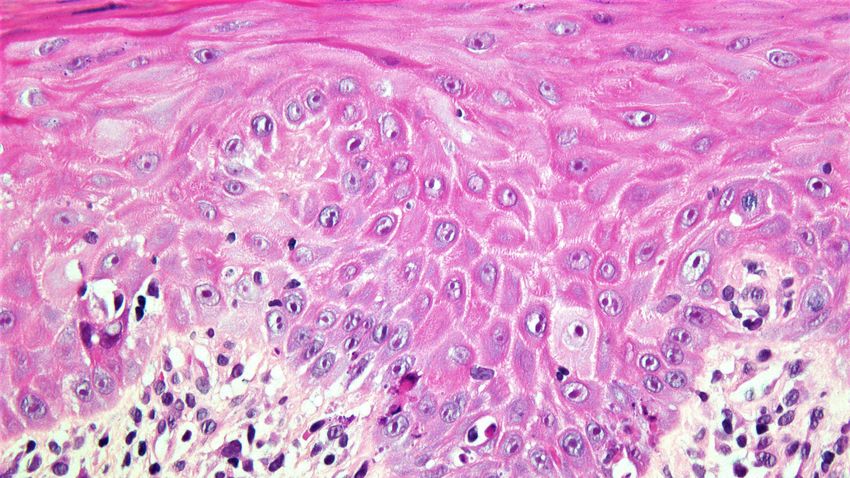
Figure
Figure2.2.(a)(a)
Histopathological
Histopathologicalsection showing
section showingparakeratotic partly
parakeratotic atrophic
partly squamous
atrophic epithelium
squamous with a
epithelium
band-like dense inflammatory infiltrate in the superficial chorion (HE stain, ×10). (b) Higher
with a band-like dense inflammatory infiltrate in the superficial chorion (HE stain, ×10). (b) Higher magnification
showing inflammatory
magnification lymphocytic
showing inflammatory infiltrate and apoptotic
lymphocytic bodiesand
infiltrate in the basal layer
apoptotic (HE in
bodies stain,
the ×40).
basal layer
(HE stain, ×40).
Given the suspected allopurinol involvement, the medication was withdrawn and not substituted
Given the
in agreement suspected
with allopurinol
his general involvement,
practitioner. the medication
Three weeks was withdrawn
after discontinuation, and notregression
significant substitutedof
in agreement
the lesions waswith his general
observed and practitioner.
the patient no Three weeks
longer hadafter
anydiscontinuation,
symptoms. Thesignificant regression of
one-year postoperative
the lesions was
examination observed
revealed a thinand the patient
keratosis of theno longer
buccal had any
mucosa andsymptoms. The one-year of
a discreet depapillation postoperative
the dorsolin-
examination
gual revealed aafter
mucosa. Follow-up thin3 keratosis
years showedof the buccal healing
complete mucosa(Figure
and a 3).
discreet depapillation of the
dorsolingual mucosa. Follow-up after 3 years showed complete healing (Figure 3).
(a)showing inflammatory lymphocytic infiltrate and apoptotic bodies in the basal layer (HE stain, ×40).
Given the suspected allopurinol involvement, the medication was withdrawn and not substituted
in agreement with his general practitioner. Three weeks after discontinuation, significant regression of
the lesions was observed and the patient no longer had any symptoms. The one-year postoperative
Dermatopathology 2020, 7 22
examination revealed a thin keratosis of the buccal mucosa and a discreet depapillation of the dorsolin-
gual mucosa. Follow-up after 3 years showed complete healing (Figure 3).
Dermatopathology 2020, 7, x (a) 5
(b)
(c)
Figure
Figure 3. (a–c)
3. (a–c) Three-year follow-up
Three-year follow-up showing
showing complete
completehealing of theof
healing right
thebuccal
right mucosa and the lower
buccal mucosa and the
lip lip
lower (a),(a),
the the
lateral border
lateral of theof
border tongue (b), and(b),
the tongue theand
left buccal
the leftmucosa
buccaland lip (c).and lip (c).
mucosa
3. Discussion
3. Discussion
Gout Gout is the
is the most
most commonform
common formofofarthritis
arthritis in
in men
men over
overthe
theage
ageofof4040and
anditsits
incidence keeps
incidence on on
keeps
increasing [4,5]. Allopurinol is an effective urate-lowering drug, working as a xanthine oxidase
increasing [4,5]. Allopurinol is an effective urate-lowering drug, working as a xanthine oxidase inhibitor. inhibi-
tor. It is considered as the first line preventative treatment of chronic gout worldwide. Some infrequent
adverse events are well described, mainly the hypersensitivity syndrome. Skin reactions may occur in
approximately 2% of patients. Most cutaneous reactions are maculo-papular eruptions; however, severe
reactions, such as toxic epidermal necrolysis, are also documented [6].
Oral manifestations in the form of lichenoid lesions, although frequently quoted, are rare, and have
only been reported twice in the literature [7,8] (see Table 1). The clinical presentation in all reportedDermatopathology 2020, 7 23
It is considered as the first line preventative treatment of chronic gout worldwide. Some infrequent
adverse events are well described, mainly the hypersensitivity syndrome. Skin reactions may occur
in approximately 2% of patients. Most cutaneous reactions are maculo-papular eruptions; however,
severe reactions, such as toxic epidermal necrolysis, are also documented [6].
Oral manifestations in the form of lichenoid lesions, although frequently quoted, are rare, and have
only been reported twice in the literature [7,8] (see Table 1). The clinical presentation in all reported
cases was painful oral ulcerations. Patients were 1 female and 3 males, and patient age ranged from 53
to 75 years. Lesions were described as white striae, white plaques, along with erosions and ulcerations.
All lesions were present bilaterally on the buccal mucosa, on the borders of the tongue, and in two
cases on the mucosa of the lower lip. Clinical impression, in all reported cases, was that of erosive
lichen planus. A biopsy was performed in only one case [7]. The latter was said to be consistent with
lichen planus without further details.
Table 1. Clinical and histopathological characteristics of allopurinol-induced oral lesions in patients
reported in 3 publications.
Allopurinol Histopathological
Clinical Features Outcomes
Indication Features
Ulcerations
M, Caucasian, Healing of ulcers
Keratotic striae and Gout No biopsy
53 y Persistence of the keratosis
plaques
Ulcerations on the
M, Caucasian, Healing of ulcers
Chau et al. lip, buccal mucosa Gout No biopsy
56 y Persistence of the keratosis
1984 [7] and tongue
Ulcerations
M, Caucasian, Keratotic striae on LP versus Persistence of minor
Gout
60 y the lip, buccal lichenoid reaction ulcerations
mucosa and tongue
Erosions and
ulcerations
Nair et al. F, Chinese, Keratotic striae and Resolution of erosions
Gout No biopsy
2005 [8] 75 y plaques on the Persistence of faint keratosis
buccal mucosa and
tongue
Buccal ulceration
M, Caucasian, Keratotic striae on LP versus
Present case Gout Complete healing
59 y the lip, buccal lichenoid reaction
mucosa and tongue
In our case, the patient complained of pain and the buccal mucosa and tongue were affected, with
erosions on the former. Histologically, our case showed a lichenoid aspect although indistinguishable
from an idiopathic lichen planus. The presence of parakeratosis is not helpful in oral lesions and
eosinophils were very rare, differently to drug-induced skin lesions.
The diagnosis of drug-induced lichenoid lesions may be difficult, as it may share a similar aspect
to those found in the idiopathic oral lichen planus, both clinically and histopathologically. It is
important to make the difference between the two conditions, because lichen planus is usually treated
by corticosteroids or immunomodulatory agents, whereas drug-induced lichenoid lesions are treated
by the withdrawal of the offending agent.
The difficulty of diagnosis also lies in the fact that cutaneous and/or oral lesions may occur after
a variable latency period from the introduction of the offending drug [3]. In the present case, a few
weeks of allopurinol therapy were sufficient to observe oral lesions, which is consistent with the two
previous reports.
Our diagnosis was conducted according to the updated French drug reaction assessment of
imputability criteria [9]. In the present case, the chronological score was 3 (C3) whereas the semiological
score was 2 (S2). Therefore, intrinsic imputability score was 5 (I5), and extrinsic imputability score was
2 (B2) considering the bibliographical data. Imputability of allopurinol on the basis of the obtained
score was consistent.Dermatopathology 2020, 7 24
The resolution after drug withdrawal seems to be spread over a variable period of time, and the
literature is evasive about the degree of regression that the clinician can expect. In the present case,
complete healing was observed at the 3-year follow-up. For other drugs, some authors suggest a time
frame of up to 24 months before full resolution, depending on the initial extension of the lesions,
their severity or the drug incriminated [3,10].
Although the World Health Organization (WHO) classifies oral lichen planus as a precancerous
disease, incidence of malignant transformation is still a matter of discussion. Discrepancies in studies
may stem from variations in diagnostic criteria, insufficient knowledge or recognition of the late stage of
the disease (post-lichen state), confusion with other keratotic and/or atrophic lesions, poor detection of
early dysplastic changes, and too short follow-up periods in prospective studies. The latest systematic
review assessed an overall transformation rate of 1.40% [11], but this was probably underestimated if
one takes into account the previously discussed limitations. Nowadays, the WHO and some authors
consider lichenoid reactions to have a malignant potential [3,12]. However, the exact transformation
rate is unknown since diagnostic criteria are not always clear-cut. Further studies are necessary to
elucidate the true premalignant role of such lesions.
4. Conclusions
The purpose of this article is to report an unusual case of drug-induced reaction of the oral
mucosa, induced by allopurinol, for which a complete healing was obtained after withdrawal of the
drug. Clinicians must be aware of the recognition and management of oral lichenoid drug reactions.
The diagnosis may be difficult, since a great overlap in clinical and histopathological presentation with
oral idiopathic lichen planus exists. Although a matter of controversy, diagnosis and follow-up of
these lichenoid lesions seems all the more important due to potential malignant transformation.
Author Contributions: Conceptualization, A.P., B.L. and T.L.; writing—review and editing, A.P., B.L. and T.L.;
patient management, J.-P.C. All authors have read and agreed to the published version of the manuscript.
Funding: This research received no external funding.
Conflicts of Interest: The authors declare no conflict of interest.
References
1. Saurat, J.-H.; Lipsker, D.; Thomas, L.; Borradori, L.; Lachapelle, J.-M. Dermatologie Et Infections Sexuellement
Transmissibles, 6th ed.; Elsevier-Masson: Paris, France, 2017.
2. Lombardi, T.; Küffer, R. Concept actuel du lichen plan oral. Le diagnostic facile au début, peut devenir très
difficile dans les lichens anciens. La Presse Médicale 2016, 45, 227–239. [CrossRef] [PubMed]
3. Cheng, Y.-S.L.; Gould, A.; Kurago, Z.; Fantasia, J.; Muller, S. Diagnosis of oral lichen pla-nus: A position
paper of the American Academy of Oral and Maxillofacial Pathology. Oral Surg. Oral Med. Oral Pathol. Oral
Radiol. 2016, 122, 332–354. [CrossRef] [PubMed]
4. Seth, R.; Kydd, A.S.; Buchbinder, R.; Bombardier, C.; Edwards, C.J. Allopurinol for chronic gout. Cochrane
Database Syst. Rev. 2014. [CrossRef] [PubMed]
5. Smith, E.U.R.; Díaz-Torné, C.; Perez-Ruiz, F.; March, L.M. Epidemiology of gout: An up-date. Best Pract. Res.
Clin. Rheumatol. 2010, 24, 811–827. [CrossRef] [PubMed]
6. Atzori, L.; Pinna, A.L.; Mantovani, L.; Ferreli, C.; Pau, M.; Mulargia, M.; Aste, N. Cutaneous adverse drug
reactions to allopurinol: 10 year observational survey of the dermatology de-partment—Cagliari University
(Italy). J. Eur. Acad. Dermatol. Venereol. 2011, 26, 1424–1430. [CrossRef] [PubMed]
7. Chau, N.Y.; Reade, P.C.; Rich, A.M.; Hay, K.D. Allopurinol-amplified lichenoid reactions of the oral mucosa.
Oral Surg. Oral Med. Oral Pathol. 1984, 58, 397–400. [CrossRef]
8. Nair, R.G.; Newsome, P.R.H.; Itthagarun, A.; Samaranayake, L.P. Severe oral erosive lichen planus due to
methyldopa and allopurinol: A case report. Hong Kong Dent. J. 2005, 2, 122–125.
9. Arimone, Y.; Bidault, I.; Dutertre, J.-P.; Gérardin, M.; Guy, C.; Haramburu, F.; Hillaire-Buys, D.; Meglio, C.;
Penfornis, C. Réactualisation de la méthode française d’imputabilité des effets indésirables des médicaments.
Therapie 2011, 66, 517–525. [CrossRef] [PubMed]Dermatopathology 2020, 7 25
10. Halevy, S.; Shai, A. Lichenoid drug eruptions. J. Am. Acad. Dermatol. 1993, 29, 249–255. [CrossRef]
11. Giuliani, M.; Troiano, G.; Cordaro, M.; Corsalini, M.; Gioco, G.; Muzio, L.L.; Pignatelli, P.; Lajolo, C. Rate of
malignant transformation of oral lichen planus: A systematic review. Oral Dis. 2019, 25, 693–709. [CrossRef]
[PubMed]
12. Van der Meij, E.H.; Mast, H.; van der Waal, I. The possible premalignant character of oral lichen planus and
oral lichenoid lesions: A prospective five-year follow-up study of 192 patients. Oral Oncol. 2007, 43, 742–748.
[CrossRef] [PubMed]
© 2020 by the authors. Licensee MDPI, Basel, Switzerland. This article is an open access
article distributed under the terms and conditions of the Creative Commons Attribution
(CC BY) license (http://creativecommons.org/licenses/by/4.0/).You can also read