Case Report Early-onset breast cancer in a woman with Graves' disease
←
→
Page content transcription
If your browser does not render page correctly, please read the page content below
Int J Clin Exp Med 2012;5(4):358-362
www.ijcem.com /ISSN:1940-5901/IJCEM1204004
Case Report
Early-onset breast cancer in a woman with Graves’ disease
James E Siegler1, Xinying Li2, Steven D Jones3, Emad Kandil4
1Tulane University School of Medicine; 2Department of General Surgery, Xiangya Hospital, Central South University,
Changsha, China; 3Department of Surgery, Tulane School of Medicine; 4Division of Endocrine and Oncological Sur-
gery, Department of Surgery, Tulane University School of Medicine, 1430 Tulane Ave. SL-22, New Orleans, LA
70112, USA.
Received April 4, 2012; accepted June 19, 2012; Epub August 22, 2012; Published September 15, 2012
Abstract: The relationship between thyroid and breast diseases has been documented, but the clinical significance of
Graves’ disease and breast cancer is unclear. We present a young patient with a history of Graves’ disease who de-
veloped a multicentric infiltrating ductal carcinoma of the breast several months after discontinuing her treatment
with propylthiouracil. Early-onset breast cancer in women without a family history of early breast cancer may be re-
lated to hyperthyroidism. The relationship between thyroid hormone and estrogen is discussed.
Keywords: Graves’ disease, thyroid hormone, estrogen, breast cancer
Introduction had been treated with propylthiouracil (PTU).
Her disease was medically controlled with PTU
The relationship between diseases of the breast during this time, but medical management does
and thyroid has been previously reported [1]. not eliminate the underlying pathology of
Thyroid hormone is known to positively influ- Graves’ disease. The patient electively discon-
ence cell growth and dysplasia in breast cancer tinued her treatment due to financial reasons
cell lines as well as patients with breast dis- three months prior to her presentation with mul-
ease. Only a limited number of cases exist in tiple breast masses. She reported no family
which patients with Graves’ disease develop an history of ovarian or colon cancer, although her
early-onset breast cancer [2, 3], however data maternal grandmother underwent a mastec-
suggests there is a higher incidence of breast tomy at 75 years of age for breast cancer.
cancer in patients with other autoimmune thy-
roid diseases, specifically Hashimoto’s thyroidi- She was afebrile and normotensive at initial
tis [1, 4]. In this brief communication, we report presentation. Physical exam confirmed three
a young female patient with a history of Graves’ mobile masses in her left breast. She displayed
disease who presented with the acute-onset no thyromegaly, tracheal deviation, or discrete
development of a multicentric infiltrating ductal palpable nodules. There was no palpable lym-
carcinoma following discontinuation of her treat- phadenopathy in central, lateral or posterior
ment for Graves’. The cellular and molecular compartments of neck. Her TSH level was low at
interactions between thyroid hormone and es- 0.280 mIU/mL (normal = 0.340-4.820 mIU/mL)
trogen are discussed in detail. and free T4 was 0.83 ng/dL (normal = 0.77-
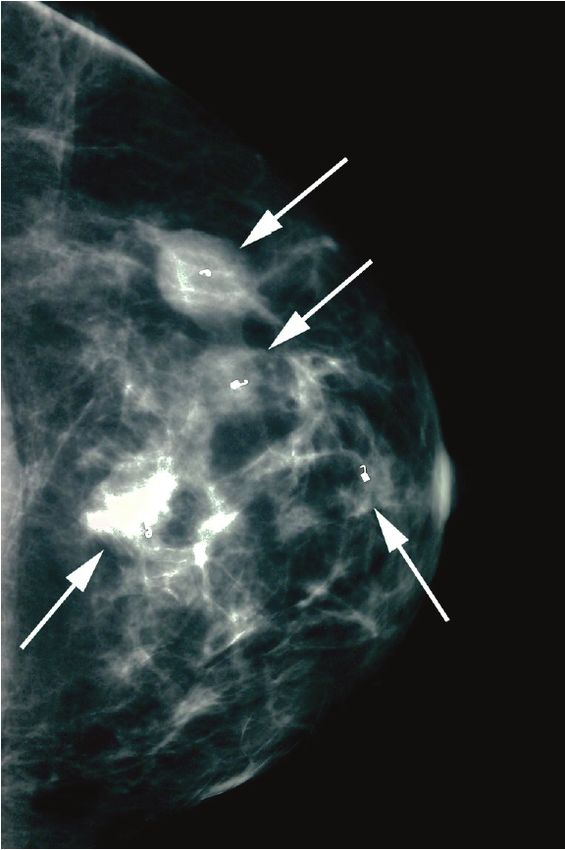
1.61 ng/dL). Mammogram demonstrated four
Case report highly suspicious masses of the left breast
(Figure 1), and ultrasound determined mass
A thirty year-old African American gravida 6, sizes ranging from 0.5 to 2.0 cm in diameter.
para 3 female was referred for evaluation of two Breast MRI confirmed these measurements
lumps in her left breast that she noticed in the (Figure 2). There was no evidence of abnormali-
past month. Her past medical history is only ties of the right breast on physical and radiologi-
significant for Graves’ disease, diagnosed four cal examinations. Additionally, there was no
years prior to her presentation, for which she palpable or radiologically suspicious axillary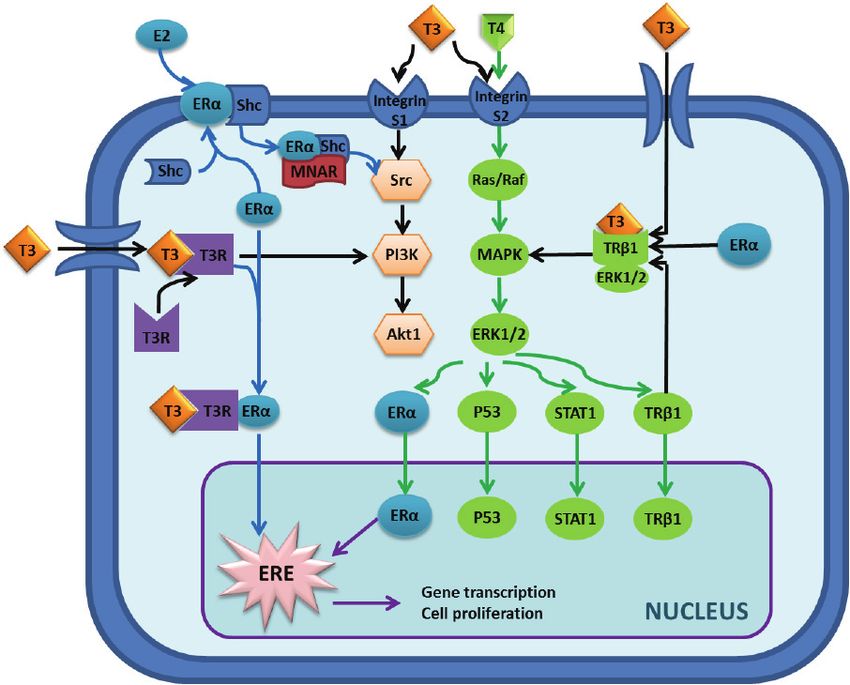
Graves’-associated breast cancer

                                                             lymphadenopathy bilaterally. Core needle biop-
                                                             sies of each of the four left breast masses re-
                                                             vealed infiltrating ductal carcinoma. A unilateral
                                                             radical mastectomy with sentinel node excision
                                                             was performed. The final pathology report dem-
                                                             onstrated a 2.3cm focus of an infiltrating ductal
                                                             carcinoma with 2 histologically identical satellite
                                                             breast lesions measuring 1.4 and 0.8 cm in
                                                             size. Three sentinel nodes were excised, 2 of
                                                             which were positive for micrometastases meas-
                                                             uring 0.4 and 0.5 mm. The overlying skin and
                                                             nipple did not show evidence of malignancy and
                                                             all surgical margins were free of tumor invasion.
                                                             Immunostaining of the excised breast tissue
                                                             was significant for human epidermal growth
                                                             factor receptor 2 (Her2 receptor) positivity. Es-
                                                             trogen and progesterone receptor staining of
                                                             this tissue sample were negative. Ki-67 prolif-
                                                             eration index was quantified at 25%. As of the
                                                             time of submission of manuscript, the patient
                                                             has completed 3 of 6 cycles of treatment with a
                                                             Taxotere, Carboplatin, and Herceptin (TCH) che-
                                                             motherapy regimen. She is expected to carry
                                                             out the remaining 3 cycles of the TCH regimen
                                                             and will be referred for radiation therapy. She
                                                             will also be considered for endocrine therapy.

Figure 1. Patient mammogram demonstrating cranio-            Discussion
caudal view of the left breast with four biopsy mark-
ers in place (arrows). Areas of concern appear whiter        The interaction between thyroid hormone and
than surrounding tissue.                                     estrogen-sensitive tissues, notably the breast,

Figure 2. Craniocaudal view of patient's breast MRI showing two independent focal densities representing malignant
processes in the left breast (arrows). The surrounding benign breast tissue appears dark in comparison.

359                                                                     Int J Clin Exp Med 2012;5(4):358-362
Graves’-associated breast cancer Figure 3. Overview of the molecular interaction between estrogen and thyroid hormone. The interaction can be bro- ken down into nuclear and non-nuclear signaling pathways. Non-nuclear pathway: Binding of estrogen to estrogen receptor α (ERα) can activate the ERα-shc complex, thereby allowing the Modulator of Nongenomic Actions of the Estrogen Receptor (MNAR) to bind and ultimately activate the src tyrosine kinase. Src can then phosphorylate/ activate the Ras/Raf g-protein or phosphoinositol 3-kinase (PI3K) protein to induce cellular proliferation pathways via Akt or Mitogen Activated Protein Kinase (MAPK), respectively. MAPK exerts its effects by phosphorylating/activating Extracellular Signal-Regulated Kinase 1/2 (ERK1/2), which amplifies its effects via a host of other signaling mole- cules—only several of which are illustrated here. At the same time, triiodothyronine (T3) and tetraiodothyronine (T4) are producing similar effects. These two hormones have been shown to stimulate different subunits of the αvβ3 in- tegrin receptor, S1 and S2, which produces the same signal cascade utilized by estrogen involving PI3K and MAPK. T3 also exerts its effects in estrogen-sensitive tissues by translocating into the cell where it can bind to its cytoplas- mic receptor and heteromerize with ERα as shown. From there, this ERα and thyroid receptor complex can either activate MAPK or translocate to the nucleus to exert its transcriptional effects. Nuclear pathway: Once inside the nucleus, the downstream effectors of both thyroid hormone and estrogen (ERα, p53, STAT1, and thyroid hormone receptor β1) can effect transcriptional changes to inhibit apoptosis as well as induce cellular proliferation. Specifi- cally, the activated ERα (which can either be phosphorylated by ERK1/2 or heteromerized with thyroid hormone re- ceptor) can stimulate the Estrogen Response Element (ERE) to alter the transcriptional levels of key proteins involved in cell proliferation and survival. has been well documented [5-8]. The associa- primary breast carcinoma [9]. This suggests that tion between breast cancer in patients with it may be simply the hyperthyroid state that in- autoimmune thyroid disease has also been de- creases the risk for a primary breast malig- scribed in case reports and larger studies [1]. nancy, rather than any underlying autoimmune While autoimmune and hyperthyroid disease thyroid process. states are associated with an increase in the risk of a primary breast cancer, hypothyroidism Poorly controlled levels of either steroid hor- has also be associated with a decreased risk of mone are known to produce malignant changes 360 Int J Clin Exp Med 2012;5(4):358-362
Graves’-associated breast cancer

in a variety of tissue subtypes. At the molecular        Although this patient has a family history of
level, the similar downstream signaling path-            breast cancer in her maternal grandmother, the
ways and heteromeric nature of steroid hor-              authors believe that the underlying pathogene-
mone receptors allow considerable cross-talk             sis of this patient’s breast cancer is not likely to
and may provide an explanation for patients              be related to the pathogenesis of her maternal
with Graves’-associated breast changes—as well           grandmother’s cancer due to the vast difference
as an explanation for estrogen-associated thy-           in age of cancer onset. Without adequate sup-
roid changes (Figure 3). It is known that estro-         pression of our patient’s hyperthyroidism, her
gen and thyroid hormones activate the same               early-onset multifocal breast cancer may have
mitogen-activated protein kinase (MAPK) and              resulted from the estrogenic effects of thyroid
other non-nuclear signaling pathways [7]. Alone,         hormone on proliferating breast tissue or from
this MAPK and its associated extracellular sig-          some other, less understood autoimmune phe-
nal-regulated kinase 1/2 (ERK 1/2) already mo-           nomenon. Despite the fact that immunostaining
bilize the cytoplasmic machinery necessary to            for estrogen receptor was negative in our pa-
stimulate cell growth and division. Additionally,        tient’s breast tissue specimen, alternative
triiodothyronine (T3) has been shown to induce           mechanisms can cause this, as discussed
a cascade of events which ultimately upregulate          above, in which thyroid hormone may induce
estrogen response element (ERE)-mediated                 downstream signals similar to the way estrogen
gene transcription in breast cancer cells dose-          signaling has been shown to modulate breast
dependently [5, 10]. Even in physiologically nor-        cancer. Whether the malignant changes in our
mal breast tissue, ERE-mediated gene transcrip-          patient began prior to her diagnosis of Graves’
tion is responsible for the majority of cell growth      disease or during the course of her treatment
and division. The response of breast cancer              cannot be determined. Perhaps earlier detec-
cells to T3 from these studies was also ob-              tion of her thyroid disease might have deterred
served to be attenuated with the co-                     this aberrant breast process. It is imperative
administration of an estrogen antagonist. These          that physicians recognize the clinical signifi-
data suggest the role of T3 as a competitive             cance of the interactions between thyroid hor-
inhibitor of the estrogen receptor. By an alterna-       mone and estrogen in these hormonally-
tive mechanism, T3 and T4 are also thought to            responsive tissues.
stimulate the αvβ3 integrin receptor, and ulti-
mately active the downstream ERα [6]. Although           Address correspondence to: Dr. Emad Kandil, Divi-
each of these results presented here are limited         sion of Endocrine and Oncological Surgery, Depart-
by the fact that they were demonstrated only in          ment of Surgery, Tulane University School of Medi-
estrogen receptor-positive cell lines, they may          cine, 1430 Tulane Ave. SL-22, New Orleans, LA
                                                         70112 Tel: (504) 988-7407; E-mail:
explain the increased risk of breast cancer in           Ekandil@tulane.edu
patients with poorly controlled hyperthyroid dis-
ease (perhaps even in estrogen receptor-                 References
negative primary breast cancers). If one consid-
ers all these points of interaction, it is likely that   [1]   Giani C, Fierabracci P, Bonacci R, Gigliotti A,
there is no singular mechanism explaining the                  Campani D, De Negri F, Cecchetti D, Martino E
interaction between thyroid hormone and estro-                 and Pinchera A. Relationship between breast
gen, but that there are multiple points of over-               cancer and thyroid disease: relevance of auto-
lap in their signaling pathways. Because of such               immune thyroid disorders in breast malignancy.
significant overlap, much of which cannot be                   J Clin Endocrinol Metab 1996; 81: 990-994.
discussed in a manuscript of this length, there          [2]   Munoz JM, Gorman CA, Elveback LR and Wentz
may be a role for thyroid hormone in the patho-                JR. Incidence of malignant neoplasms of all
                                                               types of patients with Graves' disease. Arch
genesis of estrogen receptor-negative breast                   Intern Med 1978; 138: 944-947.
cancer. This area of clinical research deserves          [3]   Radical mastectomy in a patient with coexistent
further investigation.                                         Graves' disease. Am J Surg 1976; 132: 110-
                                                               111.
Conclusion                                               [4]   Ito K and Maruchi N. Breast cancer in patients
                                                               with Hashimoto's thyroiditis. Lancet 1975; 2:
We propose that the development of a multifo-                  1119-1121.
cal breast cancer in our patient is likely an ad-        [5]   Hall LC, Salazar EP, Kane SR and Liu N. Effects
verse sequela of her history of Graves’ disease.               of thyroid hormones on human breast cancer
                                                               cell proliferation. J Steroid Biochem Mol Biol

361                                                                Int J Clin Exp Med 2012;5(4):358-362
Graves’-associated breast cancer
2008; 109: 57-66. [9] Cristofanilli M, Yamamura Y, Kau SW, Bevers T,
[6] Tang HY, Lin HY, Zhang S, Davis FB and Davis Strom S, Patangan M, Hsu L, Krishnamurthy S,
PJ. Thyroid hormone causes mitogen-activated Theriault RL and Hortobagyi GN. Thyroid hor-
protein kinase-dependent phosphorylation of mone and breast carcinoma. Primary hypothy-
the nuclear estrogen receptor. Endocrinology roidism is associated with a reduced incidence
2004; 145: 3265-3272. of primary breast carcinoma. Cancer 2005;
[7] Dinda S, Sanchez A and Moudgil V. Estrogen- 103: 1122-1128.
like effects of thyroid hormone on the regula- [10] Nogueira CR and Brentani MM. Triiodothyron-
tion of tumor suppressor proteins, p53 and ine mimics the effects of estrogen in breast
retinoblastoma, in breast cancer cells. Onco- cancer cell lines. J Steroid Biochem Mol Biol
gene 2002; 21: 761-768. 1996; 59: 271-279.
[8] Zhang X, Jeyakumar M and Bagchi MK. Ligand-
dependent cross-talk between steroid and thy-
roid hormone receptors. Evidence for common
transcriptional coactivator(s). J Biol Chem
1996; 271: 14825-14833.
362 Int J Clin Exp Med 2012;5(4):358-362You can also read