TUMOR MAPPERS THE - Ludwig Cancer Research
←
→
Page content transcription
If your browser does not render page correctly, please read the page content below
THE
     TUMOR MAPPERS
     The unique cooperative research model of the Ludwig Center
     at Harvard is being productively harnessed by the
     Tumor Atlas Project, an ambitious effort to create
     high-dimensional maps of any and all tumors.

     When Peter Sorger set out to develop a              progression and response to therapies. It
     method for mapping the different cells in           is also a sort of technological avatar of an
     tumors, he didn’t expect actual mapmakers           idea central to the structure of the Ludwig
     would be involved. So when members of               Center at Harvard: to bring together diverse
     Harvard’s Department of Architecture                biomedical disciplines and their associated
     approached him one day following a                  technologies to tackle the most intractable
     presentation, Sorger was surprised. “They           problems of cancer research and care.
     said, ‘That’s really cool. Let’s work together,’”
     recalls Sorger, an investigator at the Ludwig       It takes a village
     Center at Harvard and professor of systems          It’s no coincidence that TAP originated at
     pharmacology at Harvard Medical School.             Ludwig Harvard, which has a special focus
     “Unknown to me, Harvard was the place               on drug resistance in cancer. “The Tumor
     where the initial GIS”—geographic information       Atlas Project fits into every single project
     system—“was developed back in the 1950s.”           we have,” Ludwig Harvard Co-director Joan
                                                         Brugge observes. “The technology makes
     The Harvard cartographers’ expertise would          it feasible to follow many different proteins
     prove useful for organizing and visualizing         in real human tumors, which is key to an
     the flood of tumor data that the Ludwig             understanding of the state of individual
     Tumor Atlas Project (TAP), led by Sorger,           cells in tumor tissue prior to and after drug
     was generating. Launched in January 2019            treatment.”
     with funding from Ludwig Cancer Research,
     TAP aims to develop a multi-dimensional             TAP, Sorger expects, will not only help
     “map” that captures the locations and               transform our understanding of cancer
     identities of not just cancer cells but also        biology but drive innovations in diagnostic
     the noncancerous immune and supporting              pathology as well. The first phase of the
     cells that contribute to tumor evolution,           project will map tumor cells, unraveling their
56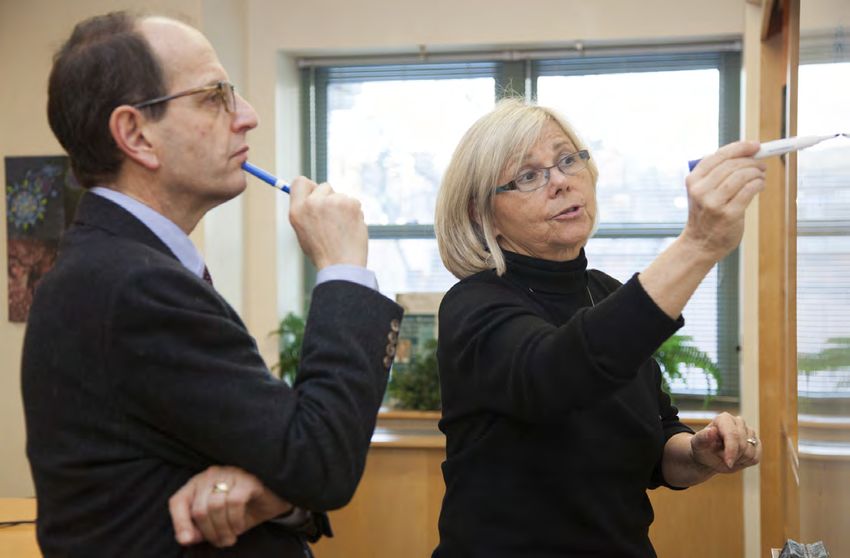
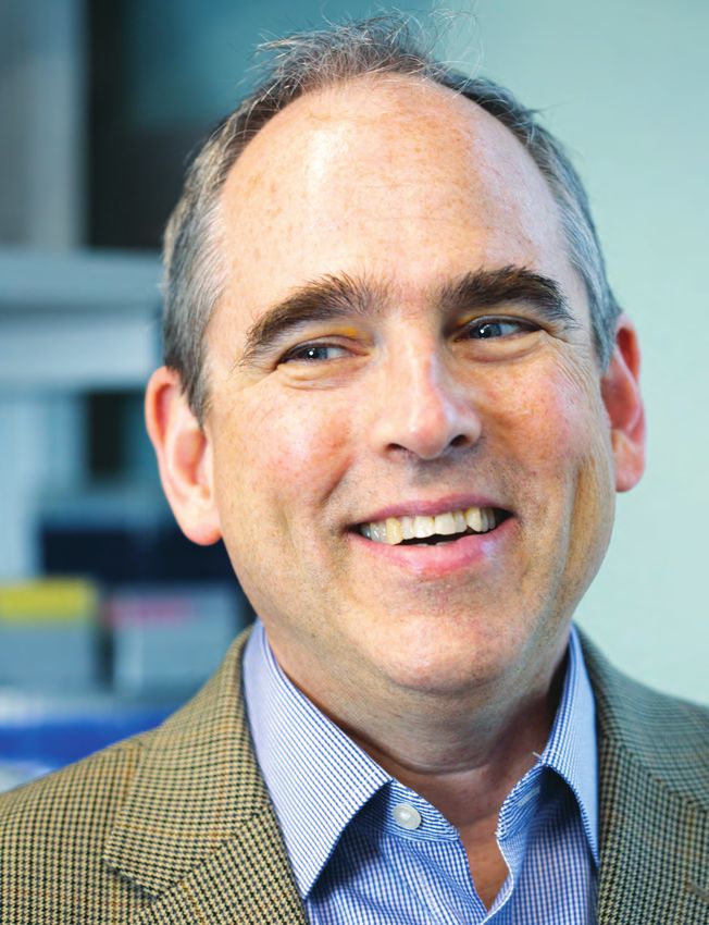
PETER
SORGER
LUDWIG
HARVARD

          Photo by Flynn Larsen
                            57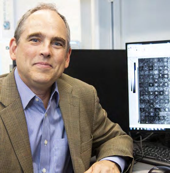
“The technology makes                                  information systems specialists.

it feasible to follow many
                                                       “The foundational technology that underlies
                                                       modern digital maps is conceptually
                                                       applicable to our Atlas,” Sorger says. “On our
different proteins in real                             website, you can zoom in and out on millions
                                                       of tumor cells from different diseases. The
human tumors, which is                                 technology behind that is the same one used
                                                       in Google Earth.”
key to an understanding                                Community building
of the state of individual                             Ludwig Harvard’s model was forged in the
                                                       earliest days of its establishment, when

cells in tumor tissue                                  Brugge and Demetri were appointed its co-
                                                       directors and had to decide how to distribute

prior to and after drug
                                                       the annual interest of the $90 million
                                                       endowment from Ludwig Cancer Research.
                                                       “George and I were in sync from the very
treatment.”                                            beginning,” says Brugge, whose own thinking
                                                       was influenced by her experience co-
                                                       founding a biotech company. “I saw how well
                                                       it can work when you have multiple people
                                                       with different expertise coming together to
                                                       help solve a problem.”

                                                       With the new funding, the co-directors
       interactions with supporting noncancerous       saw an opportunity to build a truly
       cells and immune cells, and pin down the cell   multidisciplinary model for cancer research.
       signaling pathways involved in driving tumor    “What we wanted to do was to bring the other
       growth and drug resistance. The second,         people who are really interested in a given
       Sorger says, will deploy machine vision,        problem from multiple areas of science, and
       artificial intelligence and multi-dimensional   then together develop the strategy to attack
       visualization to combine data from many         the problem, so that from the very beginning,
       specimens, facilitate expert annotation by      we would be functioning as a unit.”
       human pathologists and develop algorithms
       for predicting the responses of individual      In practice, this means that every research
       patients to specific therapies.                 team that is part of Ludwig Harvard receives
                                                       about $150,000 in seed funding annually to
       The technology benefits from a unique           pursue its research. This has helped forge
       “cooperative research model” that Brugge        a community, says Demetri, who is also the
       and Co-director George Demetri have             associate director for clinical sciences at the
       implemented at Ludwig Harvard. That             Dana-Farber/Harvard Cancer Center.
       model seeks to bring together researchers
       from multiple disciplines at the outset of      “Our pitch to faculty was, ‘If you join our
       every inquiry. The framework is vital to        community, we will have the ability to come
       TAP, which relies on contributions from         up with new ideas, intersect in different
       not just oncologists and pathologists but       ways, and provide seed money to get great
       also software developers, computational         multi-institutional, multi-investigator grants
       biologists and, of course, geographic           going forward,’” Demetri says. “Did we get
58
Photo by Flynn Larsen
Ludwig Harvard Co-directors George Demetri and Joan Brugge.
pushback? You bet we did. But in the end it says the meeting had a strong influence on
worked. Remember, this was right around her as a young scientist. “I remember sitting
the time when team science was starting there in awe and watching and listening to all
to catch fire. People were realizing that the of these senior and junior people just talking
translation from basic science to patients is about science together,” recalls Guerriero,
too complicated for any one person, and we who now directs the Breast Immunology
need to figure out how to work together.” Laboratory in the Women’s Cancer Program
at the Dana-Farber Cancer Institute. She
Brugge and Demetri also implemented a too has become a key part of the TAP and
weekly Monday meeting to which anyone tCyCIF team, with a special focus on the
associated with Ludwig Harvard research— roles of macrophages in chemotherapy and
from principal investigators to postdocs, immunotherapy.
graduate students, and clinicians—is
invited. The “Ludwig Monday meetings,” as The effect Guerriero described was by
they’ve come to be known, are a chance for design. “In some ways, it felt like part of
researchers from different disciplines to what we did was introduce people to other
come together to learn what their colleagues people,” says Demetri. “It was like a junior
are working on and determine how their high school dance, where the basic scientists
projects might intersect. were on that side of the room and the clinical
scientists were on the other side, and the two
Jennifer Guerriero, who has been attending groups were too shy or unable to talk to each
the weekly gathering since her postdoc days, other.”
59The result is a remarkably rich and nuanced
picture of tumors. “Before, we had a very
unidimensional view of individual cells,” says
Sandro Santagata, an investigator at the
Ludwig Center at Harvard, co-leader of the
TAP and an associate professor of pathology at
Brigham and Women’s Hospital. “Now we can
not only spot an immune cell, but determine
specifically which type it is and define the
functional state that it’s in, and then compare
it to a slightly different immune cell that
occupies a different space. Not only do you
get to really probe deeply the identity and the
properties of individual cells, now you also get
to see how they interface with each another.”
tCyCIF at work
tCyCIF is designed to use the kinds of
standard biopsy samples that hospitals
and researchers have been collecting from
Photo by Flynn Larsen
patients for nearly a century (it also works
with mouse models of cancer). “Our goal was
to hack directly into the standardized clinical
E pluribus unum workflow,” says Sorger, who is also a principal
At the heart of TAP is a method dubbed investigator in the U.S. National Cancer
tissue-based cyclic immunofluorescence, Institute’s Human Tumor Atlas Network, for
or tCyCIF, which is being developed at which he is mapping premalignant tissues
Ludwig Harvard under Sorger’s direction. associated with certain skin and blood
tCyCIF allows researchers to obtain images cancers. “We wanted to develop a method
containing multiple layers of protein that allows us to get deep molecular insights
information about tumors—including their from a sample that is collected from virtually
cancer cells and their associated immune every single cancer patient.” This continuity
and other noncancerous cells—at subcellular means that tumor samples collected in
resolution. It combines the output of completed clinical trials can still be analyzed,
multiple existing instruments and reagents as can accumulated samples of rare cancers
into a workflow that can scan a tissue collected over the decades.
sample dozens of times without damaging
its constituent cells. Each scan looks for Sorger envisions tCyCIF as a complement
three to five different protein markers. rather than a competitor to other cell-
When compiled, the information generates screening technologies. For example, single
a composite image of a tumor constructed cell RNA sequencing can provide a wealth of
from 40 to 60 “channels” of information. information on the gene expression profile of
individual cells. “You get detailed information
Sorger likens the process to getting to know on individual cells, but you get no information
a stranger by asking many simple yes and about their locale,” Sorger says. By contrast,
no questions. “But instead of asking just one while tCyCIF tracks only a few dozen proteins,
stranger, imagine asking these questions to a it can interrogate many square centimeters
stadium full of people simultaneously. tCyCIF of tissue—hundreds of thousands of
is very much like that,” he explains. individual cells—and determine the precise
60morphologies of cells and their spatial
relationships to each other. Combining the “Our goal was to
hack directly into the
results of RNA sequencing and other large
scale, or “omics”, assays with maps generated
by tCyCIF is a central goal of TAP.
standardized clinical
To help people expand on TAP using research
data from their own labs, Ludwig Harvard
is placing details about tCyCIF and TAP
workflow. We wanted to
in the public domain. “The patient data is
anonymized, but all of the data and any
develop a method that
insights we glean from it will be publicly
accessible,” Sorger says. “We want to make
allows us to get deep
the data and the code freely available to
the Ludwig community to demystify and molecular insights from a
sample that is collected
democratize high dimensional histology.”
Currently, TAP consists of dozens of images
of six types of tumors—including triple- from virtually every single
negative breast cancer, ovarian cancer, and
acute myeloid leukemia—but Sorger envisions
the project growing to encompass a greater
cancer patient.”
variety of cancers as other centers and
hospitals contribute samples. A key step will
be combining image data from many different Ludwig Harvard team have travelled to
patient specimens into general-purpose Ludwig Lausanne to initiate a collaboration
maps. It is not yet clear how this will be on ovarian organoid cultures. As another
accomplished, and Sorger and Santagata look step toward more open collaboration, the
forward to investigators from many Ludwig Ludwig Center at Harvard and the Lausanne
Centers becoming involved. Branch are experimenting with a workflow in
which tumor samples scanned with tCyCIF
A higher order experiment at one center are analyzed with software
The idea for TAP was partly inspired by at the other. Work on Barrett’s esophagus
Ludwig Harvard’s cooperative research with Ludwig Oxford is being planned as well,
model, and Demetri thinks the project and Ludwig Harvard is working closely with
could be a vehicle for disseminating the Ludwig MIT to apply t-CyCIF to mouse models
model to other centers. “I see it as a social of cancer.
experiment,” Demetri says. “Can we use it as
a testing ground to see how we can all work “We envision interactions with additional
better together? Everybody wants this kind of Ludwig Centers and Branches, either
information, and there are whole companies through direct collaboration or the transfer
being formed to do this, but if we can do it of technologies and methods,” Sorger says.
academically and openly distribute the tools “We’re comfortable with either approach.
and the data to people, we will engender trust Within a few years we hope that this grows
and enable people to ask better questions beyond a technical collaboration into a
and get answers faster.” Ludwig-wide effort to efficiently ask and
answer questions about shared data on drug
All tissue samples for TAP have so far come resistance and the prospects for improving
from U.S. hospitals, but members of the therapeutic responses.”
61You can also read