Morphotype diversity of Prorocentrum lima in the western part of Indonesian waters - Smujo
←
→
Page content transcription
If your browser does not render page correctly, please read the page content below
B I O D I V E R S IT A S ISSN: 1412-033X
Volume 22, Number 2, February 2021 E-ISSN: 2085-4722
Pages: 607-614 DOI: 10.13057/biodiv/d220212
Morphotype diversity of Prorocentrum lima in the western part of
Indonesian waters
RIANI WIDIARTI, NEVIATY PUTRI ZAMANI, DIETRIECH GEOFFREY BENGEN, HAWIS MADDUPPA
Graduate School of Marine Science, Department of Marine Science and Technology, Faculty of Fisheries and Marine Sciences, Institut Pertanian Bogor.
Jl. Rasamala, Kampus IPB Darmaga, Bogor 16680, West Java, Indonesia. Tel./fax.: +62-251-8623644, email: neviaty@apps.ipb.ac.id
Manuscript received: 21 November 2020. Revision accepted: 9 January 2021.
Abstract. Widiarti R, Zamani NP, Bengen DG, Madduppa H. 2021. Morphotype diversity of Prorocentrum lima in the western part of
Indonesian waters. Biodiversitas 22: 607-614. Prorocentrum lima is one of the toxic benthic dinoflagellates, known to produce various
toxins, including okadaic acid and dinophysis toxins. The species have a wide range of morphological variability, and possess morphotype
diversity, which makes it essential to have detailed morphology observation for identification and other purposes. However, such
comprehensive observation has never been reported from Indonesian waters. This study aims to determine the morphological characteristics
(including pore size and number of pores) of P. lima morphotypes, in the western part of Indonesian waters (Bintan Island, Belitung Island,
Seribu Islands, and Karimunjawa Islands). The results showed three different and unique morphotypes of P. lima, namely morphotype 1,
morphotype 2, and morphotype 3. Three clusters were presented by Cluster Analysis, corresponded to the three morphotypes, which were
Belitung Island clusters, Seribu Islands and Karimunjawa Islands cluster, and Bintan Island cluster, respectively. These findings support
distribution of P. lima and its potential risk of toxicity in Indonesian waters, which prompts the necessity of conducting future research, to
avoid the negative impact.
Keywords: Cluster Analysis, morphological variability, morphology characters, reef islands, toxic dinoflagellates
INTRODUCTION city beach waters (Dwivayana 2015; Eboni et al. 2015;
Oktavian et al. 2017; Seygita et al. 2015), North Lombok
Some benthic dinoflagellates produce toxic substances, (Gili Meno and Gili Air) (Widiarti et al. 2016a), South
which causes several poisonous syndromes experienced by Lampung waters (Pahawang Besar Island and Kelagian
humans or other mammals, which commonly occur after Kecil Island) (Widiarti and Adi 2016), and Weh Island
the consumption of various seafood products. waters-North Aceh (Rubiah Island) (Widiarti et al. 2016b).
Approximately nine species of Prorocentrum have been However, no detailed observation on cell surface
known to produce okadaic acid (OA) and dinophysis toxins morphology has been recorded.
(DTX’s) (Hoppenrath et al. 2013; Hoppenrath et al. 2014), Prorocentrum lima has a wide range of morphological
which causes Diarrhetic Shellfish Poisoning (DSP). variability, and possesses morphotype diversity
Prorocentrum lima is one of the benthic dinoflagellates, (Hoppenrath 2013; Zhang et al. 2015; Chomérat et al.
known to produce those various toxins (Nishimura et al. 2018). The morphotype diversity of P. lima has been
2019). Furthermore, P. lima associated with other toxic revealed by several studies, such as in South China Sea
benthic dinoflagellates, Gamberdiscus toxicus, also causes waters (Zhang et al. 2015) and Caribbean Sea waters
Ciguatera Fish Poisoning (CFP) in tropical areas (Chomérat et al. 2018) (Table 1). Zhang et al. (2015),
(Burkholder 1998; Lehane and Lewis 2000). showed the cell shape, cell length and width, ratio length
Prorocentrum cells show similar shape and size, and width (ratio L/W), pore shape, and the number of
making it necessary to have detailed feature observations of pores. Similarly, Chomérat et al. (2018) showed cell shape,
cell surface morphology, for identification and cell length and width, ratio L/W, and pore shape and
classification purposes (Faust 1990). Prorocentrum has dimension. However, the studies lacked information on
been reported in Indonesian waters. For instance, in Seribu both pore dimension and number of pores, which should
Islands (Penjaliran Barat Island, Pramuka Island, Panggang have been used to complete the database on morphotype
Island, Semak Daun Island, Pari Island, Air Island and diversity of P. lima. The morphotype diversity of P. lima,
Tidung Island) (Widiarti 2002; Widiarti 2011; Widiarti and has never been reported in Indonesian waters. Therefore,
Pudjiarto 2015), Belitung Island (Buyut Island, Kelayang this research is being conducted to determine the
Cape, and Keran Island) (Widiarti 2010), Bali waters morphological characteristics (including pore size and
(Kuta, Sanur, and Nusa Dua), Lombok (Gili Trawangan) number of pores) of P. lima morphotype, from several sites
(Skinner et al. 2011), west coast of South Sumatera and in the western part of Indonesian waters: Bintan Island,
Bintan Island coast-Riau Islands (Thamrin 2014), Padang Belitung Island, Seribu Islands, and Karimunjawa Islands.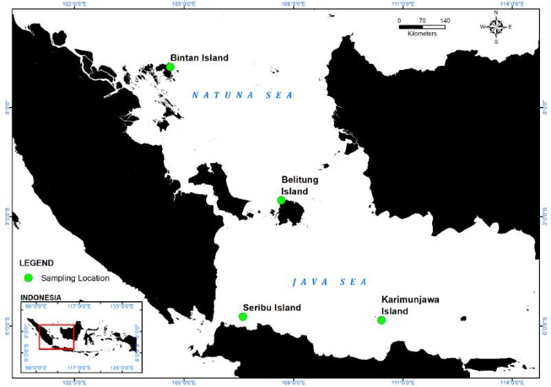
608                                      B I O D I V E R S I T A S 22 (2): 607-614, February 2021

Table 1. Prorocentrum lima characters by previous studies

                                                                                Results
       Characters
                                South China Sea Waters (Zhang et al. 2015)              Caribbean Sea Waters (Chomérat et al. 2018)
Cell Shape                     Ovate, broadly ovate, oblong oval, broadly oblong                    Oblong oval, broadly ovate
Cells Length (µm)                                   32-48.1                                                 31.1-38.2
Cells Width (µm)                                   25.2-38.2                                                27.8-32.3
Ratio L/W                                          1.06-1.61                                                1.10-1.36
Pore Size (µm)                                         -                                                     0.4-0.8
Number of Pores                                      28-94                                                      -
(valve and marginal pores)
Pore Shape                               round/ oblong/ kidney-shaped                               round/ oval/ kidney-shaped

                                                                      macroalgae growth, where the toxic dinoflagellates are
                                                                      more likely to attach (deSylva 1994; Lehane and Lewis
             MATERIALS AND METHODS                                    2000). The coral reefs in all four locations have already
                                                                      been disrupted by domestic, tourism, and local fishery
Study area                                                            activities (Estradivari et al. 2009; Yusuf 2013; Susetiono et
    Sampling was conducted from April to September 2018               al. 2016), so that the macroalgae were also found
in four islands, located in the western part of Indonesian            abundantly in the areas. Sampling locations were situated
waters, including Bintan Island, Belitung Island, Seribu              on the eastern coast of Bintan Island, the northern coast of
Islands, and Karimunjawa Islands (Figure 1). The sampling             Belitung Island, Pramuka Island on Seribu Islands, and
locations focused on coral reefs with poor conditions,                Karimunjawa Island on Karimunjawa Islands.
because it potentially provides a new surface for various

Figure 1. Sampling locations in Bintan Island (Station II), Belitung Island (Station IV), Seribu Islands (Station I), and Karimunjawa
Islands (Station III)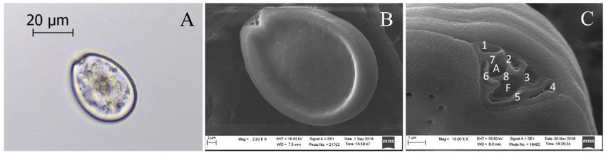
WIDIARTI et al. – Morphotype diversity of Prorocentrum lima                              609

Procedures                                                      Environmental-factors-measurement
Data-collection                                                     During sample collections within the four locations,
     The macroalgae collected, were only limited to             measurements of the environmental condition were also
Sargassum and Padina. As has been observed by Widiarti          conducted. These measurements included, salinity by
(2002), benthic dinoflagellates are commonly found              refractometer (ATAGO), water temperature and dissolved
attached to both macroalgae. Macroalgae's thallus was           oxygen by DO-meter (HANNA), light intensity by
randomly harvested on the reef flat areas with 45 cm-100        luxmeter (LX-1010B), water acidity by pH indicator
cm depth, and placed inside wide-mouthed plastic bottles        (MColorpHast 6.5-10), water current by current meter, with
containing ambient seawater (Tester et al. 2014). The           nitrate and phosphate using APHA Analysis Method
whole process was conducted underwater (Tester et al.           (2012) in the laboratory.
2014; Jauzei et al. 2018), to avoid sample disruptions due
to air and sunlight exposures.                                  Data analysis
     After collection, plastic bottles containing macroalgae        Morphological character data were tabulated, and
and seawater were shaken using vortex machine (1250 rpm         compared with morphotypes grouping suggested by Zhang
for 1 minute), to detach benthic dinoflagellates from the       et al. (2015). Character similarity of each Operational
surface of macroalgae. Furthermore, the samples were            Taxonomic Units (OTU) was analyzed by cluster analysis
filtered through a series of sieves with a mesh size of 125     methods, with the use of Hierarchical Cluster Analysis,
and 20 µm, to separate samples from sediments and other         which was further processed by R version 3.6.2. The
larger organisms. For morphological observation using LM        cluster analysis results were presented in the form of a
and SEM, samples were fixed using glutaraldehyde to             dendrogram (treelike diagram) (Sneath 2005).
reach the final concentration of 4% (v/v).

Morphological-character-observation                                           RESULTS AND DISCUSSION
    Cell shape and size were observed, by using a light
microscope (LM) (Leica DM 500). The fixed cells were                All samples collected from the four sampling locations,
isolated using modified Pasteur pipette, dripped onto the       showed that Prorocentrum lima cells possessed
object-glass, and sealed with a cover glass. The slides were    ovoid/ovate shape, visible pyrenoid at the center of the cell,
then observed under a microscope with 200 magnification.        scattered pores on both valves (except in the center of the
Furthermore, cell length and width were measured                cell), and V-shaped periflagellar areas with 8 platelet plates
digitally, using LEICA LAS EZ 2.0.                              (Figure 2).
    Cell pores (shape, size and the number of pores) and            Based on the observation using LM, the length and
periflagellar area were observed, using Scanning Electron       width of the cells ranged from 35.49-44.94 µm (mean
Microscope (SEM) (ZEISS with EVOIMA10 type). The                38.27 µm, s.d 2.77 µm, n = 20), and 25.57-31.56 µm (mean
isolated cells were dripped onto 2 x 1 cm2 filter paper         28.63 µm, s.d. 2.05 µm, n = 20), with the ratio L/W being
(Whatman 125 mm, pore size 3 µm), which was already             1.17-1.44 (mean 1.34, s.d 0.09, n = 20). Furthermore, based
placed on top of the object-glass covered with carbon tape.     on the observation using SEM, the pore length and width
The samples were air-dried and coated with gold (Au),           ranged from 0.21-0.40 µm (mean 0.30 µm, s.d 0.05 µm, n
using Sputter Coater (Quorum type Q150R ES), with 20            = 20), and 0.15-0.33 µm (mean 0.21 µm, s.d 0.05 µm, n =
(mA) Sputter Current and 60 second Sputter Time. After          20). The number of pores observed was around 40-68
the coating process, SEM samples were analyzed, and the         (mean 50.3, s.d 9.2, n = 20), with the valve shape being
images were taken using the SE (Secondary Electron)             round to kidney-shaped (Table 2). Due to the unclear
detector, with 8.0 mm Working Distance (WD) and EHT             images obtained by SEM, the observation of pores in this
16.00 kV. Pore length and width measurements were               study was based only on valve pores.
obtained using biometric program tpsUtil32 dan tpsDig232.

                                     A                                         B                                         C

Figure 2. Morphology characters of Prorocentrum lima using LM and SEM. A. Cell shape, pyrenoid in the center of the cell; B.
Scattered pores except in the center of the cell; C. V-shaped periflagellar area with eight platelets
610                                      B I O D I V E R S I T A S 22 (2): 607-614, February 2021

Table 2. Morphology characters of Prorocentrum lima from all sampling locations (n = 20)

                                                                            Sampling locations
          Characters
                                        Seribu Islands              Bintan Island       Karimunjawa Islands         Belitung Island
Cell Length (µm)                         36.05-41.16                 40.26-44.94             35.81-37.30             35.49-37.79
Cell Width (µm)                          26.32-30.05                 29.45-31.56             25.57-27.17             28.86-31.51
Ratio L/W                                 1.33-1.42                   1.34-1.42                1.35-1.44               1.17-1.23
Pore Length (µm)                          0.29-0.40                   0.28-0.37                0.23-0.31               0.21-0.38
Pore Width (µm)                           0.23-0.33                   0.19-0.21                0.15-0.19               0.16-0.32
Number of Pores (Valve Pores)               40-48                       51-68                    42-53                   48-59
Pore Shape                           round/kidney-shaped         round/kidney-shaped     round/kidney-shaped             round

    Further examination using Cluster Analysis, showed                possessed high values of pore dimensions (0.29-0.40 µm in
that the optimal number of the cluster could be obtained              length and 0.23-0.33 µm in width), while showing low
through the use of a Scree and Silhouette Plot. There were            mean values of both characters, based on Cluster Analysis.
three optimal numbers of the cluster, showed by the elbow             These occurred as a mean result of pore dimensions
and peak points at the Scree and Silhouette Plots,                    between values from Seribu Islands and from the
respectively (both showed cluster k = 3 at axes 1). Cluster           Karimunjawa Islands, which possessed the lowest values
Analysis showed three clusters, where cluster 1 consists of           from all sampling locations (0.23-0.31 µm in length and
four members, cluster 2 consists of ten members, and                  0.15-0.19 µm in width) (Table 3).
cluster 3 consists of six members. Cluster 1 represented by               Cluster 3 represented by I4, II1, II2, II3, II4 and II5,
I3, IV1, IV4 and IV5, was described as one P. lima                    was described as one P. lima specimens at station 1 and
specimens at station 1 (Seribu Islands), and three                    five P. lima specimens at station 2 (Seribu Islands and
specimens at station 4 (Belitung Island). Based on the                Bintan Island), respectively. Based on the cluster
cluster dendrogram, three specimens from Belitung Island              dendrogram, four specimens from Bintan Island, were
were closely related to each other (Figure 3). Cluster 1 was          closely related to each other (Figure 3). Cluster 3 was
characterized by a low mean value of ratio L/W. Specimens             characterized by a high mean value of ratio L/W, and the
of P. lima obtained from Belitung Island possessed the                number of pores. In this research, P. lima specimens from
lowest ratio L/W values compared to other sampling                    Bintan Island possessed the highest pore number (51-68),
locations (Table 2).                                                  compared to other sampling locations (Table 3). In
    Cluster 2 represented by I1, I2, I5, III1, III2, III3, III4,      previous studies, different pore shapes were frequently
III5, IV2 and IV3, which were described as three P. lima              observed in one cell (Hoppenrath et al. 2013; Zhang et al.
specimens at station 1 (Seribu Islands), all specimens at             2015), same as this study where both round and kidney-
station 3 (Karimunjawa Islands) and two specimens at                  shaped pores, were also found in one cell. Pore shape
station 4 (Belitung Island). Based on the cluster                     character was not used as a variable in the Cluster
dendrogram (Fig 3), three specimens from the Seribu                   Analysis.
Islands were closely related to all specimens from                        Environmental parameters data in four different
Karimunjawa Islands. At cluster 2, variables III1, III2, III3,        sampling locations showed, salinity values ranged from 30-
III4 and III5 possessed closer distance, compared to other            34.7‰, temperature from 27.5-30.8°C, dissolved oxygen
members of the cluster, which is showed by the shorter                (DO) from 7.6-13.5 ppm, light intensity from 1392-46983
height of Euclidian distance at axes 2. Cluster 2 was                 lux, acidity from 7.1-7.8, water current from 0.039-0.115
characterized by the lowest mean values of cell length                ms-1, nitrate from 0.087-0.101 mg.L-1 and phosphate from
(36.4040), cell width (26.9190), pore length (0.2740), and            0.002-0.009 mg.L-1 (Table 4).
pore width (0.1920). The P. lima cells in Seribu Islands

Figure 3. Closely related groups at each cluster based on Euclidian distance. A. Cluster 2, Seribu Islands and Karimunjawa Islands; B.
Cluster 3, Bintan Island; C. Cluster 1, Belitung Island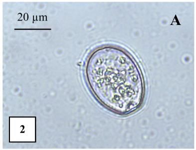
WIDIARTI et al. – Morphotype diversity of Prorocentrum lima                                   611

Table 3. Mean values of morphology characteristic from each cluster (n = 20)

   Group            Cell Length        Cell Width         Ratio L/W            Pore Length        Pore Width         Pore Number
   1 (n = 4)          37.5175           30.66500           1.225000              0.3475            0.2775000              49.25
  2 (n = 10)          36.4040           26.91900           1.356000              0.2740            0.1920000              45.20
   3 (n = 4)          41.8750           30.12667           1.388333              0.3150            0.2066667              59.50

Table 4. The average values of environmental factors measurement at four sampling stations

                                                Temperature        DO      Light             Water current      Nitrate   Phosphate
               Location              Salinity                                       pH
                                                   (°C)           (ppm)     (lux)               (ms-1)         (mg.L-1)    (mg.L-1)
Seribu Islands (Station I)             30.0        27.5            13.4     1392    7.8         0.048            0.094      0.002
Bintan Island (Station II)             32.0        30.5             7.6    37270    7.1         0.039            0.087      0.009
Karimunjawa Islands (Station III)      34.7        30.8            11.7    30850    7.2         0.115            0.101      0.003
Belitung Island (Station IV)           34.0        29.6            13.5    46983    7.2         0.055            0.098      0.003

    The cells of P. lima have specific morphological                Seribu Islands
characters, which are generally used to differentiate this              Observation on P. lima cells obtained in Seribu Islands
Prorocentrum species from others. Those characters were             waters showed ovate shape, cell length and width ranging
symmetric cells with oblong oval, ovoid (Hoppenrath et al.          from 36.05-41.16 µm (mean 38.39 µm, s.d. 2.17 µm, n = 5)
2013) or ovate shape (Zhang et al. 2015), visible pyrenoid          and 26.32-30.05 µm (mean 27.90 µm, s.d. 1.58 µm, n = 5),
in the center of the cell, scattered pores on both valves but       respectively, with the ratio L/W from 1.33-1.42 (mean
void in the center of the cell (Fukuyo 1981; Faust 1990),           1.38, s.d. 0.04, n = 5). The shape and size were within the
and wide V-shaped periflagellar areas consisting of 8               range of P. lima "morphotype 2" by Zhang et al. (2015),
platelet plates (Hoppenrath et al. 2013; Zhang et al. 2015).        with cell length and width ranging from 36.8-40.0 µm, and
In this study, observation using LM or SEM showed that              25.6-28.9 µm, respectively, and the ratio L/W at 1.33-1.45
benthic dinoflagellates obtained from Bintan Island,                (Table 2, Figure 4.1). However, observation on pores
Belitung Island, Seribu Islands, and Karimunjawa Islands,           showed different results, where the pore number with 40-
possessed all the mentioned characters, so all the                  48 (mean 44, s.d. 5.2, n = 5) is closely within the range of
specimens collected could be defined as P. lima (Figure 2).         "morphotype 1" by Zhang et al. (2015), which was 42-84.
    Based on the observation using LM, the cells showed             The shape of pores observed in this study were round to
normal and broadly ovate shape, with dimensions ranging             kidney-shaped, showing pore shape variation of
from 35.49-44.94 µm (mean 38.27 µm, s.d 2.77 µm, n =                "morphotype 2" which is round, and "morphotype 3,"
20) and 25.57-31.56 µm (mean 28.63 µm, s.d. 2.05 µm, n =            which is kidney-shaped/oblong.
20) (Table 2). The cell sizes were within the range of P.
lima length and width, which were at 31-47 µm and 22-40             Bintan Island
µm for Grzebyk et al. (1998), and 30-57 µm and 21-46 µm                 Observation on P. lima cells collected in Bintan Island
for Zhang et al. (2015). The ratio L/W from this study was          waters showed ovate shape, cell length and width ranging
1.17-1.44 (mean 1.34, s.d 0.09, n = 20), which was also             from 40.26-44.94 µm (mean 42.02 µm, s.d. 1.86 µm, n = 5)
within the range of P. lima by Zhang et al. (2015) at 1.06-         and 29.45-31.56 µm (mean 30.34 µm, s.d. 0.87 µm, n = 5),
1.61, or that of Chomérat et al. (2018) at 1.10-1.36. Based         respectively, with the ratio L/W from 1.34-1.42 (mean
on the observation using SEM, the cells showed pore                 1.38, s.d. 0.03, n =5). The shape and size were within the
length and width ranging from 0.21-0.40 µm (mean 0.30               range of P. lima “morphotype 3” by Zhang et al. (2015),
µm, s.d 0.05 µm, n = 20), and 0.15-0.33 µm (mean 0.21               with cell length and width at 37.5-48.1 µm and 27.3-38.2
µm, s.d 0.05 µm, n = 20), which were still within the range         µm, respectively, and the ratio L/W at 1.23-1.5 1 (Table 2,
of 0.37 µm by Nagahama and Fukuyo (2005), and 0.31-                 Figure 4.2). Furthermore, observation on pores showed
0.70 µm by Hoppenrath et al. (2013). The number of pores            number of pores with a count of 51-68 (mean 61.2, s.d 6.3,
obtained in this study ranged from 40-68 (mean 50.3, s.d            n = 5), which corresponds to "morphotype 2" by Zhang et
9.2, n = 20), relatively close to Zhang et al. (2015), which        al. (2015). The shape of pores observed in this study were
was 42-94.                                                          round to kidney-shaped, showing pore shape variation of
    P. lima showed five morphotypes, which differ in cell           "morphotype 2" which is round, and "morphotype 3,"
length, cell width, ratio L/W, and pore number (Zhang et            which is kidney-shaped/oblong.
al. 2015). Meanwhile, other characters such as pore shape,
pore length, and pore width, were also characters that could        Karimunjawa Islands
help differentiate morphotype (Grzebyk et al. 1998).                    Observation on P. lima cells obtained in Karimunjawa
Observation on P. lima cells obtained from four sampling            Islands waters showed ovate shape, cell length and width
locations also showed different results, and descriptions on        ranging from 35.81-37.30 µm (mean 36.43 µm, s.d. 0.57
each sampling locations are as follows,                             µm, n = 5) and 25.57-27.17 µm (mean 26.11 µm, s.d. 0.65
612 B I O D I V E R S I T A S 22 (2): 607-614, February 2021
µm, n = 5), respectively, with the ratio L/W from 1.35-1.44 47.8, s.d. 4.2, n = 5), corresponds to "morphotype 1" by
(mean 1.40, s.d. 0.04, n = 5). The shape and size were Zhang et al. (2015), which was 42-84. The shape of pores
within the range of P. lima "morphotype 2" by Zhang et al. observed in this study were round to kidney-shaped,
(2015), with cell length and width from 36.8-40.0 µm and showing pore shape variation of "morphotype 2" which is
25.6-28.9 µm, respectively, and the ratio L/W from 1.33- round, and "morphotype 3," which is kidney-
1.45 (Table 2, Figure 4.3). Furthermore, observation on shaped/oblong.
pores showed number of pores with a count of 42-53 (mean
A B C
A B C
A B C
A B C
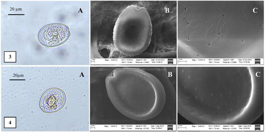
Figure 4. Morphology characters of Prorocentrum lima. 1. Seribu Islands. 2. Bintan Island. 3. Karimunjawa Islands. 4. Belitung Island.
A. Cell shape and size under LM observation; B. Cell shape and size under SEM observation; C. Pore shape and size observation using
SEMWIDIARTI et al. – Morphotype diversity of Prorocentrum lima 613
Belitung Island The results of this study prompt the necessity of
Observation on P. lima cells obtained in Belitung Island conducting an abundance survey, establishing strains,
waters showed broadly ovate shape, cell length and width toxins and molecular phylogenetic analysis, and growth
ranging from 35.49-37.79 µm (mean 36.24 µm, s.d. 0.94 experiment, to assess the negative impact in the future.
µm, n = 5) and 28.86-31.51 µm (mean 30.17 µm, s.d. 1.05
µm, n = 5), respectively, with the ratio L/W from 1.17-1.23
(mean 1.20, s.d 0.02, n = 5). The shape and size were ACKNOWLEDGEMENTS
within the range of P. lima "morphotype 1" by Zhang et al.
(2015), with cell length and width from 32.0-44.4 µm, and The authors kindly express gratitude to the Scholarship
27.3-36.5 µm, respectively, and the ratio L/W from 1.06- for Doctoral Program (BUDI-DN) by Ministry of Finance
1.36 (Table 2, Figure 4.4). Furthermore, observation on and Ministry of Research Technology and Higher
pores showed number of pores with a count of 48-59 (mean Education, the Republic of Indonesia, for funding the study
48.2, s.d. 10.4, n = 5), corresponds to "morphotype 1" by and research, and colleagues from Department of Biology,
Zhang et al. (2015), which was 42-84. The shape of pore Universitas Indonesia, for the assistance during sampling
observed in this study was round, and similar to that of collection. Also, the authors are grateful to the Bioimaging
"morphotype 1". Laboratory staffs at the Department of Biology, Universitas
Prorocentrum lima is known as a common species, Indonesia, and Nanotechnology Laboratory staff from
widely distributed in tropical, subtropical and temperate Research and Development Center of Agricultural
waters (Hoppenrath et al. 2013; Nishimura et al. 2019). In Postharvest (R. Idris Suryadi), for observation and images
this study, the cells were found in all sampling locations. collection using LM and SEM, with further gratitude to the
Previous studies showed that P. lima was also found in GIS laboratory staff (Satria Indratmoko) and Statistic
several locations in Indonesian waters, namely Seribu Clinique at FMIPA, Universitas Indonesia, Depok.
Islands (Widiarti 2002; Widiarti 2011; Widiarti and
Pudjiarto 2015), Belitung Island (Widiarti 2010), North
Lombok (Widiarti et al. 2016a), South Lampung waters REFERENCES
(Widiarti and Adi 2016) and Weh Island waters-North
Aceh (Widiarti et al. 2016b), with environmental condition Burkholder JM. 1998. Implications of harmful microalgae and
ranging from 28-33 °C for temperature, 26.0-34.5 ‰ for heterotrophic dinoflagellates in management of sustainable marine
salinity, 6.5-7.4 for water acidity, 6.17-8.30 ppm for fisheries. Ecological Applications 8 (1): S37-S62.
Chomérat N, Bilien G, Zentz F. 2018. A taxonomical study of
dissolved oxygen, and614 B I O D I V E R S I T A S 22 (2): 607-614, February 2021
Lehane L, Lewis RJ. 2000. Ciguatera: recent advances but the risk Hutauruk RM, Heltonika B, Karnila R, Windarti, Syawal H, Efriyeldi
remains. Int J Food Microbiol 61(2-3): 91-125. DOI: 10.1016/S0168- (eds). Proceeding of 3rd International Seminar of Fisheries and
1605(00)00382-2 Marine Science. University of Riau, Pekanbaru, 9-10 October 2014.
Ministry of Forestry and Environment. 2004. Decision on Sea Water Widiarti R. 2002. Epibenthic dinoflagellate on macroalgae at Penjaliran
Quality Standard No. 51. Ministry of Forestry and Environment- Barat Island reef flat, Jakarta Bay. Sains Indonesia 1 (7): 1-9.
Republic of Indonesia, Jakarta. [Indonesian] [Indonesian]
Nagahama Y, Fukuyo Y. 2005. Redescription of Cryptomonas lima, Widiarti R. 2010. Dinoflagellate which cause Ciguatera Fish Poisoning
collected from Sorrento, Italy, the basionym of Prorocentrum lima. (CFP) in Belitung Island waters. In: Nababan B (eds). Prosiding
Plankton Biol Ecol 52 (2): 107-109. Pertemuan Ilmiah Tahunan ISOI VII. Ikatan Sarjana Oseanologi
Nishimura T, Uchida H, Noguchi R, Oikawa H, Suzuki T, Funaki H, Ihara Indonesia, Pangkalpinang, 4 October 2010. [Indonesian]
C, Hagino K, Arimitsu S, Tanii Y, Abe S, Hashimoto K, Mimura K, Widiarti R. 2011. Toxic dinoflagellate, which causes Ciguatera Fish
Tanaka K, Yanagida I, Adachi M. 2019. Abundance of the benthic Poisoning in Seribu Islands waters, North Jakarta: Preliminary study
dinoflagellate Prorocentrum and the diversity, distribution, and on species distribution. In: Nababan B (eds). Prosiding Pertemuan
diarrhetic shellfish toxin production of Prorocentrum lima complex Ilmiah Nasional Tahunan ISOI VIII. Ikatan Sarjana Oseanologi
and P. caipirignum in Japan. Harmful Algae 96: 101687. DOI: Indonesia, Makassar, 25-27 September 2011. [Indonesian]
10.1016/j.hal.2019.101687. Widiarti R, Adi APW. 2016. The potentially toxic benthic dinoflagellate
Oktavian B, Thamrin, Siregar YI. 2017. Analysis of epiphytic in Pahawang Besar dan Kelagian Kecil Islands, Lampung. In:
dinoflagellates on Thalassia hemprichii at Nirwana Beach Waters, Nababan B (eds). Prosiding Pertemuan Ilmiah Nasional Tahunan
Kabung Bay District, Padang City, West Sumatera Province. Jurnal ISOI XII. Ikatan Sarjana Oseanologi Indonesia, Banda Aceh, 10-12
Online Mahasiswa. [Indonesian] December 2015. [Indonesian]
Seygita V, Thamrin, Siregar YI. 2015. Analisis kelimpahan Dinoflagellata Widiarti R, Pudjiarto RK. 2015. Dinoflagellata toksik penyebab Ciguatera
Bentik beracun di Perairan Teluk Bayur, Sumatera Barat. Dinamika Fish Poisoning di Perairan Pulau Tidung, Kepulauan Seribu. Jurnal
Lingkungan Indonesia 2 (2): 92-99. DOI: 10.31258/dli.2.2.p.92-99. Ilmiah Ilmu Biologi 1 (1): 5-8. [Indonesian]
[Indonesian] Widiarti R, Pudjiarto RK, Pratama I. 2016a. Potentially toxic benthic
Skinner MP, Lewis RJ, Morton S. 2011. The abundance of potentially dinoflagellate in Gili Meno and Gili Air waters, Lombok. In:
toxic epiphytic dinoflagellates and nutrients from Bali and Gili Setyawan AD, Sugiyarto, Pitoyo A, Sutomo, Widiastuti A, Windarsih
Trawangan, Indonesia. Mar Res Indones 36 (2): 11-23. DOI: G (eds). Prosiding Seminar Nasional Masyarakat Biodiversitas
10.14203/mri.v36i2.38 Indonesia. Universitas Sebelas Maret, Surakarta, 7 November 2015.
Sneath PHA. 2005. Numerical taxonomy. In: Brenner DJ, Krieg NR, [Indonesian]
Staley JT, Garrity GM (eds). Bergey's Manual® of Systematic Widiarti R, Pudjiarto RK, Fathoniah I, Adi APW. 2016b. Epiphytic
Bacteriology 2(Pt A): The Proteobacteria, Introductory Essay. dinoflagellate on macroalgae which potentially cause Ciguatera Fish
Springer, USA. Poisoning in Weh Island waters, Aceh. In: Setyawan AD, Sugiyarto,
Susetiono, Pratomo A, Kurniawan D, Susiana. 2016. Monitoring on the Pitoyo A, Sutomo, Widiastuti A, Windarsih G. (eds). Prosiding
Health of the Coral Reef and Related Ecosystem at Bintan District Seminar Nasional Masyarakat Biodiversitas Indonesia 2 (1).
2014. UMRAH-CRITC-LIPI, Tanjung Pinang. [Indonesian] Universitas Sebelas Maret, Surakarta, 23 April 2016. [Indonesian]
deSylva DP. 1994. Distribution and ecology of ciguatera fish poisoning in Widiarti R, Zamani NP, Bengen DG, Madduppa H. 2018. The
Florida, with emphasis on the Florida Keys. Bull Mar Sci 54 (3): 944- dinoflagellate causing ciguatera fish poisoning, Prorocentrum lima, in
954. Karimunjawa island waters-Central Java. IOP Conf Ser: Earth
Tester PA, Kibler SR, Holland WC, Usup G, Vandersea MW, Leaw CP, Environ Sci 325: 012014. DOI: 10.1088/1755-1315/325/1/012014
Teen LP, Larsen J, Mohammad-Noor N, Faust MA, Litaker RW. Yusuf M. 2013. Coral reef condition and fish potency in Karimunjawa
2014. Sampling harmful benthic dinoflagellates: Comparison of National Park waters, Jepara District. Bull Oseanografi Mar (2): 54-
artificial and natural substrate methods. Harmful Algae 39: 8-25. 60. [Indonesian]
DOI: 10.1016/j.hal.2014.06.009. Zhang H, Li Y, Cen J, Wang H, Cui L, Dong Y, Lu S. 2015. Morphotypes
Thamrin. 2014. Analysis of benthic dinoflagellate Gambierdiscus, of Prorocentrum lima (Dinophyceae) from Hainan Island, South
Ostreopsis, and Prorocentrum density on the west coast of Sumatera China Sea: Morphological and molecular characterization. Phycol 54
Island and Bintan Island coast in Riau Archipelago, Indonesia. In: (5): 503-516.You can also read