Type-II Lepra Reaction and Granulomatous Uveitis - An Unusual Presentation
←
→
Page content transcription
If your browser does not render page correctly, please read the page content below
DJO Vol. 30, No. 3, January-March 2020
Case Report
Type-II Lepra Reaction and Granulomatous Uveitis –
An Unusual Presentation
Sujit Das1, B. Pradeep2, Pushpanjali Ojha1
Department of Ophthalmology, Andaman and Nicobar Islands Institution of Medical Science (ANIIMS), Port Blair, India
1
2
Department of Dermatology, Andaman and Nicobar Islands Institution of Medical Science (ANIIMS), Port Blair, India
We describe a case of young male who presented with lepra reaction with multiple macula-papular
rash over face, forehead, ear lobules with peripheral neuropathy (Ulnar nerve thickening) and bilateral
granulomatous uveitis. Slit skin smear was negative but skin nodular biopsy showed multiple discreet
deeper dermis noncaseating well formed epitheloid cell granulomas with multinucleated giant cells
intermixed with lymphocytes. Fite Faraco stain was negative suggestive of lepra reaction. Patient
responded well with topical steroid and cycloplegic drops with disappearance of iris nodules. Systemic
Abstract
steroid was given for 12 weeks with gradual tapering to control lepra reaction and to prevent further
ocular morbidity. Since ocular involvement can be seen even after completion of anti-leprosy treatment,
the need for screening and periodic eye examination of the patient should be emphasized, for early
identification of potentially sight-threatening lesions which can be easily treated. An ophthalmologist
and a trained leprologist should preferably be included in the treatment of Hansen disease with ocular
manifestations.
Delhi J Ophthalmol 2020;30;63-66; Doi http://dx.doi.org/10.7869/djo.530
Keywords: Erythema Nodosum, Iridocyclitis, Lepromatous Leprosy, Leprosy, Uveitis
Introduction Case Report
Leprosy (Hansen disease) is a chronic granulomatous A 36-yeasr-old male presented with pain, photophobia,
multi-organ inflammatory disease caused by intracellular redness and diminution of vision in both eyes for 10 days
acid-fast gram-positive bacillus, the Mycobacterium duration. He was treated in primary health centre with
leprae. Leprosy predominantly affects the skin, peripheral antibiotics and lubricating drops. On examination, his distant
nerves, and eyes. Up to 75% of individuals with leprosy visual acuity was 6/24 in both eyes; intraocular pressure was
have ocular involvement and 40% have ocular disability.1 21mmHg in right eye and 22mmHg in the left eye. There were
Mycobacterium leprae has a strong preference for low Koppe and Busacca nodules [Figure-1(a)], koppe nodule,
temperatures, hence are mainly found in the skin, nose, synechia and pigment release [Figure-1(b)], segmental
earlobes and peripheral nerves. Within the eye, the organism synechiae [Figure-1(c)], matton fat keratic precipitates
is found only in the anterior segment which has relatively [Figure-1(d)] and hypopyon [Figure-1(e)] formation in
lower temperature. The organism has not been observed in both the eyes. There were multiple erythematous maculo-
the posterior segment or the optic nerve.2 The eye is affected papulo-nodular lesions were present in the forehead, face
via direct invasion or during lepra reaction. Ophthalmic [Figure-2(a)], forearms, arms and back. Ear lobules (Pinna)
manifestations of leprosy include lagophthalmos, corneal were grossly erythematous and thickened [Figure-2(b)].
ulceration, acute or chronic iridocyclitis, and secondary Ulnar nerves were thickened. Other features including
cataract.3 Ocular complications may also occur indirectly lagophthalmos, exposure keratopathy, corneal ulceration,
through impairment of lid closure (VII nerve) and corneal conjunctival or scleral leproma were absent. He had history of
anaesthesia (V nerve) and through damage to adnexal similar episode of ocular pain, redness, photophobia twice in
tissues.3 Most of the ocular complications may lead to visual the past and had a history of one-year treatment for Leprosy
impairment and blindness; therefore, early detection and five years back. A diagnosis of granulomatous uveitis was
appropriate treatment is essential. Typically, the systemic made and the patient was started on topical prednisolone
disease is confirmed by detection of bacilli on slit skin smear acetate 1% 1drop 1 hourly along with Homatropine 2% 1
or skin tissue biopsy.4 Histologic findings include multiple drop thrice daily. Skin specialist opinion was taken and was
bacilli with acid-fast or Fite-Faraco stain positive, along diagnosed as a case of type 2 Lepra reaction and the patient
with iris pearls. In addition to histopathology, polymerase was put on systemic prednisolone (1mg/kg/body weight)
chain reaction (PCR) can also be used to diagnose leprosy.4 slowly tapered over 12 weeks. Slit skin smear from forehead,
In typical type-II lepra reaction without active leprosy, there cheek and ear lobules was negative for acid-fast bacilli.
is erythematous maculo-papular rash along with negative Skin incision biopsy was taken from macula nodular lesion
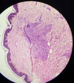
slit skin smear and Fite Faraco stain. Skin nodular biopsy of the arm which showed multiple discreet deeper dermis
shows noncaseating epitheloid cell granulomas with multi- non caseating well formed epitheloid cell granulomas with
nucleated giant cells intermixed with lymphocytes. multinucleated giant cells intermixed with lymphocytes.
E-ISSN: 2454-2784 P-ISSN: 0972-0200 63 Delhi Journal of Ophthalmology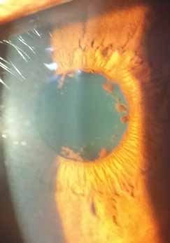
DJO                                                                                                                     Vol. 30, No. 3, January-March 2020

[Figure-3]. Fite Faraco stain was found negative suggesting                           and viral markers were al negative. Complete blood count and
a lepra reaction [Figure-3]. Mantoux test was negative                                blood sugar estimation was normal. Systemic involvement
with normal radiogram and contrast-enhanced computed                                  and iridocyclitis gradually improved with disappearance of
tomography of the chest. On haematological investigation,                             iris nodules [Figure-4]. Later on he developed complicated
angiotensin converting enzyme levels was normal [21U/L].                              posterior subcapsular cataract [Figure-5(a)] with diminution
Serological test for syphilis, human immune deficiency virus                          of vision (6/60) in both eyes. Ultrasonography –B scan was
                                                                                      performed before surgery and the retina appeared normal.
                                                                                      He then underwent cataract surgery with posterior chamber
 (a)                       (b)                        (c)                             intra ocular lens [PCIOL] with surgical peripheral iridectomy
                                                                                      [Figure-5(b)]. Post operatively his visual acuity was 6/12
                                                                                      in both eyes improving to 6/9 with correction. Intraocular
                                                                                      pressure was 17mmg in both eyes. Patient is kept under
                                                                                      regular follow-up.

(d)                                     (e)

   Figure 1: (a) koppe and bussaca nodule; (b) koppe nodule and pigment
     release; (c) segmental synechia; (d) motton fat keratic precipitates;
                          (e) hypopyon formation.                                     Figure 3: Nodular biopsy shows noncaseating well formed epitheloid cell
                                                                                        granulomas with multinucleated giant cells intermixed with lymphocytes.

 (a)

(b)

Figure 2: (a) macula papullo nodular lesion involving face; (b) Lepra reaction
                                 of pinna.                                                      Figure 4: Disappearance of iris nodules after treatment.

E-ISSN: 2454-2784       P-ISSN: 0972-0200                                        64                                                            www.djo.org.in
DJO Vol. 30, No. 3, January-March 2020
(a) (b) affecting the facial nerve, cornea and iris is characteristic
of type II reaction, which may severely damage the eye,
directly or indirectly.
Iridocyclitis is generally managed with topical steroids and
cycloplegic drugs over a prolonged duration. Evidence
strongly indicates that the most serious effects on body tissues
consequent to infection of the skin, nerves, and eyes with
Mycobacterium leprae are because of the immune response.5
In both lepromatous and non-lepromatous leprosy, adverse
immunological reactions, either cell-mediated or humoral,
may develop suddenly and have considerable severity,
necessitating treatment with steroids or immunosuppressive
drugs.9 The present case represents a clinical and histological
demonstration of iridocyclitis, resulting from lepromatous
leprosy, which could have progressed into blindness if left
undetected or treated inappropriately. A close and long
follow-up is required in these cases, as these patients are at
risk of significant ocular morbidity, despite completing the
multidrug therapy.9
Figure 5: (a) Complicated posterior subcapsular cataract (PSC); (b) posterior Conclusion
chamber intra ocular lens and surgical peripheral iridectomy. It is recommended that an ophthalmologist and a trained
leprologist, must be included in the treatment of Hansen
disease with ocular manifestations. The risk of ocular
Discussion
complication increases with increased duration of disease
Leprosy is a multi organ infectious disease affecting mainly
and with lepra reactions. Since ocular involvement can
the skin nerves and eyes. Skin lesions commonly are
be seen even after completion of anti-leprosy treatment,
macules and plaques, rarely papules or nodules. Lesions
the need for screening and periodic eye examination of all
vary from being hypopigmented with reduced sensations
the patients with leprosy should be emphasized, for early
in tuberculoid leprosy to multiple confluent nodular
identification of potentially sight threatening lesions which
lesions in lepromatous leprosy.1,2,3 Nerves are damaged in
can be easily treated.
two locations, either peripheral nerve trunks near the fibro
osseous tunnels or small dermal nerves in the skin lesions.
Leprosy is also associated with type-1 and type-2 reactions.5 References
Type-1 reactions occur in patients with borderline leprosy 1. Global leprosy update, 2013; reducing disease burden. Wkly
and never with polar leprosy. This reaction manifests with Epidemiol Rec. 2014; 89:389–400.
2. Dana MR, Hochman MA, Viana MA, Hill CH, Sugar J. Ocular
signs of inflammation within macules, papules and plaques
manifestations of leprosy in a noninstitutionalized community
with appearance of new lesions and fever.5 Erythema in the United States. Arch Ophthalmol. 1994; 112:626–629.
Nodusum Leprosum (ENL) or type 2 reaction classically 3. Khan T, Awan AA, Kazmi HS, Shah AA, Muhammad S,
presents as tender erythematous nodules on the face, Muhammad S. Frequency of ocular complications of leprosy
arms and legs. It occurs in lepromatous leprosy with skin in institutionalized patients in NWFP Pakistan. J Ayub Med Coll
infiltration and bacterial index of 4 or more.5 Abbottabad. 2002; 14:29–33.
4. 5 Ffytche TJ, McDougall AC. Leprosy and the eye: a review. J R
The present case was an outpatient case being treated for Soc Med. 1985; 78(5): 397–400
conjunctivitis elsewhere. Detailed ophthalmic evaluation 5. Jolliffe DS. Leprosal reactional states and their treatment. Br J
was performed revealing features of granulomatous uveitis. Dermatol. 1977; 97:345–352.
This finding, along with systemic features and positive skin 6. Campos WR, Orefice F, Sucena MA, Rodrigues CA. Bilateral
biopsy, confirmed the diagnosis of lepromatous leprosy iridocyclitis caused by Mycobacterium leprae diagnosed
through paracentesis. Indian J Lepr. 1998; 70:27–31.
with lepra reaction. Campos et al. reported the diagnosis of 7. Messmer EM, Raizman MB, Foster CS. Lepromatous uveitis
mycobacterium leprae through AC paracentesis in a case of diagnosed by iris biopsy. Graefes Arch Clin Exp Ophthalmol. 1998;
bilateral iridocyclitis[6].Lepromatous uveitis has also been 236:717–719.
diagnosed through skin, aqueous humor, and iris biopsy, as 8. Khan T, Awan AA, Kazmi HS, Shah AA, Muhammad S,
reported by Messmer et al.7 Muhammad S. Frequency of ocular complications of leprosy
in institutionalized patients in NWFP Pakistan. J Ayub Med Coll
The reported frequency of iridocyclitis is 7%-24%.8
Abbottabad. 2002; 14:29–33.
Iridocyclitisis a potentially blinding clinical manifestation of 9. KM Waddell, PR Saunderson. Is leprosy blindness avoidable?
erythema nodosum leprosum (type II reaction), which results The effect of disease type, duration, and treatment on eye
from antigen antibody reaction, mainly in multibacillary damage from leprosy in Uganda. BJO 1995; 79: 250-256
(lepromatous) leprosy.1,5 Acute inflammatory reaction
E-ISSN: 2454-2784 P-ISSN: 0972-0200 65 Delhi Journal of OphthalmologyDJO Vol. 30, No. 3, January-March 2020
Cite This Article as: Das S, Pradeep B, Ojha P. Type-II Lepra Reaction and
Granulomatous Uveitis – An Unusual Presentation.
Acknowledgments: Nil
Conflict of interest: None declared
Source of Funding: None
Date of Submission: 28 June 2019
Date of Acceptance: 6 October 2019
Address for correspondence
Sujit Das MS
Department of Ophthalmology,
JNU Medical College & Research Centre,
Jaipur, Rajasthan-302017
India
Email id: imdrsujitdas@gmail.com
Quick Response Code
E-ISSN: 2454-2784 P-ISSN: 0972-0200 66 www.djo.org.inYou can also read