Pectus Excavatum in Puppies and Kittens - MSPCA-Angell
←
→
Page content transcription
If your browser does not render page correctly, please read the page content below
Pectus Excavatum in Puppies and Kittens
                                 By Sue Casale, DVM, DACVS
                                 angell.org/surgery
                                 surgery@angell.org
                                 617-541-5048
                                 March 2021

Pectus excavatum, which is from Latin meaning “hollow
breast,” is a chest wall deformity seen frequently in humans
and less often in veterinary patients.1 It has been described in
dogs, cats, lambs, calves, rabbits and sea otters.2,3 It is a
congenital defect where the caudal ribs and sternum do not
grow properly resulting in a concavity starting around the 3rd
to 5th rib and continuing to the xiphoid process.1,2 This
deformity causes compression of the right side of the heart
with deviation of the heart, to the left side of the thorax.4
Pectus excavatum is the most common chest wall deformity
seen in both people and animals, accounting for 90% of
human cases.1 In people, it occurs in approximately one in 400
live births and is more common in males with as high as a 9:1
                                                                       Pectus excavatum, seen here, is a congenital
ratio reported.1,4 It is not thought to be as common in animals
                                                                       defect resulting in a chest wall deformity.
and no sex predilection has been described. Pectus excavatum
was first described in people the early 1600s and several case
reports from the late 1800s were published.1 The first surgery for pectus excavatum was reported in 1911 and
involved removal of rib cartilage.1 The first reported case of pectus excavatum in an animal was in 1968 in a
Siamese cat.5 The first report in dogs was in 1973 in three Setter-cross littermates.6 Since that time, there have been
numerous case reports of the condition, but because it is fairly rare, the literature is still limited.
The etiology of pectus excavatum is unknown. Early theories ranged from hypertension of the diaphragm, and
nutritional disturbances causing a weakness of the sternum, to an increased intrauterine pressure, causing the fetus’
chin to compress the thorax.1 Current thought is that there is a defect in metabolism in the sternocostal cartilage that
leads to biomechanical weakness and overgrowth.1 The condition has been shown to be genetic in people, with 43%
of patients having a family history.4 There is an association with scoliosis and connective tissue disorders like

 Angell Animal Medical Center • 350 S. Huntington Ave., Boston, MA 02130 • 617-522-7282 • fax: 617-989-1635
Marfan, Noonan and Ehlers-Danlos syndromes.4 Predisposition for pectus excavatum is seen in Bengal and
Burmese cats as well as brachycephalic dogs, most commonly in the Maltese and English Bulldog.7-10 Acquired
pectus excavatum has been reported in people secondary to respiratory obstruction and there is a single case report
describing acquired pectus excavatum in a dog secondary to laryngeal paralysis.11, 12 Recently, a rat model for
pectus excavatum has been reported where removal of the last four costal cartilages results in collapse of the
sternum.13
Pectus excavatum is present at birth and is detectable within days in puppies and kittens.2,12 The defect worsens with
growth and in people it is sometimes is not obvious until adolescence.1,4 Once growth ceases, the condition does not
progress. The clinical signs can be variable with most cases in humans presenting with mild signs or concern about
cosmesis.4 In severe cases, disabling cardio and respiratory compression is present and clinical signs worsen with
age. Dyspnea, chest pain, fatigue, palpitations, tachycardia, and exercise intolerance have all been reported in
people.4 Dyspnea is the most common clinical sign in animals.12 Exercise intolerance, inappetence, recurrent
respiratory infections, weight loss, cough, cyanosis, and heart murmurs are also reported.12,14
Pectus excavatum is diagnosed on physical examination
however imaging helps to determine the severity of the
condition. The anthropometric or clinical index, measures the
depth of the defect and compares it to the depth of the
thorax.15,16,17 This is a simple test that requires no additional
equipment. Radiographs show the degree of deformity and
can be used to calculate several different indices which assess
the amount of compression. The frontosagittal index (FSI)
compares the width of the thorax at its widest point with the
depth of the thorax at its most narrow point and a ratio is determined.16 The
vertebral index (VI) compares the distance between the dorsal border of the
spine and the sternum at the narrowest point, with the depth of a vertebral
body.16 In animals, two view thoracic radiographs can be used to calculate both
the FSI and the VI and when combined with clinical signs, help to determine if
surgery is recommended. The Haller index (HI), also called the pectus index,
was first described in 1987 and is calculated by dividing the transverse diameter
of the chest by the narrowest anterior-posterior distance on a computed
tomography (CT) scan axial slice.16,18 The Haller index is the most commonly
used pectus excavatum index used in people. It can also be calculated using
plain film and has been shown to correlate well with the CT calculations.16
Indices may not correlate with clinical signs in some cases. CT scan has been
shown to result in a lower vertebral index and a higher frontosagittal index in
kittens when compared to radiographic measurements.19 CT was useful in
determining if the xiphoid was midline or lateralized with the thought that midline defects cause more clinical
signs.19 CT scan also helped determine safe corridors for surgical suture placement.19
Treatment for pectus excavatum depends on the severity of the clinical signs. Pain is a common complaint in human
patients and non-steroidal anti-inflammatory medication and acupuncture have been reported to help.4 Physical
therapy has been recommended to help strengthen the chest muscles and help develop the ribcage. Daily medial
lateral compression of the chest or compression bandages have been described for neonatal puppies and kittens
however, this is unlikely to improve severe deformities.2 In people, the use of a vacuum bell has been reported with
success in milder cases and in young patients who still have a compliant thorax.20 Several hours of daily use is
required and patients wear the device for up to two years after the deformity is corrected. In the case of severe
pectus excavatum or in older patients, surgery is required. The two most commonly performed surgeries are the
Ravitch procedure and the less invasive Nuss procedure. The Ravitch procedure involves an open surgical approach
with removal of the deformed cartilage and metal bars are implanted to support the sternum. This is a long
 Angell Animal Medical Center • 350 S. Huntington Ave., Boston, MA 02130 • 617-522-7282 • fax: 617-989-1635
procedure with potentially significant morbidity. The Nuss
procedure is performed under thoracoscopic guidance and a curved
metal bar is placed and then flipped into position to raise the
sternum.21,22,23 In veterinary patients, the choice of surgery is
dependent on age. In older animals, an open approach is required to
correct the deformity. Ostectomy of a portion of the costal cartilage
is required to allow the sternum to be realigned and an internal
splint, such as a bone plate, is used to keep the sternum in this
position.24 An open approach to release the sternum, combined with
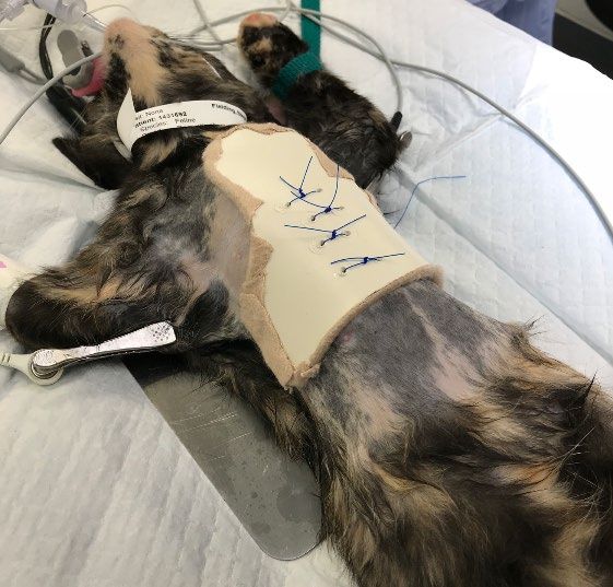
an external splint has also been described.25,26 Animals less than 4
months of age will typically have a compliant thorax and external
splints with circumsternal sutures can be used.27 The sutures are tied
to the splint to keep the thorax in a normal position and the splint is
left in place for 3 to 4 weeks.27,28,29 Complications associated with
An external splint to address pectus
surgery include inadvertent puncture of the lungs or heart, excavatum.
pneumothorax, hemorrhage and re-expansion pulmonary edema which
can be fatal.28,30 Minor complications include bandage sores and abrasions from the splint or reaction to the sutures.
Anesthesia in patients with pectus excavatum can be challenging.31 Ninety-five percent of patients present with
some degree of ventricular compression.31 Heart murmurs and arrhythmias are common.14,31 Underlying respiratory
disease including infections should be treated prior to surgery and assisted ventilation is needed during anesthesia.
Because surgery is often performed in very young puppies and kittens, their body temperature should be carefully
monitored as their thermoregulation is not fully developed and they have little fat. Their metabolism rate is higher
than adult patients and as a result, they require shorter fasting times and higher fluid rates.14 Enzymes systems
should be fully developed by 4 weeks allowing the use of injectable medications.14
Pectus excavatum is a treatable condition that has a favorable prognosis in puppies and kittens. Severe cases can do
well with surgery. A procedure with circumsternal sutures and an external splint is the most commonly performed
in young animals. Surgery is best accomplished between 8 and 12 weeks when the thorax is still compliant but the
patient is old enough to tolerate anesthesia.
References
1) Brochhausen C, Turial S, Muller FKP, et al. Pectus excavatum: history, hypotheses and treatment options.
Interac CV Thorac Surg 2012; 14: 801-806
2) Singh M, Parrah JUD, Moulvi, BA et al. A review on pectus excavatum in canines: a congenital anomaly.
Iran J Vet Surg 2013; 8(1): no 18 59-64
3) Mann N, MacLean J, et al. Pectus excavatum and swimmer puppy syndrome with concurrent congenital
cardiac anomalies in two domestic rabbits. J Ex Pet Med 2019; 29: 212-216
4) Abid I, Ewais MM, Marranca J, et al. Pectus excavatum: a review of diagnosis and current treatment
options. J Am Osteopath Assoc 2017; 117(2):106-113
5) Grenn HH, Lindo DE, Pectus excavatum (funnel chest) in a feline. Can Vet Jour 1968; 9(12):279-282
6) Pearson JL, Pectus excavatum in the dog (a case report). Vet Med 1973; 125-128
7) Charlesworth TM, Sturgess CP, Increased incidence of thoracic wall deformities in related Bengal kittens.
J Fel Med Surg 2012; 14(6): 365-368
8) Sturgess CP, Waters L, et al. Investigation of the association between whole blood and tissue taurine levels
and the development of thoracic deformities in neonatal Burmese kittens. Vet Record 1997; 141: 566-570
9) Komsta R, Osinski Z, Debiak P, et al. Prevalence of pectus excavatum (PE), pectus carinatum (PC),
tracheal hypoplasia, thoracic spine deformities and lateral heart displacement in thoracic radiographs of
screw-tail brachycephalic dogs. PLOS ONE 2019
Angell Animal Medical Center • 350 S. Huntington Ave., Boston, MA 02130 • 617-522-7282 • fax: 617-989-163510) Hassan EA, Hassan MH, Torad FA, Correlation between clinical severity and type and degree of pectus
excavatum in twelve brachycephalic dogs. J Vet Med Sci 2018; 80(5):766-771
11) Kurosawa TA, Ruth JD, et al. Imaging diagnosis- acquired pectus excavatum secondary to laryngeal
paralysis in a dog. Vet Rad Ultra 2012; 53(3):329-332
12) Fossum TW, Boudrieau RJ, Hobson HP, Pectus excavatum in eight dogs and six cats. JAAHA 1989;
25:595-605
13) David VL, Ciornei B, Horhat FG, et al. Rat model of pectus excavatum. Life 2020; 10:96
14) Boudrieau RJ, Fossum TW, et al. Pectus excavatum in dogs and cats. Compend Cont Ed 1990; 12(3): 341-
355
15) Rebeis EB, Milanez de Campos JR, et al. Anthropometric index for pectus excavatum. Clinics 2007;
62(5):599-606
16) Martinez-Ferro M, Indexes for pectus deformities. In Chest Wall Deformities and Corrective Procedures
Springer Inter Pub, Switzerland, 2016
17) Sujka JA, St. Peter SD, Quantification of pectus excavatum: anatomic indices. Sem Ped Surg 2018;
27(3):122-126
18) Poston PM, Patel SS, Rajput M, et al. The correction index: setting the standard for recommending
operative repair of pectus excavatum. Ann Thorac Surg 2014; 97:1176-1180.
19) Charlesworth TM, Schwarz T, Sturgess CP, Pectus excavatum: computed tomography and medium-term
surgical outcome in a prospective cohort of 10 kittens. J Fel Med Surg 2016; 18(8):613-619
20) Haecker FM, Sesia S, Non-surgical treatment of pectus excavatum. J Vis Surg 2016;2:63
21) Hebra A, Calder BW, Lesher A, Minimally invasive repair of pectus excavatum. J Vis Surg 2016;2:73
22) Sakamoto Y, Yokoyama Y, et al. Outcomes of the Nuss procedure for pectus excavatum in adults. J Plast
Recon Aesth Surg 2020
23) Xie Y, Ning J, Application of polydioxanone sutures in the Nuss procedure. Thorac Cardiovasc Surg 2021
24) Risselada M, de Rooster H, Liuti T, et al. Use of internal splinting to realign a noncompliant sternum in a
cat with pectus excavatum. JAVMA 2006; 228(7): 1047-1052
25) Mestrinho LA, Ferreira CA, et al. Open surgical correction combined with an external splint for correction
of a non-compliant pectus excavatum in a cat. J Fel Med Surg 2011; 14(2):151-154
26) Crigel MH, Moissonnier P, Pectus excavatum surgically repaired using sternum realignment and splint
techniques in a young cat. J Small Ani Pract 2005; 46:352-356
27) McAnulty JF, Harvey CE, Repair of pectus excavatum by percutaneous suturing and temporary external
coaptation in a kitten. JAVMA 1989; 194(8): 1065-1067
28) Fossum TW, Boudrieau RJ, et al. Surgical correction of pectus excavatum, using external splintage in two
dogs and a cat. JAVMA 1989; 195(1):91-97
29) DeGroot W, Tobias KM, External splinting for pectus excavatum in kittens. Clinician’s Brief, May 2018
30) Soderstrom MJ, Gilson SD, Gulbas N, Fatal reexpansion pulmonary edema in a kitten following surgical
correction of pectus excavatum. JAAHA 1995; 31:133-136
31) Patvardhan C, Martinez G, Anaesthetic considerations for pectus repair surgery. J Vis Surg 2016;2:76
Angell Animal Medical Center • 350 S. Huntington Ave., Boston, MA 02130 • 617-522-7282 • fax: 617-989-1635You can also read