Dermointegration in the exposed titanium cranioplasty: a possible protective phenomenon
←
→
Page content transcription
If your browser does not render page correctly, please read the page content below
Journal of Surgical Case Reports, 2021;1, 1–4

                                                                               doi: 10.1093/jscr/rjaa551
                                                                               Case Report

CASE REPORT

Dermointegration in the exposed titanium

                                                                                                                                                         Downloaded from https://academic.oup.com/jscr/article/2021/1/rjaa551/6123958 by guest on 23 February 2021
cranioplasty: a possible protective phenomenon
Steven Liben Zhang1 ,*, Hanjing Lee2 , Elijah Zhengyang Cai2 , Yan Lin Yap2 ,
Tseng Tsai Yeo3 , Thiam Chye Lim2 , Char Loo Tan4 and Jane Lim2
1
 Section of Plastic, Reconstructive and Aesthetic Surgery, Department of General Surgery, Tan Tock Seng
Hospital, Singapore, 2 Division of Plastic, Reconstructive & Aesthetics Surgery, Department of Surgery,
University Surgical Cluster, National University Hospital, Singapore, 3 Division of Neurosurgery, Department of
Surgery, University Surgical Cluster, National University Hospital, Singapore and 4 Department of Pathology,
Yong Loo Lin School of Medicine, National University Hospital, National University of Singapore, Singapore
*Correspondence address. Section of Plastic, Reconstructive and Aesthetic Surgery, Department of General Surgery, Tan Tock Seng Hospital, 11 Jln Tan
Tock Seng, Singapore 308433. Tel: (+65) 9090 5401. E-mail: zlb.steven@gmail.com

Abstract
Implant exposure is a known complication of titanium mesh cranioplasty and is usually managed by implant removal and/or
exchange. We describe a case of exposed titanium mesh cranioplasty which was managed with implant exchange and
bipedicled f lap coverage, and showcase an interesting phenomenon of full-thickness skin present beneath the exposed mesh.
This was confirmed on histopathology, which showed the presence of dermal appendages including pilosebaceous units and
eccrine glands. We postulate that the mechanism behind this phenomenon involves islands of viable skin ‘dropping’ between
holes in the mesh and coalescing beneath the exposed implant, as suggested by histopathology findings of nodular protrusions
and varying degrees of epidermal hyperplasia. This protects the underlying dura from external infection. We propose for this
phenomenon to be called dermointegration. Our findings suggest that similar cases, particularly patients who are not fit for
general anaesthesia, may potentially be managed with a more conservative approach.

INTRODUCTION                                                                       Despite its benefits, the titanium cranioplasty implant is
Cranioplasty is commonly performed following craniectomy for                   associated with complications, including thinning of the over-
various aetiologies including traumatic brain injury, stroke and               lying soft tissue and implant exposure [3]. Implant exposure is
malignancy. It is indicated to protect the brain and reduce aes-               particularly troublesome as it causes issues with hygiene and
thetic deformity associated with contour depression [1]. While                 social embarrassment, poses a risk of secondary infection, com-
a variety of materials are available [2], titanium is commonly                 promises aesthetic outcome and usually necessitates implant
used because it is hard, rigid, strong, light, resistant to infection,         removal and revision surgeries [4].
biologically inert, easily obtainable and a cheaper alternative to                 We report an interesting phenomenon noted in a case of
patient-specific implants [1, 2]. It is most frequently used in the            exposed titanium mesh cranioplasty, where full-thickness skin
form of a mesh plate.                                                          was found beneath the exposed implant and confirmed on

Received: November 18, 2020. Accepted: November 28, 2020
Published by Oxford University Press and JSCR Publishing Ltd. All rights reserved. © The Author(s) 2021.
This is an Open Access article distributed under the terms of the Creative Commons Attribution Non-Commercial License (http://creativecommons.org/
licenses/by-nc/4.0/), which permits non-commercial re-use, distribution, and reproduction in any medium, provided the original work is properly cited.
For commercial re-use, please contact journals.permissions@oup.com

                                                                                                                                                   1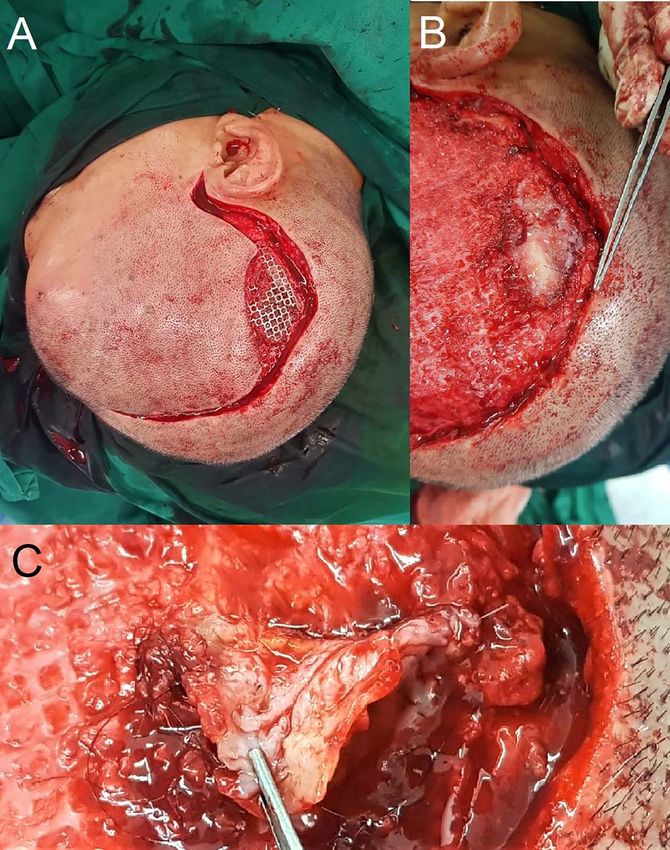
2      S.L. Zhang et al.

                                                                                                                                                                       Downloaded from https://academic.oup.com/jscr/article/2021/1/rjaa551/6123958 by guest on 23 February 2021
Figure 1: Parietal scalp wound with an exposed implant. A 54-year-old man
presented with a right parietal scalp wound measuring 5 × 2.5 cm, with exposed
titanium mesh cranioplasty implant (A). Computed tomography showed outward
tenting of the implant beneath the area of exposure (B). This was correlated
intraoperatively, due to venting cuts made from the initial surgery (C). This had
likely caused pressure on the overlying skin and resulted in skin breakdown and
implant exposure.
                                                                                    Figure 2: Intra-operative findings of hair-bearing skin beneath the exposed
                                                                                    implant. The wound was debrided and incision extended along the previous
                                                                                    scar (A). Upon removal of the implant, we found a layer of epithelium with hair
                                                                                    follicles and surrounding granulation tissue beneath the area of the exposed
histopathology to include skin appendages like pilosebaceous                        implant (B, C); this was fully excised down to the dural covering layer and sent
                                                                                    for histopathology.
units and eccrine glands. We postulate the mechanism behind
this phenomenon and discuss the implications that this may
have on the future management of similar cases.

                                                                                    and/or exchange, with the aims of preventing secondary infec-
CASE REPORT                                                                         tion and maintaining an acceptable aesthetic outcome [4]. Resul-
                                                                                    tant skin and soft tissue defects are often sizeable, and require
A 54-year-old man presented with a three-month history of a
                                                                                    reconstruction with locoregional and/or free flaps [5]. This adds
right parietal scalp wound, measuring 5 × 2.5 cm, with exposed
                                                                                    to the duration of surgery, introduces potential donor site mor-
titanium mesh (Fig. 1A). He had a post-traumatic decompressive
                                                                                    bidity, and increases surgical and anaesthetic risks for patients
craniectomy and titanium mesh cranioplasty 18 years ago. A
                                                                                    who often have concomitant neurological risk factors, including
computed tomography scan of the brain did not reveal any
                                                                                    seizures and cerebrovascular disease [6].
underlying collection and showed the configuration of the
                                                                                        From our institutional experience, we have managed patients
titanium mesh which resulted in the exposure (Fig. 1B). He
                                                                                    with exposed titanium implants who did not undergo surgery,
was counselled for and underwent wound exploration, implant
                                                                                    due to significant comorbidities which increase anaesthetic
removal and exchange, and bipedicled flap reconstruction.
                                                                                    risks. Surprisingly, these patients continue to survive for years
    Intraoperatively, venting cuts in the mesh were noted
                                                                                    without infection. Parallels can be drawn with similar situations
(Fig. 1C), which had likely caused pressure on the overlying skin,
                                                                                    such as osseointegrated dental implants and tissue expanders
resulting in breakdown and implant exposure. Most interest-
                                                                                    with external ports, where foreign bodies are exposed to the
ingly, there was a layer of epithelium with hair follicles and
                                                                                    external environment in a stable state with minimal risk of
surrounding granulation directly beneath the area of an exposed
                                                                                    infection [7, 8]. We hypothesise that the layer of full-thickness
implant (Fig. 2). This was excised and sent for histopathology,
                                                                                    skin beneath the exposed implant, as shown in our patient
which revealed dermal appendages including pilosebaceous
                                                                                    and histopathology, could be protective. Intraoperatively, there
units and eccrine glands, associated with prominent chronic
                                                                                    is macroscopic evidence of granulation tissue with islands of
inflammation (Fig. 3).
                                                                                    hair-bearing skin directly beneath the exposed implant (Fig. 2).
                                                                                    Histopathology confirmed microscopic features of full-thickness
                                                                                    skin, with dermal appendages including pilosebaceous units
DISCUSSION                                                                          and eccrine sweat glands, and even subcutaneous fat (Fig. 3).
Implant exposure is a known complication of titanium mesh                           This growth of skin has likely integrated into the dural covering
cranioplasty, occurring in about 14% of patients [3, 4]. Standard                   as would a skin graft and acts as a protective barrier against
management involves wound debridement, implant removal                              infection. A review of the literature did not reveal any published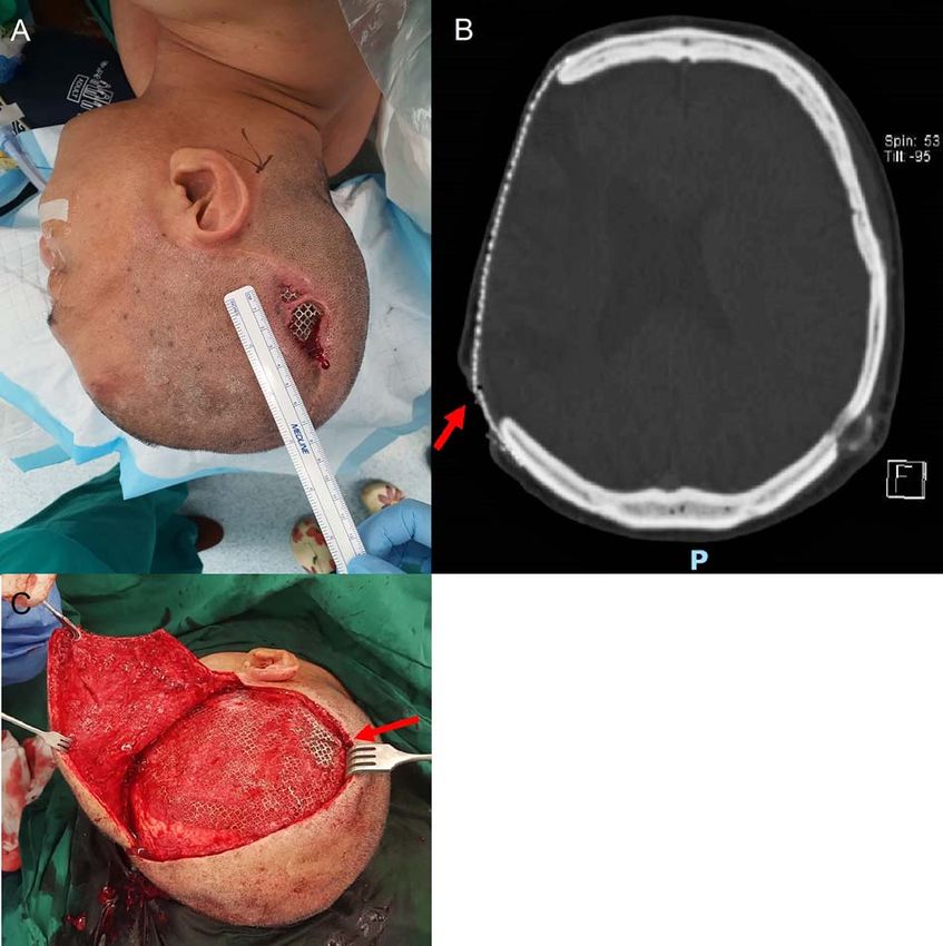
Dermointegration in exposed cranioplasty 3
Downloaded from https://academic.oup.com/jscr/article/2021/1/rjaa551/6123958 by guest on 23 February 2021
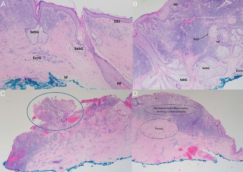
Figure 3: Histopathology findings. (A, B) Haematoxylin–Eosin staining (×40): micrographs showing skin with pilosebaceous units and prominent chronic inflammation
in the superficial dermis with exocytosis. SebG = sebaceous gland; EccG = eccrine glands; DEJ = dermo-epidermal-junction; HF = hair follicle; PsU = pilosebaceous unit
(black arrow). (C) Haematoxylin–Eosin staining (×20): Nodular protrusions of skin (blue circle), corresponding to gaps/holes within titanium mesh. (D) Haematoxylin–
Eosin staining (×20): marked dermal inflammation with lichenoid bands and fibrosis, with varying degrees of epidermal hyperplasia (black double-arrows) lending
support to our hypothesis of re-epithelialisation between skin islands. Epidermis is relatively thinner towards the edge of the specimen (yellow double-arrow),
corresponding to the interface with the edge of the exposed implant.
Figure 4: Proposed mechanism of dermointegration. In the normal patient, the titanium plate lies between the dura and overlying scalp skin (top left). Local factors such
as abnormal plate contour exert pressure on the skin and causes thinning and pressure necrosis (top right). The plate extrudes through the skin, and the mesh plate
design results in islands of the skin ‘dropping’ through holes within the mesh (middle left). Islands of skin coalesce beneath the exposed implant, with re-epithelialisation
and granulation tissue formation (middle right). The eventual result is a layer of full-thickness skin overlying and protecting the dura beneath the exposed plate (bottom
left).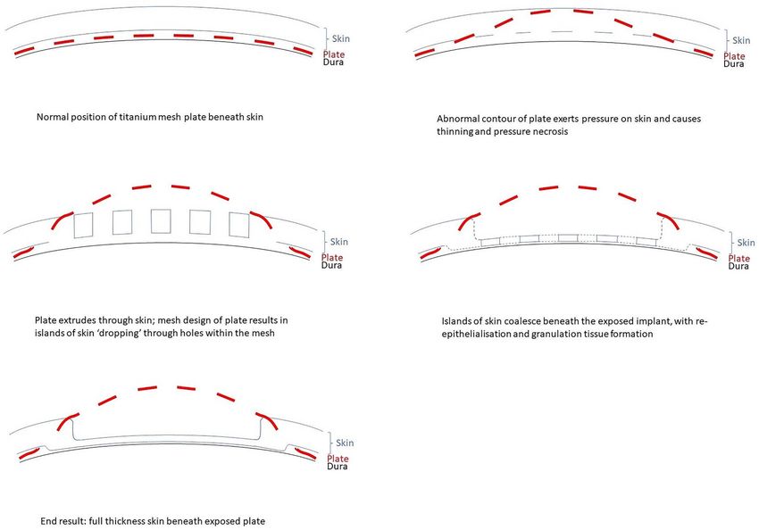
4     S.L. Zhang et al.

reports of such a phenomenon. We propose for this phenomenon          as functional soft tissue coverage (while accepting some degree
to be called ‘dermointegration’.                                      of suboptimal aesthetic contour). Further studies are required
    While the exact mechanism behind this phenomenon                  to better understand the mechanism behind this phenomenon,
remains unclear, we can understand the pathophysiology by             and to investigate the long-term outcome of patients who are
reviewing a crucial step in the proliferative process of wound        managed conservatively.
healing—epithelialisation, which is marked by proliferation and
migration of keratinocytes [9]. Keratinocytes are regenerated
from stem cells within the pilosebaceous units, eccrine sweat
glands and outer root sheath of the hair follicle [10], which         REFERENCES
differentiate into keratinocytes and repopulate the stratum            1. Hill CS, Luoma AM, Wilson SR, Kitchen N. Titanium cran-
basale [9]. These keratinocytes migrate across the wound,                 ioplasty and the prediction of complications. Br J Neurosurg
proliferating at its edges until they meet in the middle [10].            2012;26:832–7.
This epithelial layer protects the wound from infection and            2. Zanotti B, Zingaretti N, Verlicchi A, Robiony M, Alfieri A,

                                                                                                                                           Downloaded from https://academic.oup.com/jscr/article/2021/1/rjaa551/6123958 by guest on 23 February 2021
desiccation [10].                                                         Parodi PC. Cranioplasty: review of materials. J Craniofac Surg
    We postulate that re-epithelialisation plays an essential role        2016;27:2061–72.
in the phenomenon of dermointegration. The initial skin break-         3. Thien A, King NK, Ang BT, Wang E, Ng I. Comparison
down occurs as a result of pressure from a focal area of the              of polyetheretherketone and titanium cranioplasty
poorly contoured titanium mesh, more commonly seen in older               after decompressive craniectomy. World Neurosurg
plates where less attention might have been given to proper               2015;83:176–80.
moulding. While the skin overlying the protruding metal under-         4. Maqbool T, Binhammer A, Binhammer P, Antonyshyn OM.
goes necrosis, small islands of viable skin or keratinocytes may          Risk factors for titanium mesh implant exposure following
‘drop’ through the holes of the titanium mesh (Fig. 4). These skin        Cranioplasty. J Craniofac Surg 2018;29:1181–6.
islands are visible on histological slides as nodular protrusions      5. Mikami T, Miyata K, Komatsu K, Yamashita K, Wanibuchi M,
(Fig. 3C). Re-epithelialisation occurs as these keratinocytes coa-        Mikuni N. Exposure of titanium implants after cranioplasty:
lesce beneath the exposed implant and over the underlying dura,           a matter of long-term consequences. Interdiscip Neurosurg
repopulating and forming a continuous epithelial layer which              2017;8:64–7.
acts as a protective barrier between the dura and the environ-         6. Walcott BP, Kwon CS, Sheth SA, Fehnel CR, Koffie RM, Asaad
ment. This is in contrast to healing by contraction, which results        WF, et al. Predictors of cranioplasty complications in stroke
in a scar without dermal appendages [10]. In addition, varying            and trauma patients. J Neurosurg 2013;118:757–62.
degrees of epidermal hyperplasia on histopathology (Fig. 3D)           7. Fugazzotto PA. Success and failure rates of osseointegrated
lends support to the hypothesis of re-epithelialisation between           implants in function in regenerated bone for 72 to 133
islands of viable skin.                                                   months. Int J Oral Maxillofac Implants 2005;20:77–83.
    In conclusion, this case provides evidence of full-thickness       8. Abdali H, Hadilou M. Finding of a clinical trial on symp-
skin growth beneath an exposed titanium mesh cranioplasty                 toms and patients satisfaction under surgery with tissue
implant, a phenomenon which we describe as ‘dermointegra-                 expander with external port. J Res Med Sci 2015;20:37–9.
tion’. This has a potential impact on the management of similar        9. Gantwerker EA, Hom DB. Skin: histology and physiology
cases in the future, specifically in patients who are poor can-           of wound healing. Facial Plast Surg Clin North Am 2011;19:
didates for long surgery due to concomitant neurological risk             441–53.
factors, individual preference, or cost concerns. In such patients,   10. Sorg H, Tilkorn DJ, Hager S, Hauser J, Mirastschijski U. Skin
one may consider the option of simply removing the portion                wound healing: an update on the current knowledge and
of an implant that is exposed and leaving the underlying skin             concepts. Eur Surg Res 2017;58:81–94.
You can also read