Removal of a Bronchial Foreign Body by Bronchoscopic Cryotherapy: A Case Study
←
→
Page content transcription
If your browser does not render page correctly, please read the page content below
Case Report
Med Laser 2021;10(1):55-59

                                                                                                                                                            Case Report
https://doi.org/10.25289/ML.2021.10.1.55
pISSN 2287-8300ㆍeISSN 2288-0224

Removal of a Bronchial Foreign Body by Bronchoscopic
Cryotherapy: A Case Study

Hyoyeon Kim                                               A foreign body in the airway can be a potentially life-threatening event.
Gwanghyun Byun                                            The diagnosis and treatment of foreign bodies in the airway are a
Sang Joon Lee                                             challenge for otolaryngologists. Despite the improvements in medical
Seung Hoon Woo                                            care and public awareness, approximately 3,000 deaths occur each year
                                                          from foreign body aspiration. A high degree of vigilance is required to
                                                          ensure prompt treatment and avoid the complications of foreign body
Department of Otolaryngology-Head and Neck
                                                          aspiration. The author encountered a case of a 77-year-old female
Surgery, Dankook University College of Medicine,          patient who had aspirated an unknown foreign body that was fixed in her
Cheonan, Korea                                            main bronchus. An initial attempt was made to remove it with a flexible
                                                          bronchoscope but failed due to the patient’s hypoxemic state during the
                                                          procedure. Under general anesthesia, a rigid bronchoscopic examination
                                                          was performed, but it was difficult to approach the object due to the
                                                          bronchus curvature. Instead, a cryotherapy instrument of bronchoscopy
                                                          was applied. The foreign body was frozen and removed to the carina,
                                                          where a laryngoscope and laryngeal forceps were used to remove it.

                                                          Key words
                                                          Cryotherapy; Foreign body; Bronchoscope; Laryngoscope

Received August 24, 2020
Accepted November 4, 2020

Correspondence
Seung Hoon Woo
Department of Otorhinolaryngology-Head and
Neck Surgery, Dankook University School of
Medicine, 201 Manghyang-ro, Dongnam-gu,
Cheonan 31116, Korea
Tel.: +82-41-550-1781
Fax: +82-41-550-7837
E-mail: lesaby@hanmail.net

C   Korean Society for Laser Medicine and Surgery

CC This is an open access article distributed under the

terms of the Creative Commons Attribution Non-
Commercial License (http://creativecommons.org/
licenses/by-nc/4.0) which permits unrestricted non-
commercial use, distribution, and reproduction in any
medium, provided the original work is properly cited.

                                                                               Medical Lasers; Engineering, Basic Research, and Clinical Application   55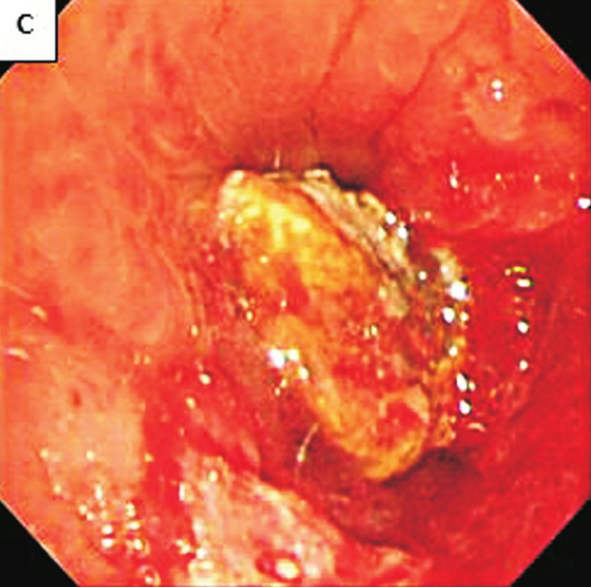
INTRODUCTION by flexible bronchoscopy, cryoprobe, and laryngeal for-
ceps.
Adult foreign body aspiration (FBA) typically presents
with a choking event followed by persistent coughing but CASE REPORT
not uncommonly, it can mimic more chronic diseases
such as chronic obstructive pulmonary disease, asthma, A 77-year-old woman was admitted to our emergency
and obstructive pneumonia when the initial event goes room with dyspnea. She had a history of marginal resec-
unnoticed (for example in elderly patient with an altered tion and colostomy due to sigmoid perforation in 2010, a
mental status).1 When an accurate diagnosis is not estab- takedown operation for colostomy repair in 2011, multi
lished immediately, entrenched foreign bodies (FBs) may infarct dementia from 2014, and most recently, percu-
lead to recurrent pneumonias, bronchiectasis, recurrent taneous coronary intervention at the mid left anterior
hemoptysis, pneumothorax, lung abscesses, pneumo- descending coronary artery and asthma aggravation
mediastinum and other complications.2,3 Extraction of as- and pneumonia, in August, 2019. After discharge, she
pirated FBs should be undertaken as soon as possible in remained on in bed rest with her respiration assisted by
order to alleviate acute symptoms and prevent long term oxygen (2 L/min) via nasal prong.
complications.4 A week before, she had a fever up to 38.5°C and inter-
This is a report of a 77-year-old female, who initially mittent thick sputum. These symptoms worsened and
manifested dyspnea, fever and sputum. These are com- eventually she had dyspnea. On examination, dyspnea
mon symptoms of pneumonia but she turned out to have was aggravated, and a steady increase of oxygen was
a FB in her left main bronchus. While examining and needed to maintain saturation. Chest X-rays (Fig. 1A, B)
attempting to remove it, we applied several steps to ap- and computed tomography (CT) scan (Fig. 1C, D) taken
proach it and in the end, we carefully removed it through after admission showed signs of left pneumonia, but her
A B
Fig. 1. Simple chest X-ray, anterior-
posterior view, (A) Initial X-ray on
Admission day and (B) Diffuse pneu
monic infiltration in left after 2 days
of admission. Chest computed tomo
graphy (CT), axial view showing
ground-glass opacity at multiple
sites as signs of pneumonia, unspe
cified, (C) CT obtained with media
stinal window setting (D) CT obtained
C D
with lung window setting.
56 Medical Lasers; Engineering, Basic Research, and Clinical Application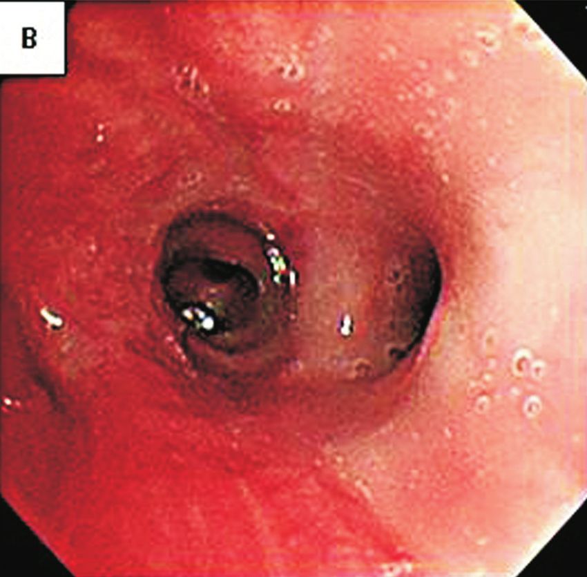
Aspirated Foreign Body Removal by Cryotherapy
Hyoyeon Kim, et al.
Case Report
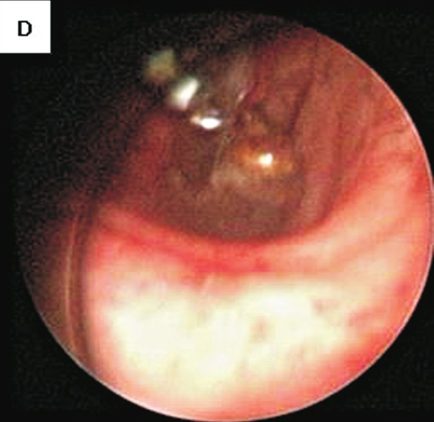
symptoms did not improve even after changing intrave- to freeze it (Fig. 3B). The FB material was carefully pulled
nous antibiotics. She was moved to the intensive care unit out and disconnected from the cryoprobe at the carina.
from the general ward for close monitoring. Removal was completed with using laryngeal forceps (Fig.
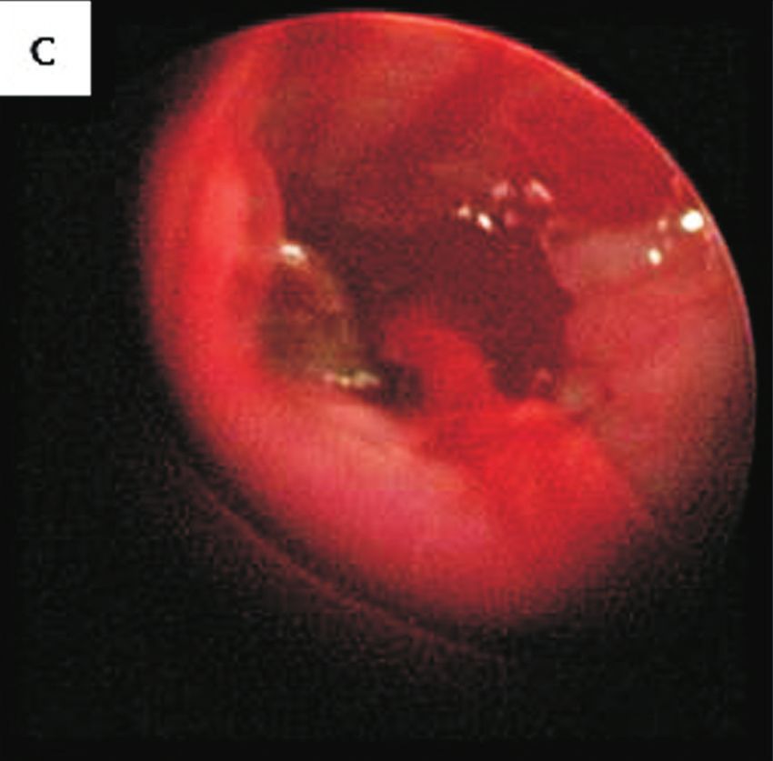
On the third day of admission, flexible bronchoscopy 3C, D).
was done (Fig. 2A, B). A moderate amount of mucopuru- Four days later, the patient’s vital signs were stabilized
lent secretion was found in the right bronchial tree. Mul- and follow-up rigid bronchoscopy was done. Multifocal
tifocal hyperemic edematous mucosal change was found hyperemic edematous mucosal change with erosion was
in the right bronchial tree. We found a stone like FB at the found in the right bronchial tree. Similarly, multifocal hy-
orifice of left main bronchus, but failed to remove it, due peremic mucosal change was found in the left bronchial
to patient’s immediate hypoxemic state (SpO2 88%, via tree, as a sign of acute bronchitis. There were no other
HFNC FiO2 0.9) (Fig. 2C, D). She was referred to the oto- complications.
rhinolaryngology department for exploration and removal
of the FB. DISCUSSION
Under general anesthesia, a rigid bronchoscopic exam
was done. There was FB material and pus at the left main Aspiration of a FB is a potentially life-threatening emer-
bronchus as was seen in at previous flexible broncho- gency and 75% of cases occur in children younger than
scopic exam (Fig. 3A), but we found it difficult to approach 3 years of age.5 FBA does happen in adults and elderly
the FB due to the bronchus curvature. We then applied people as well.
a flexible bronchoscope to fit in the bronchial curvature. Most adult patients with FBA have obvious risk factors
Using a cryoprobe guided by the pulmonologist, an ad- for aspiration including neurological deficits with swallow-
equate approach to the lower tracheal level was achieved ing difficulties or altered mental status, neuromuscular
and the tip of the cryoprobe touched the surface of the FB disease, intoxication, or an iatrogenic cause. FB aspiration
A B
Fig. 2. Bronchoscopy, (A) Mucopu
rulent secretion in the right bron
chial tree, (B) Multifocal hyperemic
edematous mucosal change in the
right bronchial tree, partial exposure
of foreign body (FB) fixed in the
right bronchial tree Rigid bron
choscope, (C) Exposure of FB fixed
C D in the right bronchial tree, (D) Trials
of FB removal with laryngeal forcep.
VOLUME 10 NUMBER 1 March 2021 57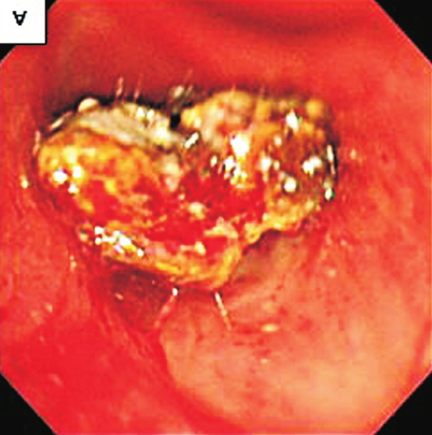
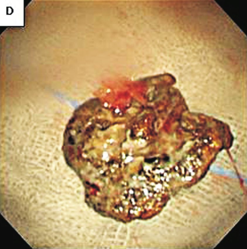
A B
Fig. 3. Flexible bronchoscope and
cryoprobe, (A) FB stuck at left bron
chial tree, (B) Successful cryoadhe
sion of the probe and the surface of
FB, (C) Detached FB from cryoprobe
at carina, (D) Completely removed
FB, which later turned out to be an
aspirated old dental metal filling
C D
material.
risk appears to increase with age. Iatrogenic FB aspira- lobar consolidation in the majority of patients who have
tion in adults is most commonly related to dental proce- FBA.9
dures requiring local anesthesia and supine positioning, Chest CT is more sensitive for identification of FBs,
and is also associated with tracheostomy care.6 helps with procedural planning, and has become the gold
After FBA occurs, the FB can settle into 3 anatomic standard of imaging studies when a FBA is suspected.
sites, the larynx, trachea, or bronchus. Of aspirated for- The most important aspect of successful FB extraction
eign bodies, 80 to 90% become lodged in the bronchi. In is pre-procedural planning, considering the character-
adults, bronchial FBs tend to be lodged in the right main istics of the FB, its size, location in the airway, and the
bronchus because of its lesser angle of convergence patient’s comorbid conditions. In general, rigid bronchos-
compared with the left bronchus and because of the copy is preferred in cases of acute respiratory distress,
location of the carina left of the midline.7 Larger objects while flexible bronchoscopy allows for a more compre-
tend to become lodged in the larynx or trachea. In adults hensive airway survey and has an overall 90% success
approximately 40% of FBs are found in the left bronchial rate for retrieval.10,11 In our case, the use of the flexible
tree and only 5 to 11% remain in the trachea.8 cryoprobe was shown to be very effective, for removing
For examination, standard posteroanterior and lat- FB, especially as the FB had an organic portion with sig-
eral chest radiographs should be obtained in all patients nificant granulation tissue around it.12-14 In this technique,
where FBA is suspected. Radiographs directly identify FB the cryoprobe is advanced towards the FB through the
in only 25% of patients, because only a small proportion of working channel. After contact, the probe is activated
objects such as coins, nails, teeth, and dental appliances to rapidly freeze the object and cause it to adhere to the
are radiopaque. Nonetheless, chest radiographs dem- cryoprobe tip.15,16 Once cryoadhesion has occurred, the
onstrate indirect findings of atelectasis, hyperinflation, or object can be gently removed from the airway. Care must
58 Medical Lasers; Engineering, Basic Research, and Clinical Application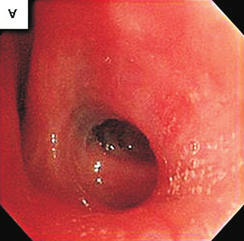
Aspirated Foreign Body Removal by Cryotherapy
                                                                                                                        Hyoyeon Kim, et al.

                                                                                                                                              Case Report
be taken not to touch the bronchial walls with the probe,                9. Wiseman NE. The diagnosis of foreign body aspiration in child-
as this can result in removal of bronchial tissue, bleeding,                hood. J Pediatr Surg 1984;19:531-5.
and rarely, airway perforation. The adherent ability of FBs             10. David AP, Xu MJ, Rosbe KW, Meyer AK, Gesthalter YB, Chan
is primarily based on their water content.10                                DK. Cryoprobe retrieval of an airway foreign body: a case
   After removal of the FB, it is imperative that the bron-                 report and literature review. Int J Pediatr Otorhinolaryngol
choscopist performs a second airway survey for additional                   2019;125:79-81.
FBs and other side effects (such as granulation or bron-                11. Park MW, Baek SK, Jung KY. Laser epilation for unwanted hair
chial stenosis.17-21                                                        in the larynx. Med Laser 2012;1:31-3.
   In summary, for patients with FBs in tricky positions in             12. Kim J, Lee YI, Lee JH, Oh SH, Lee SE, Kim YK. Successful treat-
the trachea, a flexible cryoprobe can be an alternative way                 ment of post-operative keloid with combined cryotherapy and
to remove them. We would like to report this rare case of                   ablative fractional CO2 laser. Med Laser 2020;9:58-61.
using a cryoprobe for FB removal with the bibliography                  13. Jung SY, Hwang NH, Yoon ES, Park SH. Erbium:yttrium alu-
attached.                                                                   minum garnet laser treatment for xanthelasma palpebrarum.
                                                                            Med Laser 2017;6:24-8.
REFERENCES                                                              14. Lee SJ, Cho S, Kim YK, Cho SB. Use of a 595-nm pulsed-dye la-
                                                                            ser to treat post-procedure ecchymoses. Med Laser 2014;3:87-
 1. Hewlett JC, Rickman OB, Lentz RJ, Prakash UB, Maldonado F.              9.
    Foreign body aspiration in adult airways: therapeutic approach.     15. Kim J, Lee YI, Lee JH, Oh SH, Lee SE, Kim YK. Successful treat-
    J Thorac Dis 2017;9:3398-409.                                           ment of post-operative keloid with combined cryotherapy and
 2. Oliveira CF, Almeida JF, Troster EJ, Vaz FA. Complications of           ablative fractional CO2 laser. Med Laser 2020;9:58-61.
    tracheobronchial foreign body aspiration in children: report of 5   16. Sehgal IS, Dhooria S, Behera D, Agarwal R. Use of cryoprobe
    cases and review of the literature. Rev Hosp Clin Fac Med Sao           for removal of a large tracheobronchial foreign body during
    Paulo 2002;57:108-11.                                                   flexible bronchoscopy. Lung India 2016;33:543-5.
 3. Mun IK, Ju YR, Lee SJ, Woo SH. A case of bilateral tension          17. Rafanan AL, Mehta AC. Adult airway foreign body removal.
    pneumothorax after the successful CO2 laser-assisted removal            What's new? Clin Chest Med 2001;22:319-30.
    of a bronchial foreign body in a child. Med Laser 2020;9:65-70.     18. Lee SJ, Chung PS, Chung SY, Woo SH. Transoral laser excision
 4. Cho JG, Park MW, Baek SK, Kwon SY, Jung KY, Woo JS. CO2                 of a pyriform sinus cyst. Med Laser 2019;8:84-6.
    laser in the diagnosis and treatment of early cancer of the vocal   19. Lee SJ, Chung PS, Chung SY, Woo SH. Respiratory protection
    fold. Med Laser 2012;1:21-7.                                            for LASER users. Med Laser 2019;8:43-9.
 5. Blanco Ramos M, Botana-Rial M, García-Fontán E, Fernández-          20. Hong JC, Lee KD. Transoral laser surgery for supraglottic car-
    Villar A, Gallas Torreira M. Update in the extraction of airway         cinoma. Med Laser 2013;2:1-7.
    foreign bodies in adults. J Thorac Dis 2016;8:3452-6.               21. Cho JG, Park MW, Baek SK, Kwon SY, Jung KY, Woo JS. CO2
 6. Wolkove N, Kreisman H, Cohen C, Frank H. Occult foreign-                laser-induced endotracheal fire. Med Laser 2013;2:36-9.
    body aspiration in adults. JAMA 1982;248:1350-2.
 7. Wu TH, Cheng YL, Tzao C, Chang H, Hsieh CM, Lee SC. Long-
    standing tracheobronchial foreign body in an adult. Respir Care
                                                                         How to cite this article: Kim H, Byun G, Lee SJ, Woo SH.
    2012;57:808-10.
                                                                         Removal of a bronchial foreign body by bronchoscopic
 8. Hada MS, Samdhani S, Chadha V, Harshvardhan RS, Prakash
                                                                         cryotherapy: a case study. Med Laser 2021;10:55-59. https://doi.
    M. Laryngeal foreign bodies among adults. J Bronchology In-
                                                                         org/10.25289/ML.2021.10.1.55
    terv Pulmonol 2015;22:145-7.

                                                                                                     VOLUME 10 NUMBER 1 March 2021      59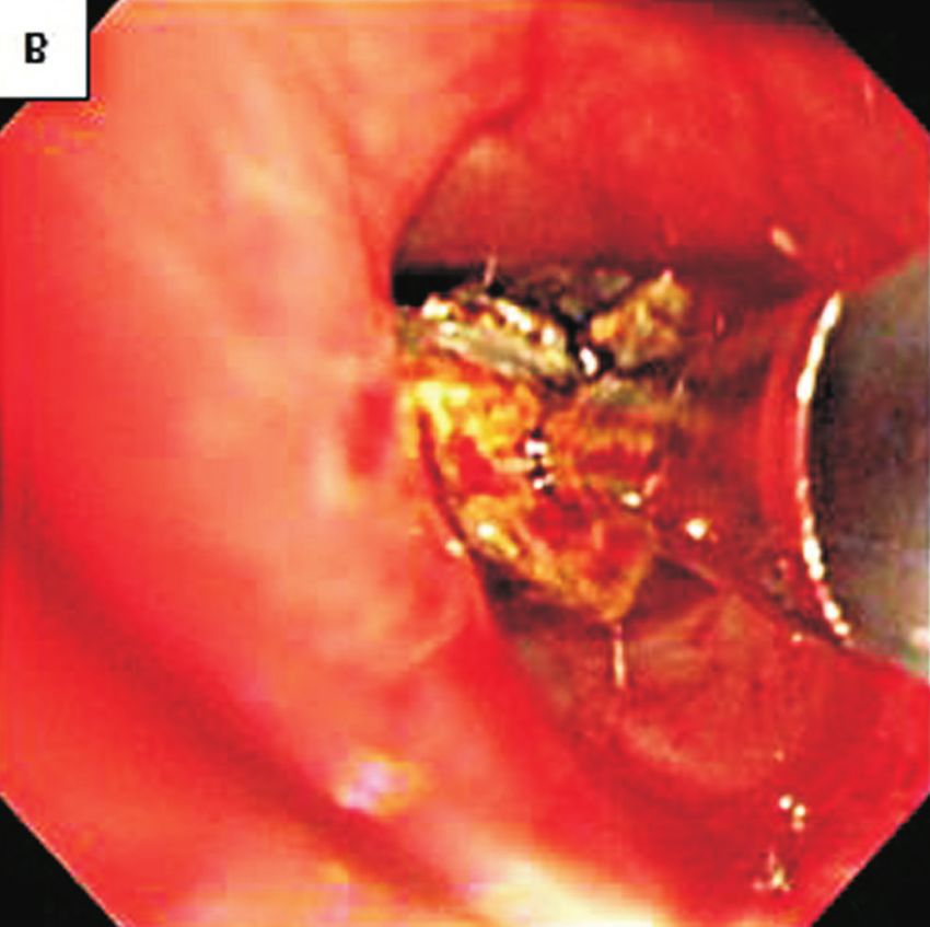
You can also read