The Fetal Study on Cranial and Spinal Dysraphism of Neural Tube
←
→
Page content transcription
If your browser does not render page correctly, please read the page content below
Quest Journals
Journal of Medical and Dental Science Research
Volume 8~ Issue 4 (2021) pp: 33-38
ISSN(Online) : 2394-076X ISSN (Print):2394-0751
www.questjournals.org
Research Paper

    The Fetal Study on Cranial and Spinal Dysraphism of Neural
                               Tube
                              *
                                Dr.A.Himabindu1, Dr.N.Bhimaidevi2
        1.Professor & HOD Department of Anatomy, GITAM Institute of Medical Sciences &Research,
                                      Rushikonda, Visakhapatnam.
     2. Professor & HOD Department of Anatomy, Maharajah’s Institute of Medical Sciences ,Nellimarla,
                                             Vizianagarum
                               *Corresponding Author: Dr.A.Himabindu

ABSTRACT:
Neural tube defects (NTD) are more common congenital birth defect affecting the babies all over the world. .It
is one of the causes of fetal mortality and morbidity .Defective neurulation process during embryonic period
leads to this condition. Based on the level of neural tube involvement the defects are divided as cranial
dysraphism and spinal dysraphism.Cranial dysraphism include lethal anencephaly ,encephalocele and
iniencephaly .In spinal dysraphism ,failure in the fusion of caudal end of neural tube leads to meningocele and
myelomeningocele.These conditions are associated with mesodermal involvement leading to bony defects .The
present study was done on 50 human fetuses that were collected for development of museum in Anatomy.In
this processmore number of fetuses with neural tube defects were identified. The fetuses were separated based
on the level of neural tube involvement. In anencephaly,encephalocele and rare iniencephaly in which the
cranial end of neural tube is involved and in meningocele and myelocele the caudal end neural tube is involved.
All these fetuses were studied further. Manifestation of NTD is multifactorial that have genetic or
environmental basis with high recurrence rate. Neural tube defects are usually diagnosed in early weeks of
intrauterine life by ultrasound examination .In the various forms of neural tube defects,anencephaly is
incompatible with life and depending on the level of exposure of neural tissue ,open spinal defects will survive
with permanent disability.Folic acid is given as a supplement to prevent the neural tube defects and the role of
folic acid in NTD’s was studied by authors and they expressed that folic acid is important in nucleic acid
formation and in the metabolic activity of enzymes. The deficiency of folic acid blocks all this process and leads
to formation of neural tube defect. So pregnant woman with previous history of neural tube defect are advised to
take folic acid before planning the pregnancy and continue for three months after conception to prevent the
recurrence of neural tube defects..
KEY WORDS: Neural tube,Mesoderm, Anencephaly, Meningocele, Folic acid

Received 29 Mar, 2021; Revised: 10 Apr, 2021; Accepted 12 Apr, 2021 © The author(s) 2021.
Published with open access at www.questjournals.org

                                          I.     INTRODUCTION
         Neural tube defects(NTD) are most common congenital anomalies .It affects approximately 300,000
babies
          every year worldwide.NTD are most serious congenital anomaly involving the nervous system
leading to fetal mortality and morbidity.Its incidence is 1-5 /1000 live births. Development of nervous system
starts in the embryonic period of intrauterine life. Under the influence of notochord, neural groove is formed
from neuroetoderm.The two ends of neural groove forms neural tube that closes in the middle and extends
cranially and caudally leaving anterior neuropore and posterior neuropore .The anterior neuropore closes by
25th day and posterior neuropore closes by 27th day ,that closes the neural tube .Neural tube formation and
closure occur at 4th week of intrauterine life.Failure in the closure of the neural tube disrupts the differentiation
of central nervous system and formation of vertebral arches that derives from mesoderm. This leads to
developmental malformations along the neuroaxis and manifest as neural tube defects .Neural tube defects are in
the form of open type if the neural tissue is exposed and closed type if the defect is covered by skin. Failure of
cranial neural tube closure leads to cranial dysraphism which manifests as anencephaly with defective brain
development ,encephalocele with midline skull defects and iniencephaly with defective folding of
*Corresponding Author: Dr.A.Himabindu                                                                      33 | Page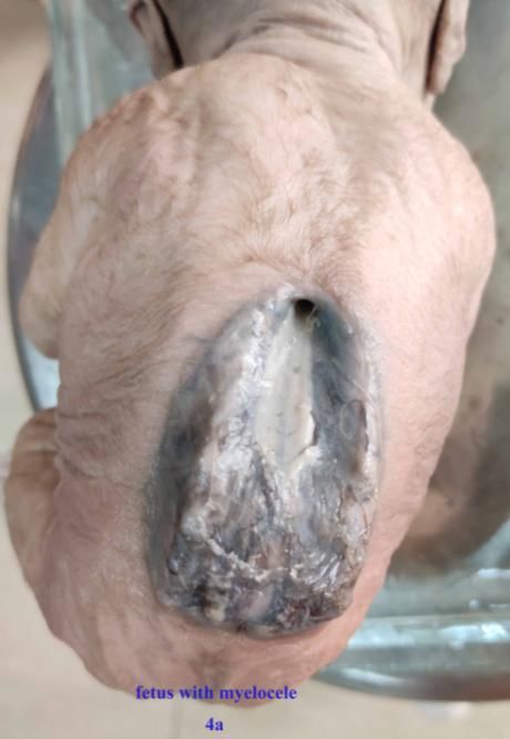
The Fetal Study on Cranial and Spinal Dysraphism of Neural Tube

embryo.Failure of caudal neuropore closure leads to spinal dysraphism that manifest as myelocele and
myelomeningocele.. A rare form of NTD is craniorachischisis that results from failure of the neural tube closure
over the entire body axis. The two most common neural tube defects are spina bifida and anencephaly (1).
Spina bifida is an open form of spinal dysraphism in which the fetal spinal column does not close completely
with exposed neural tissue and anencephaly is most common cranial dysraphism with absence of parts of brain
and the overlying skull.
          Anencephaly is a cephalic neural tube defect associated with absence of the mesodermal tissue dorsal
to the neural elements leads to failure in the bony skull development . The condition is lethal and the baby dies
within few hours .It can be diagnosed easily in the antenatal period by ultrasound imaging.
          Encephalocele with midline skull defects ,can be termed as crania bifida. Crania bifida is less
common compared to spina bifida.Encephaloceles are two types -anterior and posterior .The anterior
encephalocele is more common with protrusions from the face as proboscis and posterior encephalocele
present in the occipital region as occipital encephalocele .It may have present with infratentorial or supra
tentorial brain contents.Iniencephaly is a very rare neural tube defect , characterized by an occipital bone defect
,cervical dysraphism ,fixed retroflexion of the fetal head and severe lordosis of cervicothoracic spine.
          Spinal dysraphisms is due defective caudal neural tube closure associated with mesenchymal
defects.They can be diagnosed at birth or in early infancy.Open spinal dysraphisms include meningocele
,meningomyelocele with exposed neuronal structures without skin covering and accounts for 95% of spinal
dysraphism. This caudal neural tude defect may present at any region but lumbosacral involvement is more
common.Closed spinal dysraphisms are minor defects that do not have any exposed neural structures and it is
covered by skin.

                                 II.   MATERIALS AND METHODS
         Fetuses were collected from Victoria General Hospital,Visakhapatnam for the development of
Anatomy museum. During this process anomalous fetuses of various system were found. Of these fetuses with
neural tube defects were separated and studied further .The fetuses were separated based on the level of neural
defect that was present in the cranium and spinal column.The fetuses that were presented with cranial defects
were studied to know the level of defect and involvement of nervous system and the same was observed in the
fetuses of spinal defects.The extent of involvement of nervous system and level of bony involvement was
studied and noted.

                                        III.    OBSERVATIONS
          The present study identified fetuses with cranial dysraphism like anencephaly,encephalocele and rare
iniencephaly .This study found more number of fetuses with spinal dysraphism that manifestated as
meningomyelocele at various regions, more commonly at lumbar region.
          Out of 50 fetuses collected, two fetuses with anencephaly ,one fetus with encephalocele and a fetus
with a rare neural tube defect of iniencephaly were observed . Fifteen fetuses presented with spinal and bony
defects at various vertebral levels .The defect may be at cervical ,thoracic ,lumbosacral region, more commonly
at lumbosacral region.
          A male fetus was presented with anencephaly with absent cranial vault . The face was presented with
typical frog eye appearance of anencephaly. The cranial fossae appeared empty with macerated brain tissue
.Few contents of hind brain were observed close to the foramen magnum and it continued in the vertebral canal
. (fig.1)

*Corresponding Author: Dr.A.Himabindu                                                                    34 | Page
The Fetal Study on Cranial and Spinal Dysraphism of Neural Tube
Fig.1Showing Anencephalic fetus
Encephalocele was observed in a full term female fetus with a bulky mass at occipital region .No other
external anomaly was found in this fetus. On dissecting the mass ,at occipital region,brain tissue of infratentorial
components werefound. The spinal cord was exposed in the cervical region due to defective vertebral bodies. In
this defect , the skeletal elements were not formed properly due to defective mesoderm. So the skeletal defect
leads to herniation of brain tissue.
Fig.2 fetus showing posterior encephalocele
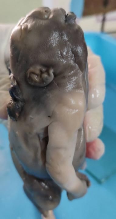
A rare neural tube defect of this study was iniencephaly.This was found in a male fetus which was
presented with huge face with retroflexion of head ,the face pointed towards the roof. The neck was absent
and the skin of chest is continuous with face. Normally iniencephalic fetus presents with huge head with normal
cranial vault,but this study presented an iniencephalic fetus with anencephaly and craniorachisis a rare
association. .(fig.3)
*Corresponding Author: Dr.A.Himabindu 35 | Page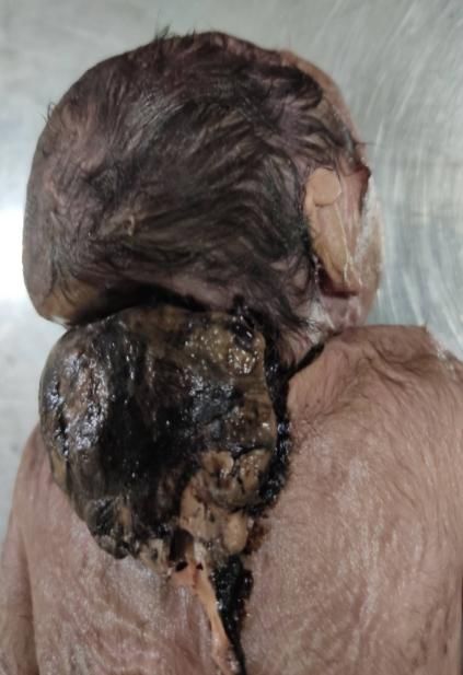
The Fetal Study on Cranial and Spinal Dysraphism of Neural Tube
Fig.3 Iniencephaic fetus-A Rare neural tube defect
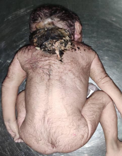
Spinal dysraphism was observed in the form of spina bifida,myelocele and myelomeningocele.More
number of fetuses were identified with myelocele with bifid spine with exposed spinal cord to the surface. This
defect was observed in cervicothoracic and more commonly at lumbosacral region. These fetuses showed
unfused lamina of vertebra with exposed vertebral canal leading to exposed spinal cord and spinal nerves.
(fig.4a)
Meningomyelocele was also observed in this study.In these fetuses the contents of vertebral canal were
exposed and covered by meningeal layers .so a sac with neural structures was observed through the vertebral
defect.(fig.4b)
Fig.4 fetuses with spinal dysraphism
*Corresponding Author: Dr.A.Himabindu 36 | Page
The Fetal Study on Cranial and Spinal Dysraphism of Neural Tube

                                               IV.DISCUSSION
          Neural tube defect is a multifactorial disorder that occurs due genetic or environmental factors with
high recurrence rate. Monogenic inheritance of NTD shows that the disease may be produced by single genes,
particularly genes present in the X gene. Environmental factors such as socio-economic status, nutritional
deficiency, hypothermia, gestational diabetes, use of anticonvulsant drugs and insulin are well known risk
factors.(2)
          Neural tube defects are usually diagnosed in early weeks of intrauterine life through imaging
techniques. Of all NTDs, anencephaly is incompatible with life and the fetus will die few hours after birth and
the child with open spinal defects will survive with permanent disability
          Neurulation is the process by which neural tube is formed around 22-23 days (2). In mammals,
neurulation occurs in two phases: primary and secondary neurulation. Primary neurulation occurs at 3rd and 4th
week of intrauterine life and forms the entire neural tube leaving cranial and the caudal neuropore. Secondary
neurulation occurs at tail bud region (3). Earlier it was proposed that the fusion of neural tube starts from a
single initiation site and progresses both cranially and caudally towards the cranial and caudal neuropores. Later
multiple sites of neural tube fusion were proposed and extensive studies were made in rats and mice. According
to Sakai (4) in mice there are four sites at which fusion of neural tube takes place. Site 1 appears in the cervical
region at the caudal part of hindbrain and proceeds bidirectionally. Site 2 appears at the junction of
prosencephalon and mesencephalon and proceeds bidirectionally. Site 3 appears at the cranial end of neural
plate and proceeds caudally. Site 4 appears at the caudal end of neural plate and proceeds cranially.Van Allen et
al., (5) proposed multisite closure model of neural tube with five sites of closures in humans based on the
observation of types and occurrence of NTDs. Of the five sites 3 are present in the cranial region and 2 are
present in the spinal region
          Anencephaly occurs in 3 phases,initially the cranial part of the neural tube fails to close followed by
protusion of the developing brain parts and its exposure to the amniotic fluid and at last degeneration of the
exposed part results in anencephaly (6). Occipital encephalocele occurs when the surface ectoderm does not
separate from the developing neural tube at the occipital region. This leads to defects in the formation of
occipital bone. Encephalocele is associated with other syndromes like Meckel – Gruber syndrome, Walker –
Warburg syndrome, Dandy – Walker syndrome, Joubert syndrome, Trisomy 13 and Amnion rupture sequence
(6).Iniencephaly was derived from greek word inion that means back of the neck and encephalos to
brain.Persistence of the embryonic cervical lordosis at the third week leads to failure of closure of the neural
tube and abnormal development of the rostral portion of the notochord and somites of the cervicooccipital
region is the cause for iniencephaly .(7)
          Open neural folds in the spinal region prevent the sclerotome-derived vertebral arches from covering
the neuroepithelium, the consequent opening in the vertebral column giving rise to the term spina bifida (8). The
neural tissues may be contained within a meninges-covered sac that protrudes through the open vertebrae or
exposed directly to the amniotic fluid. Babies born with open spina bifida usually survive with appropriate
medical care but suffer with neurological impairment, the severity depends on the level of the lesion.A less
well-defined group of closed spinal NTDs in which the vertebral arches are malformed but covered by skin may
lead a normal life.
          Open defects affecting the brain like anencephaly, craniorachischisis are lethal before or at birth.
Encephalocele can also be lethal depending on the extent of brain damage during herniation.
          Open spina bifida is compatible with life leaving neurological impairment below the level of the lesion.
          Closed spinal lesions are generally less severe and can be asymptomatic, as with spina bifida occulta
which is considered a variant of normal. However, lumbosacral spinalcord tethering can lead to lower limb
motor and sensory deficits, and a neuropathic bladder.
          Clinical observations suggest a higher frequency of NTDs in offspring of consanguineous couples as
          well as in twin pregnancies(9), pointing towards a strong genetic contribution to the etiology of
NTD.Consanguinity is thought to contribute to Mendelian autosomal recessive conditions by inheritance of
pathological mutations and also to polygenic multifactorial disorders such as NTDs by increasingthe load of risk
alleles.(10)
          Folic acid deficiency during antenatal period is thought to be a cause of NTD.The relation between
folic acid deficiency and NTDs in humans was first reported by Hibbard in1964. (11) He observed higher
incidence of aberrant folate metabolism in women who had pregnancies associated with foetal malformations.
Smithells and co-workers in 1976 observed that maternal red blood cell folate levels were significantly lower in
fetus with NTDs in comparison to those who did not have NTDs.(12) This observation led to the possibility of
preventing neural tube defects by periconceptional vitamin supplementation(13).
          Folic acid is present in the blood as tetrahydrofolate .THFA has an important role in the metabolism of
one carbon (1C) groups and THFA is the carrier of one carbon groups which are involved in metabolic
pathways and nucleic acid synthesis. So genetic defects of the enzymes involved in folate and 1C metabolism

*Corresponding Author: Dr.A.Himabindu                                                                     37 | Page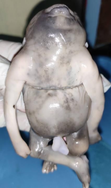
The Fetal Study on Cranial and Spinal Dysraphism of Neural Tube

have been identified in NTDs in clinical and experimental settings. Supplementation of folic acid may overcome
these metabolic blocks and reduce the risk of NTDs.(14)
          A study conducted by the British Medical Research Council on women with earlier neural tube defects
and the effect of supplementing folic acid in subsequent pregancies.This study showed that supplementation of 4
mg folic acid significantly reduced the risk of recurrence of NTDs.(15) In our country , women are more prone
to infections, anemia and other deficiency conditions, so it is preferable to recommend the folic acid on a
regular basis to women of child bearing age especially to those who are at high risk state.

                                                        V.CONCLUSION
          Neural tube defects are most common congenital anomaly of defective neural tube formation during
embryonic period of intrauterine life. It is one of the causes of fetal mortality and morbidity with high
recurrence rate. It may have either genetic and environment factors in causation of this condition. Folic acid
deficiency was identified in the manifestation of various forms of neural tube defects and supplementation of
folic acid during pre and post conceptional period will help in the prevention of this anomaly

                                                 ACKNOWLEDGEMENT:
 We acknowledge the dissection hall attendants, Mr.Simhadri,Mr.Srinivasarao and Mr.Visweswararao for their
timely help during this study.

                                                         REFERENCES
[1].    Cragan JD, Roberts HE, Edmonds LD. Surveillance for anencephaly and spina [3] bifida and impact of prenatal diagnosis in United
        States. 1985-1994. Mortal Morb Wkly Rep. 1995;44:113
[2].    Moore KL, Persaud TVN. The Developing Human. 8th ed. Saunders Elsevier; 2008
[3].    Eric RD, Timothy MG, Heather CE, John RG, Michel V, Marcy CS. Human Neural [6] Tube Defects: Developmental Biology,
        Epidemiology, and 7.Genetics. Neurotoxicol Teratol. 2005;27(3):515–24.
[4].    Sakai Y. Neurulation in the mouse: manner and timing of neural tube closure. [7] Anat Rec. 1989;223:194–203.
[5].    Van Allen MI, Kalousek DK, Chernoff GF, Juriloff D, Harris M, Mc Gillivray BC, et [8] al. Evidence for multi-site closure of the
        neural tube in humans. Am J Med Genet. 1993;47:723–43.
[6].    Hans JTD, Martin L, Akira H. Clinical Neuroembryology: Development and Developmental Disorders of               the Human Central
        Nervous System. 2nd ed. Heidelberg: Springer; 2014
[7].    R. Romero, P. Jeanty, G. Pilu, A. Ghidini, and J. C. Hobbins, “Iniencephaly,” in Prenatal Diagnosis of Congenital Anomalies, p. 65,
        Appleton & Lange, Norwalk, Conn, USA, 1988.
[8].    Copp AJ, Stanier P, Greene ND. Neural tube defects: recent advances, unsolved questions, and controversies. Lancet Neurol. 20 13;
        12(8):799–810
[9].    Budhiraja S, Dahiya P, Ghei M, Gathwala G. Neural tube defect in dizygotic twins. Pediatr Surg Int 2002; 18:211-212
[10].   Kavitha Sargunam, Dr. Renu Boora Nutri-genetic determinants of neural tube defects in India International Journal of Universal
        Science and Engineering 2016, Vol. No. 2, Jan-Dec pg-120-135
[11].   Hibbard BM. The role of folic acid in pregnancy: With particular reference to anaemia, abruption and abortion. J Obstet Gynaecol
        Br Commonwlth 1964;71:529-542.
[12].   Smithells RW, Sheppard S, Schorah CJ. Vitamin deficiencies and neural tube defects. Arch Dis Child 1976;51:944-950.
[13].   Smithells RW, Sheppard S, Schorah CJ, Seller MJ, Nevin NC, Harris R, Read AP, Fielding DW. Possible prevention of neural-tube
        defects by periconceptional vitamin supplementation. Lancet 1980;1:339-340.
[14].   Scaria T Pulikkunnel*, SV Thomas Neural Tube Defects : Pathogenesis and Folate Metabolism JAPI • VOL. 53 • FEBRUARY
        2005 127-135
[15].   MRC Vitamin Study Research Group. Prevention of neuraltube defects: Results of the Medical Research Council vitamin study.
        Lancet 1991;338:131-137

*Corresponding Author: Dr.A.Himabindu                                                                                          38 | Page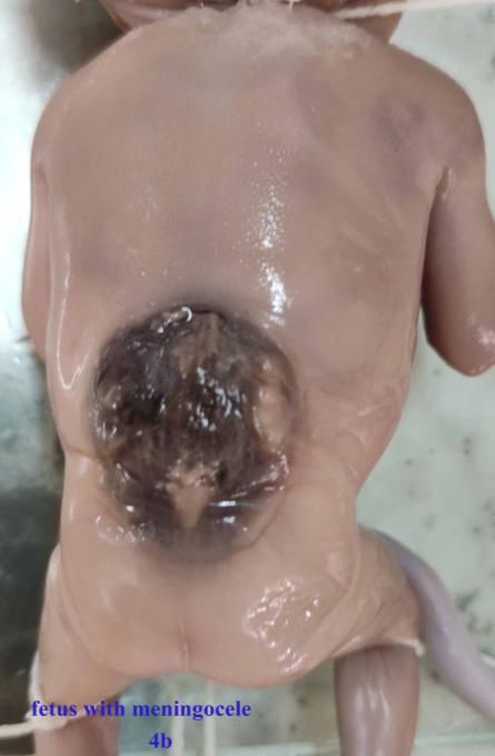
You can also read