Efficacy of Chitosan Immune Response Against - Book_IJFMT_April-June 2020.indb
←
→
Page content transcription
If your browser does not render page correctly, please read the page content below
Indian Journal of Forensic Medicine & Toxicology, April-June 2020, Vol. 14, No. 2   2113

                 Efficacy of Chitosan Immune Response Against
                    Listeria Monocytogenes Infection in Mice

             Sura Ayed Radam1, Inam Badr Faleh1; Osama Faid allah Atshan2, Mustafa Salah Hasan3
  1
      Department of pathology, 2Department Microbiology, College of Veterinary Medicine, University of Baghdad,
        3
          Department of Internal and Preventive Medicine, College of veterinary medicine, university of Fallujah

                                                          Abstract
       The present research aimed to study the effect of dietary chitosan supplementation against murine
       experimentally infection by Listeria monocytogenes.

       forty mice were divided equally into 4 groups. The 1st and 2nd groups fed on diet supplement with chitosan
       (1mg/kg diet) and (1.5mg /kg diet) for (4) weeks respectively, While 3rd and 4th groups considered as control
       positive and negative groups. At (4) weeks the first three groups were inoculated intraperitoneally i/P with
       (0.2) ml (1×10⁹) CFU/ml, while the 4th group (control negative) inoculated with (0.2) sterile normal saline.

       At (7) days post infection, the result revealed diet one of mice in each control positive and treated group at
       (24hrs.) post infection with heavy bacterial isolation from brain, spleen and liver of infected positive group
       and mild to absent bacterial isolation in the 1st and 2nd group respectively.

       Grossly presence of severe congestion in the internal organs with necrotic foci seen on the splenic surface of
       infected positive control while the characteristic feature in the treated infected group was hepatosplenomegaly.

       Sever pathological changes were noticed in the infected positive control group characterized by suppurative
       inflammation with necrosis accompanied with lymphoid depletion and amyloid like substance deposition
       while the main lesion in treated infected groups showed granulomatous lesion, lymphoid hyperplasia and
       mononuclear cells infiltration with heavy bacterial isolation from brain, spleen and liver of infected positive
       group and mild to absent bacterial isolation in the first and second group respectively, We concluded that
       chitosan stimulated and improve the immune responses in mice against Listeria monocytogenes infection.

       Key word: chitosan, Listeria monocytogenes, immunized, mice, pathology.

                       Introduction                                    Chitosan is a modified natural carbohydrate polymer
    Listeria monocytogenes is regular Gram-positive               derived from chitin, it have many medical uses because
motile from, rod with rounded ends, its cells found               their ability to reduce bleeding also help deliver drugs
as single units or short chains or may be arranged                through the skin also in limiting of fat absorption(6),
in V, L and Y forms or in palisades (1). Listeria                 also has been bio adhesive property for that used as a
monocytogenes does not produce spores and capsules                safe excipient formulations of drug, it has been used in
are not formed (2). Spread in nature where, exists                dentistry because adhere ability to hard and soft tissues
largely in decaying vegetation, soil, animal feces,               also uses in orthopedics, ophthalmology and in surgical
feed and water as make it one of the major pollutants of          procedures, it adheres to epithelial tissues and mucus
food and play essential role in transmitted of                    coat present on tissues surface also has a antifungal
infection between humans and animals (3) also infection           or antibacterial, antineoplastic and anticholestermic
by Listeria monocytogenes can be haematogenous                    action(7).
spread directly from the mother to fetus (4, 5)
                                                                                    Material and Method
                                                                        Chitosan was obtained from university of Al-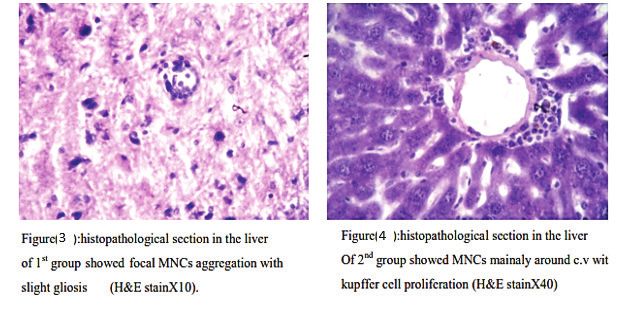
2114 Indian Journal of Forensic Medicine & Toxicology, April-June 2020, Vol. 14, No. 2
Bahasra, collage of veterinary medicine. Commercial The characteristic lesion in hepatic tissue of control
assorted pellets were grinded by food grinder and positive show aggregation of PMNCs cells in liver
weighed(1) gm and (1.5)gm of Chitosan was added paranchyma (suppurative foci) mainly in portal area
to each kilogram of grinded pellets mixed well and accompanied with atrophy of some hepatic cords together
converted into paste which passed through meat grinder with sinusoidal dilation and cellular infiltration in their
to mould the paste into the original pellets from, left lumen, The splenic tissue showed destructive changed
exposed to dry in room temperature (8). The Listeria with variable degree of lymphoid depletion in the white
monocytogenes isolate was obtained from the unit of pulp, other section showed formation of multiple cystic
Zoonotic diseases in the College of Veterinary Medicine, cavities containing cellular debris together with focal
the isolate confirmed by some biochemical tests and amyloid like substances deposition, The brain tissue
gram stain according to (9). expresses sever neuronal degeneration and apoptosis
accompanied with nuclear pyknosis and appearance of
A total number(n=40) male white Swiss BALB/C hypertrophic swelling astrocytes (gamistocyte), another
mice which obtain from the (National Center of section showed irregular cystic cavities with neuronal
Researches and Drugs Monitor in Baghdad); then vaculation.
divided into fourth groups. The 1st group (n=5) mice
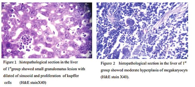
were fed on diet supplement with chitosan (1mg/kg diet) While the characteristic lesion in the liver of treated
and (1.5mg /kg diet) for (4) weeks respectively, While 1st group (fed on diet with 1gm\kg of chitosan) were
3rd and 4th groups considered as control positive and development of early small granulomatous lesion
negative groups. At (4) weeks the first three groups were seen in dilated sinusoids together with proliferation of
inoculated intraperitoneally i/P with (0.2) ml (1×10⁹) kupffer cells (figure:1), the microscopic examination in
CFU/ml, while the 4th group (control negative) inoculated the spleen revealed mild white pulp hyperplasia with
with (0.2) sterile normal saline, histopathological proliferation of megakaryocyte (figure:2), together with
examination of internal organs(liver, spleen and brain) slight vacuolar changes in some neurons also the results
were taken from both control and infected groups about showed moderate gliosis (figure:3).
(1cm3) was taken and fixed in 10% formalin saline for
histopathological section which was done according The pathological lesion in liver of treated 2nd group
to(10). (fed on diet with 1.5gm\kg of chitosan) characterized
by focal mononuclear cells (MNCs) aggregation mainly
Result and Discussion around central vein (figure:4) while presence of follicular
hyperplasia in the white pulp was the main lesion
1) Gross pathological changes: observed in splenic tissue (figure:5), while the main
The main gross feature in control group was severe brain lesion in the treated infected mice characterized
congestion in the visceral organs specially in the liver, by focal aggregation of MNCs in brain tissue, associated
spleen and kidney with presences necrotic foci at the with no clear lesion in the neurons seen mainly in the
edge of spleen, while treated groups show hepato- brain section.
splenomegaly was the characteristic gross lesion in the The present study shown sever pathological lesion
treated groups. in the internal organs (liver, spleen and kidney) of the
2) Bacterial isolate and clinical signs: control positive groups these results indicate that
exposed to highly virulent microorganisms overcome
No clear clinical signs noticed on experimental the innate immune system and disseminates to internal
animals specially the treated groups were appeared organs induce tissue damage, these observation were
healthy and well feeding. The result showed heavy in consistent with (11) who explained that virulent
bacterial isolation mainly from brain, spleen and liver of Listeria monocytogenes was one of intracellular bacteria
control positive groups, while mild growth to absences disseminated via blood stream to internal organs and
in other treated groups. Also the isolate was confirmed induce nonspecific inflammatory reaction by production
again on blood agar then we made smear from isolate listeriolysin O which destroyed the endothelial cells
and stained with grams stain. of blood vessels to induce necrosis and suppurative
inflammation (12). In addition, survival and proliferation
3) Histopathological examination: of microorganisms in the hepatic and splenic cells willIndian Journal of Forensic Medicine & Toxicology, April-June 2020, Vol. 14, No. 2 2115
lead to the formation of infection foci that result the appearance of granulomatous lesion mainly in liver tissue
infiltration of alarge number of WBCs and activate this evidence was agreement with (16) Where noted that
neutrophil phagocytic cells to work on other resist the the granulomatous reaction was considered the strongest
invading germs (13). We also recorded depletion of body defense against virulent microorganism’s infection,
white pulp of spleen of control positive group these furthermore there are numerous response indicate that
observations may indicate that Listeria monocytogenes chitosan improve the immune response (17). Our results
induced reduction in acquired immune response via showed lymphoid hyperplasia in splenic tissue mainly
depletion of lymphocytic cells (14), Neuronal necrosis in mice feeding with (1.5gm\kg) chitosan this indicate
and microcavites formation may due to excess of nitric that chitosan elicited both humeral and cell mediated
oxide generation literal infection which is important immunity and activated immune cells to secret cytokines
for intracellular signaling of new transmission both that play essential role in initiated mature granuloma in
inducible and constitute nitric oxide synthase (NoS) the liver and this evidence was in agreement with (18)
are expressed in brain cells include neural lesion, Who demonstrated that feeding of chitosan increase
further more inflammatory cells include neutrophils, OX62+ percentage and DCs which up regulate the
macrophages express both (NoS and iNoS) may play an major histocompatibility complex class-II Ags. without
important role in elimination Listeria monocytogenes expression changing of co-stimulatory (CD80 or CD86)
(15)
. Also the present study explain that feeding infected molecules and Ag presenting cells produced TNFα
mice showed mild to moderate pathological lesion in the and IL-12 and activation T-lymphocytes, lymphoid
spleen, liver and brain tissue post challenge with Listeria tissue hyperplasia in animals fed diet supplement with
monocytogenes and these lesion characterized by chitosan may due to chitosan stimulated proliferation of
lymphocytic cells.2116 Indian Journal of Forensic Medicine & Toxicology, April-June 2020, Vol. 14, No. 2
Ethical Clearance: The Research Ethical 10. Luna GL. Manual of Histologic staining methods of
Committee at scientific research by ethical approval of the armed forces Institute of Pathology. McGraw-
both environmental and health and higher education and Hill Book Company, 1968; New York, USA. 3rd
scientific research ministries in Iraq. edition.
11. Dunn P L and North R J. Early gamma interferon
Conflict of Interest: The authors
production by natural killer cells is important in
declare that they have no conflict of interest.
defense against murine listeriosis. Infect. Immun.
Funding: Self-funding
1991; 59:2892–2900.
References 12. Vazquez-Boland J, Kuhn M, Berche, P, Chakroborty
T, Domanguez- Bernal G, Gonzalez-Zorn B,
1. Elliot T, Ryser Elmer H and Marth. Listeria,
Wehland J and Kreft J. Listeria pathogenesis and
Listeriosis and food Safety.3 Ed. Taylor & Francis
molecular virulence determinant. Clin. Microbiol,
Group. 2007; LLC, CRC Press.
Rev. 2001; 14: 584-640.
2. Seelige R and Bockemühl J. Kritische
13. Portnoy D A, Auerbuc U and Glomski J. The cell
Untersuchungen zur Frage einer Kapselbildung bei
biology of Listeria monocytogene infection:the
Listeria monocytogenes. Zbl. Bakteriol. Parasit.
intersection of bacterial pathogensis and cell
Infekt. Hyg., I. Orig. 1968; 206:216–227.
mediated immunity. J. Cell. biol. 2002; 3: 409-414.
3. Allerberger F and Wagner M. Listeriosis: a
14. Zenewicz LA and Shen H. Innate and adaptive
resurgent foodborne infection. Clin. Microbiol.
immune responses to Listeria monocytogenes: a
Infect. 2010; 16:16-23.
short overview .J Micro and Infect, 2007; 9:1208-
4. McLauchlin J and Low J C. Primary cutaneous 1215.
listeriosis in adults: an occupational disease of
15. Shin T, Weinstock D, Castro MD, Acland H, Walter
veterinarians and farmers. Vet. Rec. 1994; 135:615-
M, Kim H, Ahn M and Purchase H G. Neuronal
617.
constitutive and inducible Nitric oxide synthase
5. McLauchlin, J.; Mitchell,T .;merdon, J. and Jewell, expression in the brain of Listeria monocytogenes-
K. Listeria monocytogenes and listeriosis: a review infected cattl. ACTA VET. BRNO, 2001; 70: 43–
of hazard characterisation for use in microbiological 47.
risk assessment of foods. Int. J. Food Microbiol.
16. Enurah L U, Aboaba O O, Nwachukwu SCU and
2004; 92:15-33.
Nwosuh CI. Histopathological changes in the liver
6. Hadwiger LA. Multiple effects of chitosan on of mice challenged with Listeria monocytogenes
plant systems: Solid science or hype. Plant Scienc. in six zones of Nigeria. Journal of Experimental
2013; 208: 42–49. Biology and Agricultural Sciences, 2013; 1:1-15.
7. Dutta, P.K.; Dutta, J. and Tripathi, V.S. Chitin and 17. Porporatto C, Ismael D, Bianco A, Cabanillas M
Chitosan: Chemistry, properties and Application. and Correa SG. Early events associated to the oral
J. Scientific and Industrial Res., 63: 20-31.dv. co‐administration of type II collagen and chitosan:
Immunol.2004; 84:131–179. induction of anti‐inflammatory cytokines Int.
8. Salwa A S. Effect of hypercholesterolemia , Immunol. 2004;16 : 433-441.
chitosan and whole sonicated E.coli Ags on 18. Carrian P, Bianco ID and Correa SG. Local and
immune response and pathological changes in mice systemic activity of the polysaccharide chitosan at
infected with E.coli (O:127) isolated from children lymphoid tissues after oral administration. J Leukoc
suffering from diarrhea. Thesis faculty of veterinary Biol. 2005; 78:62-69.
medicine ,Baghdad university.2012.
9. Quinn P J , Markey B and Carter GR.Clinical
Veterinary Microbiology. 2004; 2nd
ed.,MosbyInt.,USA.You can also read