Mavacamten Efficacy in Mutation-specific Hypertrophic Cardiomyopathy: an In-silico Approach to Inform Precision Medicine
←
→
Page content transcription
If your browser does not render page correctly, please read the page content below
Mavacamten Efficacy in Mutation-specific Hypertrophic Cardiomyopathy:
 an In-silico Approach to Inform Precision Medicine

 Francesca Margara1, Blanca Rodriguez1, Christopher N Toepfer1,2*, Alfonso Bueno-Orovio1*
 1
 University of Oxford, Oxford, United Kingdom
 2
 Department of Genetics, Harvard Medical School, Boston, USA

 Abstract away from the actin thin filament. Broadly myosin SRX
 exists in a balance with the disordered relaxed state (DRX)
 Hypertrophic cardiomyopathy (HCM) is a common which defines myosins that are available to drive
genetic heart disease characterised by hyperdynamic contraction. Certain HCM variants decrease myosin SRX
contraction and slowed relaxation. It has been proposed thereby increasing DRX myosin heads, which are able to
that cellular hypercontractility can derive from mutations interact with actin and drive the clinical hypercontractile
that destabilise the energy-conserving myosin super phenotype [5]. Mavacamten has been shown to re-stabilise
relaxed state, SRX. A new drug, Mavacamten, has been myosin SRX and reverse the cellular hallmarks of HCM
shown to re-stabilise myosin SRX. Here we develop a [6].
human-based in-silico model to investigate how disease Here we extend a coupled human electromechanical
and drug-induced SRX changes alter cardiac contractility. cardiomyocyte model [7] to incorporate myosin
We do this to mechanistically investigate how Mavacamten SRX/DRX. We do this to investigate how HCM and
restores function in a HCM causing mutation. Our Mavacamten-induced changes in SRX alter cellular
simulations show that hypercontractility is accounted for contractility. We benchmark our model using human
by an increased availability of crossbridges due to a experimental data and conduct a simulation study to
reduced abundance of myosin SRX, but cellular diastolic mechanistically interrogate how Mavacamten restores
dysfunction is only recapitulated if there is a direct cellular function in the HCM-causing β-myosin heavy
crossbridge contribution to thin filament activation. Our chain variant MYH7R403Q/+.
model replicates reduced cellular contractility with
Mavacamten treatment, which also rescues the 2. Methods
hypercontractile phenotype in HCM. Our model
demonstrates that Mavacamten is effective in correcting 2.1. Baseline model
HCM abnormalities caused by mutations that destabilise
SRX. However, genotypes that cause HCM via other We extended our coupled ToR-ORd+Land model [7] of
molecular pathways may be incompletely salvaged by human electrophysiology, excitation-contraction coupling,
Mavacamten. and active contraction (Figure 1A) to include an explicit
 dependence of crossbridge formation on the myosin
1. Introduction DRX:SRX ratio (Figure 1B). Our model is based on two
 key assumptions: 1) changes in DRX:SRX ratio modulate
 Hypertrophic cardiomyopathy (HCM) is a common the number of functionally accessible myosin heads for
genetic heart disease, which can cause arrhythmias and interaction with actin; 2) Mavacamten shifts the myosin
heart failure [1]. Pharmacological therapy plays a key role DRX:SRX ratio towards SRX [6].
in the clinical management of HCM, as of yet there are no A new parameter R (Figure 1B) was introduced in the
therapies that specifically target disease mechanisms [2]. model to represent the ratio (DRX:SRX)/(DRX:SRX)control.
Mavacamten is the first drug developed to target HCM In our modified model, R modulates the transitions
pathophysiology, with demonstrated efficacy for between the states that represent: (i) unblocked myosin
obstructive HCM [2,3]. The exact mechanisms that govern binding sites on actin without crossbridges bound (U), and
Mavacamten’s mode of action are incompletely (ii) bound crossbridges in the pre-powerstroke state (W).
understood, and a deeper understanding is needed to guide This means that crossbridges now enter in the pre-
effective and safe patient-specific therapy. powerstroke state W based on both the actin binding sites
 Recently a new state of myosin that regulates contractile available for crossbridge formation due to calcium
function by tuning myosin head availability was observed activation U, and myosin heads functionally available for
in myocardium [4]. This myosin state was termed the super interaction with actin, modelled through the new parameter
relaxed state (SRX) where myosin heads are sequestered
 1
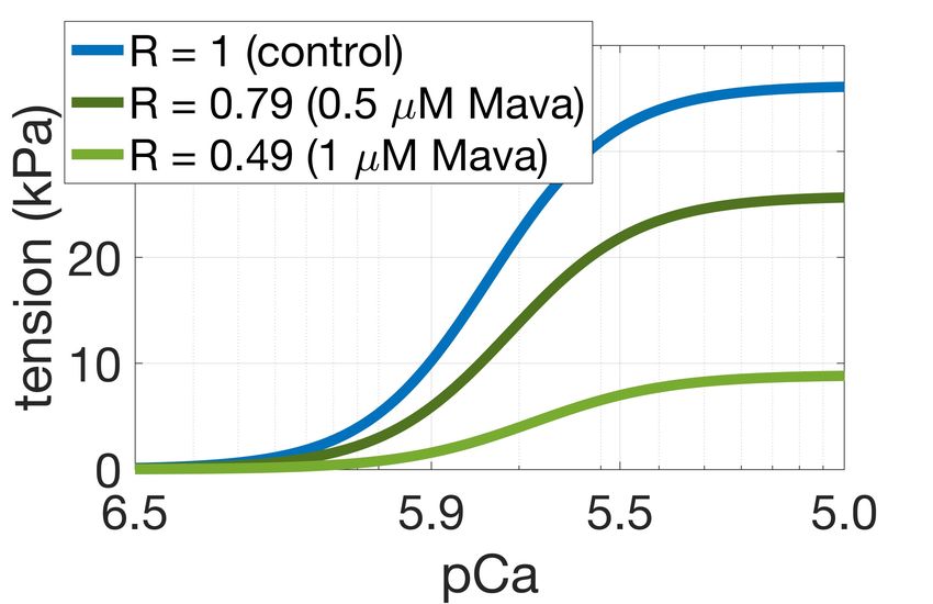
R as follows: calibrated as in [9] with additional constraints on active
 tension duration at 50% and 95% decay.

 = !" ∗ # ( ) ∗ − "! ∗ $ ( ) ∗ − "% ∗ − "! ∗

 2.3. MYH7R403Q/+ population
We impose the constraint ! (1) = " (1) = 1, so that =
1 at control conditions where To investigate the pathogenic mechanisms of the HCM
(DRX:SRX)=(DRX:SRX)control. We then quantified how causing MYH7R403Q/+ variant we constructed an in-silico
changes in R affected simulated isometric twitch tension in population of MYH7R403Q/+ cardiomyocytes by
intact cardiomyocytes and steady-state tension-calcium remodelling R to R = 1.3, as observed in experimental data
relationships in skinned cardiomyocytes. Optimal " ( ) [6]. The mechanical behaviours of the control and
and ! ( ) were chosen based on their ability to replicate MYH7R403Q/+ populations were compared to control and
changes in contractility observed in experimental data. In MYH7R403Q/+ hiPSC-CMs data [6]. Modulating R alone did
particular we aimed to replicate the ~25-30% decrease in not fully capture the MYH7R403Q/+ phenotype of impaired
maximal tension with 0.5 µM Mavacamten observed in cellular relaxation. Therefore, we incorporated a positive
 feedback mechanism from crossbridge cycling to thin
human myocardium [8], and the ~40% increase in
contractility caused by a 17% increase in DRX due to the filament activation to account for the significant
pathogenic MYH7R403Q/+ mutation observed in human prolongation of relaxation observed in-vitro. Specifically,
 calcium unbinding from troponin was slowed due to larger
induced pluripotent stem cell derived cardiomyocytes
 populations of active crossbridges. This has been shown
(hiPSC-CMs) [6].
 experimentally where calcium unbinding from Troponin C
 was found to be slowed by increased crossbridge
 Model Development abundance [10], and that calcium unbinding is essential for
 A. Electro-mechanical Model Diagram

 A INa INaL INaK INaCa INab IK1 IKr IKs
 IKb
 appropriate and timely muscular relaxation [11].
 I(Ca)Cl

 INaCa
 CaMKII
 Main Calmodulin
 Ito 2.4. In-silico trial of Mavacamten
 Jrel CSQN Cytosolic Pool IpCa
 Jtr NSR
 ICaL Troponin Ca2+
 IClb
 We simulated the administration of 0.5 µM
 RyR
 JSR
 SS
 Jup Tropomyosin ICab
 BSR Jleak SERCA
 Actin
 Mavacamten on the MYH7R403Q/+ population, and
 BSL
 ICaL
 Jdiff Myosin
 evaluated the contractile response to the simulated drug
 B. Model Outputs and Biomarkers intervention. Simulations results were then compared to
 B B1 troponin
 B2 B3
 experimental data [6].
 EADs
 B ACs The simulation of 0.5 µM Mavacamten was achieved by
 calcium further remodelling R to 0.79, after simulating the
 (DRX: SRX)

 Ca -
 (DRX: SRX)control remodelling induced by the mutation.
 troponin U
 B = blocked myosin binding sites
 U = detached state, myosin binding
 sites on actin are unblocked S W
 2.5. Simulation design
 W = pre-powerstroke
 S = post-powerstroke, force producing
 All the simulations conducted in this study were run in
Figure 1. ToR-ORd+Land model of human cardiomyocyte MatLab (Mathworks Inc. Natwick, MA, USA) using the
electromechanical function with embedded description of ordinary differential equation solver ode15s. A stimulus
myosin DRX:SRX. A: model schematic [7]. B: diagram of current of -53 µA/µF with 1 ms duration was used and for
the active tension model. In pink the modifications each simulation steady-state was reached at 1 Hz pacing.
introduced to simulate myosin DRX:SRX.
 3. Results
2.2. Control population
 3.1. Cellular active tension generation is
 From our newly generated model incorporating myosin modulated by myosin availability
DRX:SRX ratios we constructed a control population of
human cardiomyocytes. We first generated 2000
 Figure 2 shows that our phenomenological model of
electromechanical models to account for cell heterogeneity
 SRX abundance appropriately describes changes in twitch
due to variable expression of ion channels, pumps, and
 and steady-state tension generation due to crossbridge
exchangers [9] as well as different levels of protein kinase
 availability. It generated a ~40% increase in twitch tension
A-mediated phosphorylation. The population was then
 2
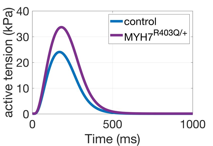
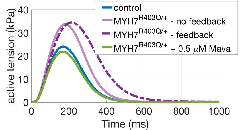
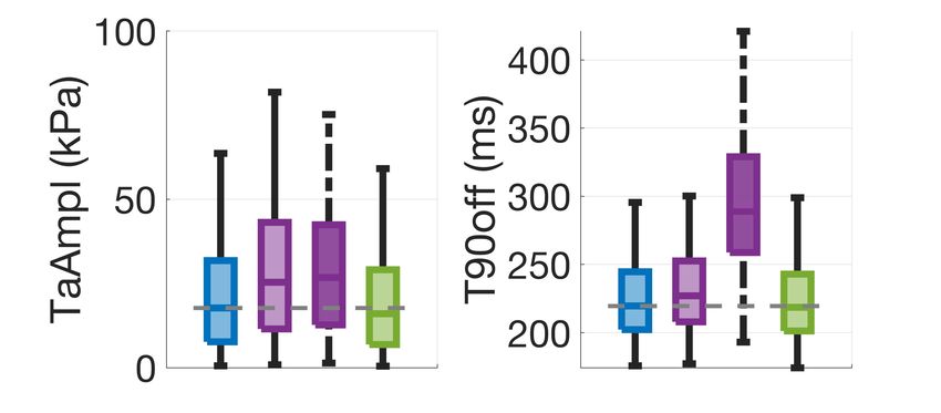
amplitude for R = 1.3 (Figure 2A) and a ~30% decrease in crossbridge cycling to thin filament activation.
steady-state maximal tension for R = 0.79 (Figure 2B), Accordingly, a larger number of crossbridges favour thin
meeting our calibration targets. For R = 0.79 the chosen filament activation and slows the release of calcium from
formulation predicted a shift in calcium sensitivity of force troponin. When we incorporated this thin filament
production of ~0.05 units from control, compared to feedback into our model, we found significantly prolonged
experiments reporting a shift of ~0.1 units in human tension relaxation in our simulations in agreement with the
myocardium at 0.5 µM Mavacamten [8]. For R = 0.49 ~35% change in relaxation time observed in experiments
simulations predicted a decrease in maximal tension of (Figure 3A, dark purple dashed line). Simulation of
~70%, consistent to animal experiments [3] at 1 µM Mavacamten in this system lowers the amount of
Mavacamten. crossbridge cycling and the myosin-based contribution to
 thin filament activation restoring cardiac relaxation.
 Model Calibration
 A B
 Mavacamten corrects hypercontractility and abnormal relaxation
 in simulated and experimental pathogenic MYH7R403Q/+
 A. Simulations

Figure 2. Active tension generation is modulated by
myosin availability. A: active tension increase under the
MYH7R403Q/+ mutation. B: maximal steady-state tension feedback

and calcium sensitivity reduction under simulated action of
Mavacamten.

3.2. Mavacamten corrects
hypercontractility and abnormal relaxation
in simulated MYH7R403Q/+ cardiomyocytes B. Experiments

Next, we conducted a simulation study to investigate the
mechanisms that cause HCM in the MYH7R403Q/+ variant.
For this, we first constructed a control population and
applied a remodelling of R = 1.3 on the 348 control models
that satisfied the calibration criteria. Figure 3A illustrates
the changes in contractility (tension amplitude, TaAmpl)
and relaxation (time to 90% decay, T90off) that follows Figure 3. Explanation of disease phenotype through
from R = 1.3 in our simulated population of human modelling and simulation. A: simulated active tension
cardiomyocytes (light purple). We observed a median changes under the MYH7R403Q/+ mutation and
increase in contractility (active tension peak) of 43% and a Mavacamten. The feedback from crossbridge cycling to
median increase in relaxation time of 3% under thin filament activation allows to recover the mutation-
MYH7R403Q/+ compared to control. Tension abnormalities induced prolongation in relaxation. B: experimental
were corrected by applying R = 0.79 as representative of sarcomere shortening and relaxation time dose-dependent
0.5 µM Mavacamten (Figure 3A, in green). This correlates responses of hiPSC-CMs to Mavacamten [6].
well with the experimental dose-dependent responses of
sarcomere shortening and relaxation time obtained in 4. Discussion
hiPSC-CMs (Figure 3B, [6]).
The model without thin filament feedback had fidelity in We conducted a modelling and simulation study to
simulating mutation-induced percentage change in mechanistically unravel how Mavacamten restores tension
contractility when compared to experimental data. generation in a β-myosin heavy chain mutation that causes
However, the model did not well describe the ~35% HCM. Our results contribute to a better mechanistic
prolongation of relaxation observed in-vitro in hiPSC- understanding of Mavacamten efficacy in a specific HCM
CMs. We therefore hypothesised that the prolongation of subgroup, which is needed to guide effective and safe
relaxation observed experimentally in MYH7R403Q/+ could patient-specific therapy. To investigate how HCM and
be explained by a positive feedback mechanism from Mavacamten alter cellular contractility, we extended a
 3
human cardiomyocyte electromechanical model to include C.N.T. (206466/Z/17/Z), and a University of Oxford BHF
representation of myosin sequestration and release from CRE Intermediate Transition Fellowship to C.N.T.
the energy-conserving state SRX. (RE/18/3/34214). We would like to acknowledge the use
 Our simulations show that hypercontractility is a of the University of Oxford Advanced Research
consequence of increased crossbridge cycling due to Computing (ARC) facility in carrying out this work.
reduced myosin SRX abundance. We also show that this
mechanism in isolation cannot explain the significantly References
impaired relaxation observed experimentally. For this
reason, we modelled a crossbridge-based contribution to [1] B.J. Maron, M.S. Maron, “Hypertrophic cardiomyopathy”,
thin filament activation that favours a prolonged calcium The Lancet, 2013;381(9862):242–55.
binding to troponin, thereby slowing cellular relaxation. [2] I. Olivotto et al., “Mavacamten for treatment of symptomatic
 We also show that our phenomenological model of obstructive hypertrophic cardiomyopathy (EXPLORER-
myosin sequestration is able to replicate the reduction in HCM): a randomised, double-blind, placebo-controlled,
maximal active tension and reduction in calcium phase 3 trial”, The Lancet, 2020;396(10253):759–69.
 [3] E.M. Green et al., “A small-molecule inhibitor of sarcomere
sensitivity of force production observed with 0.5 and 1 µM contractility suppresses hypertrophic cardiomyopathy in
Mavacamten treatment. When Mavacamten exposure was mice”, Science, 2016;351(6273):617–21.
simulated in human adult cardiomyocytes with the [4] P. Hooijman, M.A. Stewart, R. Cooke, “A New State of
MYH7R403Q/+ variant, our simulations showed that Cardiac Myosin with Very Slow ATP Turnover: A Potential
Mavacamten rescued the hypercontractile phenotype and Cardioprotective Mechanism in the Heart”, Biophys. J.,
impaired relaxation by reducing the proportion of 2011;100(8):1969–76.
crossbridges that can interact with actin. This suggests that [5] J.A. Spudich, “Three perspectives on the molecular basis of
Mavacamten can be very effective in correcting HCM hypercontractility caused by hypertrophic cardiomyopathy
abnormalities caused by mutations that destabilise SRX, mutations”, Pflugers Arch Eur J Physiol, 2019;471:701–17.
 [6] C.N. Toepfer et al., “Myosin sequestration regulates
but careful consideration should be taken in extrapolating sarcomere function, cardiomyocyte energetics, and
these results to different HCM genotypes that could act metabolism, informing the pathogenesis of hypertrophic
through different pathways. According to our simulations, cardiomyopathy”, Circulation, 2020;141(10):828–42.
the modulation of contractility by reduction of myosin [7] F. Margara et al., “In-silico human electro-mechanical
head availability provides a robust mechanism to reduce ventricular modelling and simulation for drug-induced pro-
hyperdynamic contraction, a common pathogenic arrhythmia and inotropic risk assessment”, Prog.
mechanism in HCM. This supports the effectiveness of Biophys. Mol. Biol., 2021;159:58-74.
Mavacamten in clinical trials for obstructive HCM [2]. [8] P.O. Awinda et al., “Effects of mavacamten on Ca2+
However, specific genotypes may remain untreated if the sensitivity of contraction as sarcomere length varied in
 human myocardium”, Br J Pharmacol, 2020;177:5609–21.
observed hypercontractile phenotype does not occur
 [9] E. Passini et al., “Drug‐induced shortening of the
through SRX destabilization [12]. We suggest that future electromechanical window is an effective biomarker for in
mechanistic investigations, like the ones conducted here, silico prediction of clinical risk of arrhythmias”, Br. J.
on mutation-specific disease pathways will be key to Pharmacol., 2019;176:3819–3833.
advance our understanding of HCM and to inform [10] P.A. Hofmann, F. Fuchs, “Effect of length and cross-bridge
effective patient-specific pharmacological interventions. attachment on Ca2+ binding to cardiac troponin C”, Am. J.
 Physiol. Cell Physiol., 1987;253(1):C90–6.
 [11] B.J. Biesiadecki et al, “Tri-modal regulation of cardiac
Acknowledgments muscle relaxation; intracellular calcium decline, thin
 filament deactivation, and cross-bridge cycling kinetics”,
 This work was funded by the European Union’s Biophys. Rev., 2014;6(3–4):273–89.
Horizon 2020 research and innovation programme [12] M. Schmid, C.N. Toepfer, “Cardiac myosin super relaxation
(Personalised In-Silico Cardiology project, grant (SRX): a perspective on fundamental biology, human
agreement 764738; CompBioMed 1 and 2 Centre of disease and therapeutics”, Biology Open, 2021;10:
Excellence in Computational Biomedicine, grant bio057646.
agreements 675451 and 823712; TransQST project,
Innovative Medicines Initiative 2 Joint Undertaking, grant
 Address for correspondence:
agreement 116030), a Wellcome Trust Fellowship in Basic
Biomedical Sciences to B.R. (214290/Z/18/Z), a British Francesca Margara
Heart Foundation (BHF) Intermediate Basic Science Department of Computer Science, Wolfson Building, Parks
Fellowship to A.B. (FS/17/22/32644), an NC3Rs Road, Oxford, OX1 3QD (UK)
Infrastructure for Impact Award (NC/P001076/1), the francesca.margara@keble.ox.ac.uk
Oxford BHF Centre of Research Excellence
(RE/13/1/30181), a Sir Henry Wellcome fellowship to
 4
You can also read