Protecting Children Against Commotio Cordis in Baseball by Understanding the Effects of Impact Locations Over the Heart
←
→
Page content transcription
If your browser does not render page correctly, please read the page content below
Protecting Children Against Commotio Cordis in Baseball by
    Understanding the Effects of Impact Locations Over the Heart
                      G. J. Dickey1, K. Bian1, H. R. Khan2 and H. Mao1,3
     1
       Department of Mechanical and Materials Engineering, University of Western Ontario
  2
    Department of Cardiology, London Health Sciences Center, University of Western Ontario
            3
              Department of Biomedical Engineering, University of Western Ontario

                                            ABSTRACT

Commotio Cordis is the second leading cause of sudden cardiac death in young athletes. Currently
available chest protectors on the market are shown ineffective in preventing cases of commotio
cordis in young athletes that play baseball. The new NOCSAE (National Operating Committee on
Standards for Athletic Equipment) regulations specify force limits at three loading cells over the
chest, with one upper and one lower loading cell, and one over the heart. This study focused on
understanding the effects of baseball impact locations to the heart. By understanding such effects
and then identifying vulnerable impact locations, we may design and develop chest protectors that
can effectively provide protection to prevent commotio cordis in young athletes. Simulation cases
were run using the validated CHARM-10 model, a detailed finite element model representing an
average 10-year-old child. A baseball model was developed and then used to impact the chest at
different locations. An 8x7 impact location matrix was designed with 56 unique simulations. Eight
locations in the transverse plane, seven locations in the sagittal plane. The baseball was moved by
half of the ball’s radius (18.75 mm) in the transverse and/or sagittal plane in each individual case.
An initial velocity of 17.88 m/s was used for baseball impacts. The baseball was of regulation
stiffness. Left ventricle strain and pressure, contact force between the baseball and chest and rib
deformations were analyzed. Left ventricle strain was highest from baseball impacts directly over
the left ventricle (0.34) as well as impacts slightly lateral and superior to the cardiac silhouette
(0.34). Left ventricle pressure was highest with impacts directly over the left ventricle (82.9 kPa).
For impacts close to the upper and lower positions as specified by NOCSAE, left ventricle strain
and pressure were 0.24 and 29.5 kPa (upper loading cell) and 0.04 and 4.0 kPa (lower loading
cell). This study systematically analyzed the effects of impact locations on left ventricle response.
We have identified the most dangerous impact locations not only to the left ventricle but also to
the upper, left side of the heart. Impacts to the NOCSAE lower-loading-cell position was of
minimal concern in inducing left ventricle strain or pressure. This novel study provided evidence
through computational modeling of where to emphasize protective materials for establishing
effective chest protectors that will minimize instances of commotio cordis in young baseball
athletes.

                                                   1
                      2021 The Ohio State University Injury Biomechanics Symposium
                                *This paper has not been peer-reviewed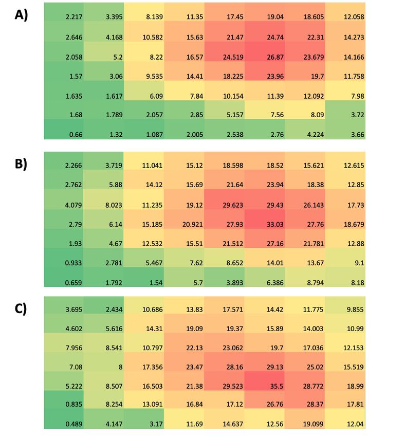
INTRODUCTION

         Commotio cordis is a rare but lethal mechanism that is the second leading cause of sudden
cardiac death in young athletes (Maron, 2003). Upwards of 75% of cases of commotio cordis occur
in competitive and recreational youth sports (Maron & Estes, 2010). It is the result of the heart
going into ventricular fibrillation from a non-penetrating impact such as a baseball over the chest
(Link, 2012; Link et al., 1998; Maron & Estes, 2010). To induce commotio cordis, a combination
of 3 specific factors much occur simultaneously: the impact must occur over the precordium, the
impact must have a velocity of approximately 40 mph, and the impact must occur during the
upslope of the T-wave during the cardiac cycle (Link et al., 1998). As of 2010, the commotio
cordis registry has recorded 224 deaths since it’s inauguration in 1995 (Maron et al., 2009; Maron
et al., 2002; Maron et al., 1995).

         Despite the use of chest protectors in youth sports, sudden death in young athletes from
impacts over the chest still occur (Doerer et al., 2007). Current chest protector brands on the market
claim their chest protectors are safe for children, but these companies may give a false sense of
safety for parents of young children. An experimental animal model commonly discussed in the
literature analyzed popular baseball and lacrosse chest protectors and their ability to decrease
ventricular fibrillation occurrence when compared to a control group (no chest protector). They
found that none of the baseball chest protectors were able to significantly reduce the rate of
ventricular fibrillation, with the frequency of ventricular fibrillation ranging between a low of 22%
and a high of 49% (Weinstock et al., 2006). Furthermore, another study found that approximately
40% of commotio cordis instances occurred across various sports despite the athlete wearing a
commercially available chest protector (Doerer et al., 2007).

        As of 2017, NOCSAE (National Operating Committee on Standards for Athletic
Equipment) introduced safety standards for commotio cordis prevention in baseball and lacrosse
chest protectors (NOCSAE, Revised July, 2019). These standards are a significant step in
developing safer chest protector designs for young athletes. Currently, the NOCSAE surrogate
model uses an upper loading cell (ULC), lower loading cell (LLC) and a cardiac loading cell (CLC)
to measure the amount of force (N) impacting the surrogate model. Reaction force is the only
parameter that NOCSAE has chosen to determine whether a chest protector is able to prevent
instances of commotio cordis. Mechanical responses of the heart remain unknown using this
criterion for evaluation.

         Commotio cordis is difficult to study due to the nature of the incident, and challenges arise
due to the inability to test on live subjects or PMHSs. To date, predominately swine studies have
been conducted in the literature (Link et al., 2001; Link et al., 2002; Link et al., 1998). Finite
element modeling can help us to understand this sudden-death mechanism through the use of a
validated finite element child model. In particular, it may allow us to analyze the left ventricle of
the heart and any deformations that occur upon impact. This analysis is a much-needed alternative
to the conventional swine studies, as the chest in swine is rounded and therefore impacts during
testing make contact directly with the left ventricle. Meanwhile, in humans the heart is positioned
so that the right ventricle is most superficial to the chest. These anatomical differences may play a
key role in the results from these swine studies.

                                                   2
                      2021 The Ohio State University Injury Biomechanics Symposium
                                *This paper has not been peer-reviewed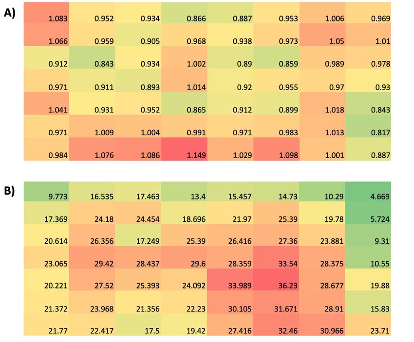
The current void in the literature, alongside the ineffectiveness of current chest protectors,
inspired us to research and explore ways to identify the most dangerous impact locations over the
chest wall. This study focused on using contour maps to identify these impact locations and create
a visual representation that is legible to everyone. By identifying the most threatening impact
locations, we may design and develop a chest protector that would provide maximum protection
without sacrificing mobility and comfort.

                                            METHODS

Finite element simulations and post processing

         Finite element simulations were run using the CHARM-10 child model developed at
Wayne State University (Shen et al., 2016), a detailed finite element (FE) model representing an
average 10-year-old child. This detailed FE model contains all major anatomical structures based
on detailed clinical scans of 10-year-old children (Mao et al., 2014) including 12 pairs of ribs,
scapula, sternum, clavicle, humerus, the entire spinal column, cartilage and ligaments, all internal
organs and major arteries (e.g., aorta). The model consists of 742,087 elements and 504,775 nodes
and contains 8-node hexahedral elements. The model was developed with a multi-block approach;
selectively reduced integration was used with hourglass control type 4 and a parameter of 0.1 for
soft tissue.

         The CHARM-10 model has been validated based on data collected through
cardiopulmonary resuscitation on live subjects (Jiang et al., 2014) and impact data gathered from
cadavers (Jiang et al., 2013). We placed an internal pressure of 9.3 kPa to the heart to replicate
blood pressure. A baseball model with a radius of 37.5 mm and material properties based on the
literature was developed to be used for impacts with the validated chest model (Vedula, 2004). All
impacts to the chest were given an initial velocity of 17.88 m/s (40 mph). Previous research has
shown that impact speeds any higher than this may cause severe cardiac damage, which is not
uniform with commotio cordis instances (Link et al., 2003). Simulations were run using the Ls-
Dyna solver. Once completed, simulations were the analyzed and data was collected using Ls-
PrePost2.4.

Matrix design

        An 8x7 impact location matrix was designed with 56 unique simulations. Eight locations
in the transverse plane, seven locations in the sagittal plane, and an initial baseball velocity of
17.88 m/s aimed towards the chest. The baseball moved by half of the ball’s radius (18.75 mm) in
the transverse and/or sagittal plane in each individual case.

Impact response

       Impact responses were analyzed using the CHARM-10 computational model on Ls-
PrePost2.4. Impact responses included: Left ventricle strain and pressure, reaction force between
                                               3
                      2021 The Ohio State University Injury Biomechanics Symposium
                                *This paper has not been peer-reviewed
the baseball and chest, max rib deformation, and rib deformation at the left ventricle which
includes rib 3 (ULV), rib 4 (MLV) and rib 5 (LLV).
Left ventricle strain and pressure were analyzed and calculated by selecting all of the
elements that make up the left ventricle. No filter was needed when analyzing strain; meanwhile,
a CFC 600 filter was used when calculating left ventricle pressure.
Reaction force was measured by analyzing the force between the baseball and chest. This
was calculated using *Contact_Automatic_Surface_to_Surface in Ls-Dyna, which is a penalty-
based contact. A CFC 1000 filter was used to collect peak values of force from impacts.
Max rib deformation was calculated by measuring the displacement (mm) between the
anterior rib that experienced direct contact from the baseball and the rib that sits directly posterior
to it (e.g., anterior rip 3, posterior rib 5).
Contour maps
Contour maps were designed on Microsoft Excel. The data was collected and then input
into an 8x7 matrix where it was color coded with a green-yellow-red color scale.
4
2021 The Ohio State University Injury Biomechanics Symposium
*This paper has not been peer-reviewedRESULTS
Left ventricle mechanical responses
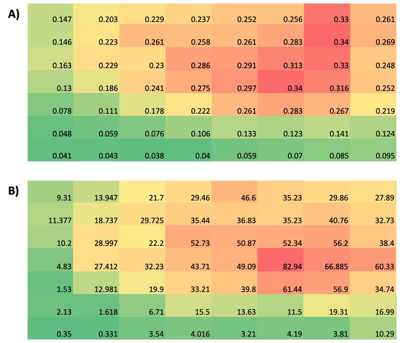
Left ventricle strain was highest from impacts directly over the left ventricle and impacts
slightly lateral and superior to the cardiac silhouette (0.34) (Figure 1. A). Left ventricle pressure
(kPa) was highest when impacts were aimed directly over the left ventricle, reaching a peak
internal pressure of 82.94 kPa (Figure 1. B).
Figure 1. A) Left ventricle strain. Left ventricle strain ranges from 0.041 (green) to 0.34 (red).
B) Left ventricle pressure. Left ventricle pressure ranges from 0.35 kPa (green) to 82.94 kPa
(red).
5
2021 The Ohio State University Injury Biomechanics Symposium
*This paper has not been peer-reviewedChest external mechanical responses
Reaction force between the baseball and chest had no visible correlation between impact
location and reaction force. Reaction force was highest when impacts were inferior to the sternum
(1.1 kN) and lowest over some areas directly over the heart (0.86 kN) (Figure 2. A). Max rib
deformation was highest with impacts directly over the left ventricle (36.2 mm) and slightly
inferior (Figure 2. B).
Figure 2. A) Reaction force between the baseball and chest. Reaction force ranges from 0.817 kN
(green) to 1.149 kN (red). B) Max rib deformation ranged from 4.669 mm (green) to 66.23 mm
(red).
6
2021 The Ohio State University Injury Biomechanics Symposium
*This paper has not been peer-reviewedRib deformations at the left ventricle
ULV, MLV and LLV displayed large rib deformations when impacts were aimed directly
over their respective rib. LLV had the highest rib deformations at 35.5 mm (Figure 3. A), while
MLV had a peak deformation of 33.0 mm (Figure 3. B), and ULV at 26.9 mm (Figure 3. C).
Figure 3. A) Rib deformation at ULV with a peak deformation of 26.87 mm (red). B) Rib
deformation at the MLV with a peak deformation of 33.03 mm (red). C) Rib deformation at the
LLV with a peak deformation of 35.5 mm (red).
7
2021 The Ohio State University Injury Biomechanics Symposium
*This paper has not been peer-reviewedDISCUSSION
This study used a detailed finite element model to analyze baseball impacts to the chest
and understand the effect of impact locations over the heart with respect to left ventricle strain, left
ventricle pressure, reaction force, max rib deformation, ULV, MLV and LLV. The left ventricle
strain and pressure contour maps illustrate where to place more emphasis on chest protectors and
may allow future researchers and baseball chest protector manufacturers to design safer products.
Meanwhile, other contour maps, such as reaction force between the baseball and the chest illustrate
why we believe reaction force is not the most effective commotio cordis injury metric, whereas
contour maps of rib deformation over the left ventricle region support our recommendation of
using it as an effective injury metric.
We have found that impacts aimed slightly lateral and superior to the left ventricle caused
very high left ventricle strain and require more intensive protection in these regions. After careful
examination of these cases producing higher strain, it was concluded that the baseball impacts the
chest wall and produces a contact force on the rib cage, specifically ribs 3 (ULV) and 4 (MLV),
increasing left ventricle strain as a result. Due to the chest cavity not being completely flat, this
caused the baseball to have a unique impact on the ribs, resulting in the baseball creating a
downward force pushing into the left ventricle.
Researchers in this field are commended for exploring new materials and thickness levels
to be incorporated into the design of their commercially available chest protectors (Kumar et al.,
2017). Meanwhile, based on our results, we believe that past and current designs may have
overlooked areas that require more protection in order to mitigate incidences of commotio cordis.
We believe that the combination of studies in which materials and thickness levels are analyzed,
alongside our study determining where extra protection is needed, we can develop a chest protector
capable of reducing instances of ventricular fibrillation.
Currently, the NOCSAE chest protector testing requires the chest protector to endure a 30-
mph (13.41 m/s) and 50-mph (22.35 m/s) condition over the chest. For 30-mph conditions, the
CLC must not exceed 400 N of force, while the ULC or LLC must not exceed 498 N of force. In
the 50-mph condition, the CLC must not exceed 800 N of force, and the ULC and LLC shall not
exceed 1001 N of force. Our data suggests that this model may be missing a potentially critical
area superior and to the left of the cardiac silhouette (Figure 1. A). For impacts close the ULC and
LLC, left ventricle strain was 0.24 (ULC) and 0.04 (LLC), while left ventricle pressure was 29.5
kPa (ULC) and 4.0 kPa (LLC). These findings suggest that impacts to the NOCSAE LLC location
are of minor concern in terms of inducing left ventricle strain and pressure. Considering this
surrogate model and our results, we believe the addition of the area we have identified in this study
may improve the protective effect of a chest protector.
A limitation to our study is that the CHARM-10 finite element model does not contain
blood flow throughout the heart. Due to our data being collected in 20 ms we do not believe that
this limitation would influence our results. However, to compensate for this limitation, we placed
an internal pressure of 9.3 kPa to the heart to replicate blood pressure.
8
2021 The Ohio State University Injury Biomechanics Symposium
*This paper has not been peer-reviewedCONCLUSIONS
This novel study used finite element modelling to help analyze the effects of baseball
impacts locations over the heart in order to reduce the incidence of commotio cordis in young
baseball athletes. Our results have identified the most dangerous baseball impact locations
regarding left ventricle strain and pressure. Strain was highest when impacts were located directly
over the left ventricle, as well as impacts slightly lateral and superior to the cardiac silhouette. Left
ventricle pressure was highest with impacts directly over the left ventricle. Additionally, we
addressed the reaction force between the baseball and chest wall, max rib deformation, and rib
deformations at the left ventricle. Reaction force did not have a strong correlation with impact
location, showing peak impact force when the impact was situated inferior to the sternum. Max rib
deformation showed the largest deformations when impacts were placed directly over the left
ventricle and impacts inferior to that position. Moreover, rib deformations of the ULV, MLV and
LLV were highest when impacts were directly over the corresponding rib, as expected. Overall,
we have described a computational approach that helps analyze the effects of baseball impact
locations over the heart and provides unique evidence that identifies where to emphasize protective
materials for establishing effective chest protectors that will minimize the risk of commotio cordis.
ACKNOWLEDGEMENTS
We acknowledge the support of NSERC and the Canada Research Chairs Program.
9
2021 The Ohio State University Injury Biomechanics Symposium
*This paper has not been peer-reviewedREFERENCES
Doerer, J. J., Haas, T. S., Estes, N. A., Link, M. S., & Maron, B. J. (2007). Evaluation of chest
barriers for protection against sudden death due to commotio cordis. Am J Cardiol, 99(6),
857-859. https://doi.org/10.1016/j.amjcard.2006.10.053
Jiang, B., Mao, H., Cao, L., & Yang, K. (2013). Experimental validation of pediatric thorax finite
element model under dynamic loading condition and analysis of injury. SAE Technical
Paper, 2013-01-0456. https://doi.org/https://doi.org/10.4271/2013-01-0456
Jiang, B., Mao, H., Cao, L., & Yang, K. (2014). Application of an anatomically-detailed finite
element thorax model to investigate pediatric cardiopulmonary resuscitation techniques on
hard bed. Computers in Biology and Medicine, 52, 28-34.
https://doi.org/10.1016/j.compbiomed.2014.05.014
Kumar, K., Mandleywala, S. N., Gannon, M. P., Estes, N. A., Weinstock, J., & Link, M. S. (2017).
Development of a Chest Wall Protector Effective in Preventing Sudden Cardiac Death by
Chest Wall Impact (Commotio Cordis). Clin J Sport Med, 27(1), 26-30.
https://doi.org/10.1097/JSM.0000000000000297
Link, M. S. (2012). Commotio cordis: ventricular fibrillation triggered by chest impact–induced
abnormalities in repolarization. Circulation: Arrhythmia and Electrophysiology, 5(2), 425-
432. https://doi.org/10.1161/CIRCEP.111.962712
Link, M. S., Maron, B. J., VanderBrink, B. A., Takeuchi, M., Pandian, N. G., Wang, P. J., & Estes,
N. M. (2001). Impact directly over the cardiac silhouette is necessary to produce ventricular
fibrillation in an experimental model of commotio cordis. Journal of the American College
of Cardiology, 37(2), 649-654. https://doi.org/10.1016/s0735-1097(00)01142-6
Link, M. S., Maron, B. J., Wang, P. J., Pandian, N. G., VanderBrink, B. A., & Estes, N. M. (2002).
Reduced risk of sudden death from chest wall blows (commotio cordis) with safety
baseballs. Pediatrics, 109(5), 873-877.
Link, M. S., Maron, B. J., Wang, P. J., VanderBrink, B. A., Zhu, W., & Estes, N. M. (2003). Upper
and lower limits of vulnerability to sudden arrhythmic death with chest-wall impact
(commotio cordis). Journal of the American College of Cardiology, 41(1), 99-104.
https://doi.org/https://doi.org/10.1016/s0735-1097(02)02669-4
Link, M. S., Wang, P. J., Pandian, N. G., Bharati, S., Udelson, J. E., Lee, M.-Y., Vecchiotti, M.
A., VanderBrink, B. A., Mirra, G., & Maron, B. J. (1998). An experimental model of
sudden death due to low-energy chest-wall impact (commotio cordis). New England
Journal of Medicine, 338(25), 1805-1811.
Mao, H., Holcombe, S., Shen, M., Jin, X., Wagner, C. D., Wang, S. C., Yang, K. H., & King, A.
I. (2014). Development of a 10-year-old full body geometric dataset for computational
modeling. Ann Biomed Eng, 42(10), 2143-2155. https://doi.org/10.1007/s10439-014-
1078-5
Maron, B. J. (2003). Sudden death in young athletes. N Engl J Med, 349(11), 1064-1075.
https://doi.org/10.1056/NEJMra022783
Maron, B. J., Doerer, J. J., Haas, T. S., Estes, N. A., Hodges, J. S., & Link, M. S. (2009). Commotio
cordis and the epidemiology of sudden death in competitive lacrosse. Pediatrics, 124(3),
966-971. https://doi.org/10.1542/peds.2009-0167
10
2021 The Ohio State University Injury Biomechanics Symposium
*This paper has not been peer-reviewedMaron, B. J., & Estes, N. A. (2010). Commotio cordis. N Engl J Med, 362(10), 917-927.
https://doi.org/10.1056/NEJMra0910111
Maron, B. J., Gohman, T. E., Kyle, S. B., Estes, N. A., & Link, M. S. (2002). Clinical profile and
spectrum of commotio cordis. JAMA, 287(9), 1142-1146.
https://doi.org/10.1001/jama.287.9.1142
Maron, B. J., Poliac, L. C., Kaplan, J. A., & Mueller, F. O. (1995). Blunt impact to the chest leading
to sudden death from cardiac arrest during sports activities. New England Journal of
Medicine, 333(6), 337-342.
https://doi.org/https://doi.org/10.1056/NEJM199508103330602
NOCSAE. (Revised July, 2019). Standard Test Method and Performance Specification Used in
Evaluating the Performance Characteristics of Protectors for Commotio Cordis. N. O. C.
O. S. F. A. Equipment. https://nocsae.org/wp-content/uploads/2018/05/ND200-19-
Commotio-Cordis-Test-Method-002-1.pdf
Shen, M., Mao, H., Jiang, B., Zhu, F., Jin, X., Dong, L., Ham, S., Palaniappan, P., Chou, C., &
Yang, K. (2016). Introduction of two new paediatric finite element models for pedestrian
and occupant protections., SAE Technical Paper, 2016-01-1492, 10.
https://doi.org/https://doi.org/10.4271/2016-01-1492
Vedula, G. (2004). Experimental and finite element study of the design parameters of an aluminum
baseball bat [Doctoral dissertation, University of Massachusettes Lowell].
Weinstock, J., Maron, B. J., Song, C., Mane, P. P., Estes, N. M., & Link, M. S. (2006). Failure of
commercially available chest wall protectors to prevent sudden cardiac death induced by
chest wall blows in an experimental model of commotio cordis. Pediatrics, 117(4), e656-
e662. https://doi.org/10.1542/peds.2005-1270
11
2021 The Ohio State University Injury Biomechanics Symposium
*This paper has not been peer-reviewedYou can also read