Cyclosporine reduces the spleen dimensions in rabbits - SciELO
←
→
Page content transcription
If your browser does not render page correctly, please read the page content below
ACTA CIRÚRGICA BRASILEIRA                                       https://doi.org/10.1590/ACB360402

                                                                                             ORIGINAL ARTICLE
                                                                                            Experimental Surgery

Cyclosporine reduces the spleen dimensions in rabbits
Luiz Ronaldo Alberti1,*     , Leonardo de Souza Vasconcellos2           , Andy Petroianu3
1.Associate Professor. Universidade Federal de Minas Gerais – Medical School – Department of Surgery – Belo Horizonte
  (MG), Brazil.
2.Associate Professor. Universidade Federal de Minas Gerais – Medical School – Department of Propedeutics – Belo Horizonte
  (MG), Brazil.
3.Full Professor. Universidade Federal de Minas Gerais – Medical School – Department of Surgery – Belo Horizonte (MG), Brazil.

     ABSTRACT
     Purpose: To assess the influence of prolonged cyclosporine use on the macro- and microscopic morphology
     of the spleen. Methods: 16 adult rabbits were divided into two groups (n = 8): group 1 – a placebo group,
     which was followed-up over a period of nine months; group 2 – which had taken an oral dose of cyclosporine
     (10 mg·kg–1·day–1) over nine months. At the end of this period, the splenic histoarchitecture of all animals was
     evaluated and the splenic corpuscles were measured. Results: The spleens of the first group presented normal
     characteristics and dimensions. The second group, however, had a reduction in all dimensions and its tissue
     texture had become soft. The white pulp and the perivascular sheath had become reduced in size and the
     number of lymphoid follicles had also fallen (p = 0.002), manifesting less splenic corpuscles (p = 0.0012) and
     lymphocyte nuclear pigments (p = 0.03). Conclusion: Prolonged use of cyclosporine reduces the spleen size,
     transforming it into a soft organ associated with a decrease in white pulp, perivascular sheath, lymphoid follicles
     and nuclear pigments in rabbits.
     Key words: Spleen. Cyclosporine. Morphology. Histology. Immunology. Rabbits.

*Corresponding author: luizronaldoa@yahoo.com.br | (55 31)99955-0400
Received: Dec 21, 2020 | Review: Feb 19, 2021 | Accepted: Mar 18, 2021
Conflict of interest: Nothing to declare.
Research performed at Bioterium, Department of Complementary Propaedeutics and Department of Surgery, Medical
School, Universidade Federal de Minas Gerais (UFMG), Belo Horizonte (MG), Brazil. Part of Master degree thesis,
Postgraduate Program in Health Science and Medicine, Institute of Education and Research of Santa Casa. Tutor: Luiz
Ronaldo Alberti.

                                                                                       Acta Cir Bras. 2021;36(4):e360402
Cyclosporine reduces the spleen dimensions in rabbits

      Introduction                                                                Group 1 – The placebo group, which was followed-up
                                                                               over nine months.
    The spleen has relevant immune functions, including                           Group 2 – Was given a daily dose of cyclosporine
the removal of antigens from the bloodstream and the                           (10 mg·kg–1·day–1) diluted in 10 mL of milk, administered
production of antibodies, immunoglobulins, amino                               through an orogastric catheter (12 Fr), over a nine-
acids, lymphocytes, monocytes and opsonins. It is                              month period.
also particularly important for complement factors,
                                                                                  At the end of the follow-up period, under intravenous
fibronectin, C-reactive protein, tuftsin and properdin1-4.
                                                                               anesthesia using ketamine (25 mg·kg –1), all animals
    Cyclosporine has an immunosuppressive effect based                         from the two groups were submitted to a median
on cellular immune response and causes a reduction of                          laparotomy and the spleen was removed for macroscopic
antibody-dependent T lymphocytes5. In terms of side                            assessment. Its longitudinal and transverse dimensions
effects, this drug interferes with the endocrine system,                       were measured, the spleen volume was calculated, and
causing changes in gonadal function and prolactin levels,                      the consistency was assessed.
decreasing fertility and fetal development5-7. The drug                            The splenic tissue was soaked in Bouin’s solution and
is also nephrotoxic and is associated with systemic                            processed for histological examination. Five µm thick
arterial hypertension, neurotoxicity and sepsis. Some                          sections were mounted and stained with hematoxylin
clinical disorders, such as nausea, vomiting, diarrhea                         and eosin, whereas Masson’s trichrome technique was
and abdominal discomfort, have also been frequently                            applied in order to examine the collagen. The splenic
described7-12.                                                                 histoarchitecture was measured with a micrometer at
   However, the side effects of cyclosporine have not                          intervals of 1000 µm, the histological assessment was
been entirely understood in relation to organs like the                        carried out under light microscopy for morphometric
spleen, which have not been studied after exposure to                          analysis. The diameters of 30 splenic corpuscles and
immunosuppressants. The purpose of this study was to                           their germinal centers were measured and their area
assess the influence of prolonged cyclosporine use on                          (µm2) and perimeter (µm) calculated.
the macro- and microscopic morphology of the spleen.                              The results were compared using the chi-squared test
                                                                               and Student’s t test. The differences were considered
      Methods                                                                  significant for values corresponding to p < 0.05.
   The study has been conducted at the Vivarium of
                                                                                    Results
the Medical School from the Universidade Federal de
Minas Gerais (UFMG) after approval from the Animal                                 All animals survived during the nine months of
Research Ethics Committee (CEUA/UFMG), protocol                                the experiment without complications. Their weight
185∕12.                                                                        increased uniformly and the amount of cyclosporine
    Sixteen male rabbits (Oryctolagus cuniculus) with                          administered to group 2 was weekly adjusted according
an initial weight between 2500 and 3000 g were kept                            to the weight of each rabbit.
in individual cages and received water and feed ad                                The mean dimensions of the spleens in group 2 were
libitum. The animals were randomly divided into two                            lower and the tissue was much softer than those from
groups (n = 8):                                                                group 1 (p = 0.002) (Table 1).

Table 1 - Values of mean ± standard error of mean of spleens and their white pulp corpuscles.
             Variables                              Group 1                               Group 2                                   p

      Dimensions (L × T) (cm)              6.12 ± 0.9 × 2.23 ± 0.45              4.53 ± 0.8 × 1.85 ± 0.37                        0.002

             Area (µm2)                     234.519.34 ± 4.150.73                 132.853.72 ± 2.454.94                         0.0012

      Maximum diameter (µm)                     702.53 ± 14.19                        450.55 ± 85.99                             0.003

      Minimum diameter (µm)                     552.32 ± 39.41                        310.69 ± 25.86                             0.004

          Perimeter (µm)                       2193.62 ± 112.05                       1234.59 ± 95.05                            0.001
Group 1: normal rabbits; without medication followed-up over nine months. Group 2: rabbits after using cyclosporine (10 mg·kg–1·day–1) followed-up over
nine months. L – longitudinal size, T – transverse size.

  2                                                                                                      Acta Cir Bras. 2021;36(4):e360402
Alberti LR et al.
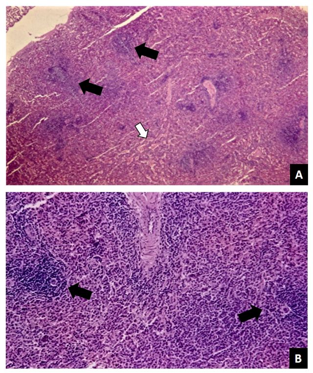
The splenic parenchyma of all rabbits from group 1
presented a normal white and red pulp, with numerous
lymphoid follicles at different stages of maturation,
as well as perivascular sheaths of varying size (Fig. 1).
On the other hand, the spleens taken from group 2
were significantly reduced in size and exhibited a white
pulp, lymphoid follicles, perivascular sheath and nuclear
lymphocyte pigmentation (Fig. 2). No signs of inflammation,
ischemia or necrosis were identified in the spleens of
either group.
(a)
(a) (b)
Figure 2 - Photomicrograph of an aplastic splenic
parenchyma of a group 2 rabbit after nine months use
of cyclosporine (10 mg·kg–1·day–1). (a) Observe reduction
in the white pulp (arrows); (b) Reduced cellularity in the
lymphoid follicles of the white pulp (arrow) (Hematoxylin
and Eosin, 400×).
Discussion
(b)
This experimental study aimed to investigate
the effects of the prolonged use of cyclosporine on the
Figure 1 - Photomicrograph of splenic parenchyma in spleen morphology. After nine months, there was a
a group 1 rabbit (without having taken cyclosporine). substantial reduction in the splenic size, its consistency
(a) Observe the normal aspect of red (white arrow) and these macroscopic parameters were associated with
and white (black arrows) pulps; (b) Normal vascularity a significant decrease in lymphoid follicle cellularity and
numerous lymphoid follicles (arrows) at different perivascular sheaths. These results are in accordance
stages of maturation as well as a perivascular sheath with other studies, which have shown the inhibitory
manifesting an abnormal size (Hematoxylin and Eosin, effect of cyclosporine on T lymphocyte activation
400×). and proliferation associated with the cytoplasmic
heterodimeric complex receptor 8-11 . This reduces
The morphometric results of the measurements made the movement of the nuclear factor of activated T
on the splenic corpuscles and the germinal centers of the lymphocytes and impairs cytokine gene induction,
white pulp are shown in Table 1. There was a reduction principally interleukins 2 and 4, as well as tumor
in the splenic corpuscles in group 2 compared to group 1 necrosis factors α and γ 12-15.
(p = 0.03). All dimensions of splenic corpuscles in group The prolonged use of cyclosporine A did not cause
2 were smaller than those in group 1: area (p = 0.0012), inflammation, damage, necrosis or vascular alteration
maximum diameter (p = 0.003), minimum diameter in the spleens 16. On the other hand, the drug was
(p = 0.004) and perimeter (p = 0.001). associated with a reduction of 43% in the white pulp,
Acta Cir Bras. 2021;36(4):e360402 3Cyclosporine reduces the spleen dimensions in rabbits
without manifesting any morphological disorder in Authors’ contribution
the red pulp. These results differ from the studies
carried by Armas et al. 9, who did not identify any Substantive scientific and intellectual contributions
splenic change in rats submitted to prolonged use to the study: Alberti RL, Vasconcellos LD and Petroianu
of cyclosporine. On the other hand, in a study with A; Conception and design: Alberti RL and Petroianu A;
24,939 transplanted patients who had been using Technical procedures: Vasconcellos LD; Critical revision:
cyclosporine for more than a year, 110 (0.45%) of Vasconcellos LD; Final approval: Petroianu A.
them showed splenomegaly proportionally higher in
males and elders in 61% of these records. However, Data availability statement
there is no record of the action of cyclosporine in the
other 99.55% of patients in this group 14. Data will be available upon request.
Pharmacodynamic studies have investigated
ultramicronized cyclosporine (atopic) in normal Funding
animals. An initial in vitro investigation demonstrated Fundação de Amparo à Pesquisa do Estado de Minas Gerais
cyclosporine-mediated suppression of T-lymphocyte
[https://doi.org/10.13039/501100004901]
activation-related molecules and cytokines16. Peripheral
blood mononuclear cells were isolated and activated, Grant No. APQ-00718-12
with half of the cells incubated while exposed to Conselho Nacional de Desenvolvimento Científico e
cyclosporine, and the other half not exposed to the Tecnológico
drug. Cells were then analyzed using flow cytometry,
[https://doi.org/10.13039/501100003593]
with T-cell expression of the intracellular cytokines
interleukin- 2 (IL)-2, IL-4, and interferon (IFN-c) evaluated Grant No. 308663/2013-6
after drug exposure. All cytokines demonstrated a Pró-Reitoria de Pesquisa da Universidade Federal de
time-dependent suppression profile. The T-cell surface Minas Gerais
molecules CD25 and CD95, which have roles in T-cell
activation and development, were evaluated after drug Acknowledgments
exposure, and there was also significant suppression
of expression of both biomarkers in the presence of Not applicable.
cyclosporine. In a subsequent in vivo study, activated
T-cell expression of IL-2, IL-4, and IFN-c was investigated References
by flow cytometry when animals were treated with
two different oral cyclosporine dosages. With high- 1. Kierdorf K, Masuda T, Jordão MJC, Prinz M. Macrophages
dose cyclosporine, activated T-cell expression of IL-2 at CNS interfaces: ontogeny and function in health and
and IFN-c was significantly suppressed, but IL-4 was disease. Nat Rev Neurosci. 2019;20:547–62. https://doi.
org/10.1038/s41583-019-0201-x
not similarly affected. Even with this low dosage of
cyclosporine, however, T-cell expression of IFN-c was 2. Hsiao H-M, Fernandez R, Tanaka S, Li W, Spahn JH, Chiu S,
still significantly suppressed. Mean T-cell expression Akbarpour M, et al. Spleen-derived classical monocytes
of IL-2 also was decreased 16. mediate lung ischemia-reperfusion injury through IL-
1β. J Clin Invest. 2018;128(7):2833‐47. https://doi.
No studies have focused on the effect of cyclosporine org/10.1172/JCI98436
on human spleens, which must be investigated using
3. Leroy C, Rigot J-M, Leroy M, Decanter C, Le Mapihan
imaging methods and studying autopsies of patients who K, Parent A-S, et al. Immunosuppressive drugs and
have undergone transplants. Other immunosuppressants fertility. Orphanet J Rare Dis. 2015;10:136. https://doi.
should be studied in order to verify their impact on org/10.1186/s13023-015-0332-8
the spleen morphology and function.
4. Ponnaiyan D, Jegadeesan V. Cyclosporine A: Novel concepts
in its role in drug-induced gingival overgrowth. Dent Res
Conclusion J. 2015;12(6):499–506. https://doi.org/10.4103/1735-
3327.170546
The prolonged use of cyclosporine in rabbits softens
and reduces the size of the spleen, is associated with 5. Colombo S, Sartori R. Ciclosporin and the cat:
Current understanding and review of clinical use. J
a decrease in white pulp, the perivascular sheath, Feline Med Surg. 2018;20(3):244–55. https://doi.
lymphoid follicles and nuclear pigmentation. org/10.1177/1098612X17748718
4 Acta Cir Bras. 2021;36(4):e360402Alberti LR et al.
6. Kaçmaz RO, Kempen JH, Newcomb C, Daniel E, Gangaputra MC, Blanco-Prieto MJ. Reformulating cyclosporine A (CsA):
S, Nussenblatt RB, et al. Cyclosporine for Ocular More than just a life cycle management strategy. J Control
Inflammatory Diseases. Ophthalmology. 2010;117(3):576– Release. 2016;225:269–82. https://doi.org/10.1016/j.
84. https://doi.org/10.1016/j.ophtha.2009.08.010 jconrel.2016.01.056
7. Barbarino JM, Staatz CE, Venkataramanan R, Klein TE, Altman
13. Badihi A, Frušić-Zlotkin M, Soroka Y, Benhamron S,
RB. PharmGKB summary: cyclosporine and tacrolimus
pathways. Pharmacogenet Genomics. 2013;23(10):563– Tzur T, Nassar T, et al. Topical nano-encapsulated
85. https://doi.org/10.1097/FPC.0b013e328364db84 cyclosporine formulation for atopic dermatitis
treatment. Nanomedicine. 2020;24:102140. https://doi.
8. Andreassen AK, Broch K, Eiskjær H, Karason K, Gude E,
org/10.1016/j.nano.2019.102140
Mølbak D, et al. Blood Pressure in De Novo Heart Transplant
Recipients Treated with Everolimus Compared with a 14. Xing J, Xiao Y, Tang X, Sheng X, Zhan W. Inhibition of
Cyclosporine-based Regimen: Results from the Randomized Cyclosporine A or rapamycin on T lymphocyte counts and
SCHEDULE Trial. Transplantation. 2019;103(4):781–8. the influence on the immune responses of B lymphocytes
https://doi.org/10.1097/TP.0000000000002445
in flounder (Paralichthys olivaceus). Fish Shellfish
9. Armas OA, Astarita RW, Wolf PL, Moossa AR, Scott Immunol. 2017;66:78–85. https://doi.org/10.1016/j.
MH, Haghighi P, et al. Effects of cyclosporin A on the fsi.2017.05.017
splenic tissue of rats: A histomorphometric analysis.
Exp Mol Pathol. 1989;50(1):92–103. https://doi. 15. Cridge H, Kordon A, Pinchuk LM, Wills RW, Thomason
org/10.1016/0014-4800(89)90059-2 JM, Mackin AJ, et al. Effects of cyclosporine on
10. Tedesco D, Haragsim L. Cyclosporine: A review. J Transplant. feline lymphocytes activated in vitro. Vet Immunol
2012;2012:230386. https://doi.org/10.1155/2012/230386 Immunopathol. 2020;219:109962. https://doi.org/10.1016/j.
vetimm.2019.109962
11. Lallemand F, Schmitt M, Bourges J-L, Gurny R, Benita
S, Garrigue J-S. Cyclosporine A delivery to the eye: A 16. Archer TM, Fellman CL, Stokes JV, Pinchuk LM, Lunsford
comprehensive review of academic and industrial efforts. KV, Pruett SB, et al. Pharmacodynamic Monitoring of
Eur J Pharm Biopharm. 2017;117:14–28. https://doi. Canine T‐Cell Cytokine Responses to Oral Cyclosporine.
org/10.1016/j.ejpb.2017.03.006 J Vet Intern Med. 2011;25(6):1391–7. https://doi.
12. Guada M, Beloqui A, Kumar MNVR, Préat V, Dios-Viéitez org/10.1111/j.1939-1676.2011.00797.x
Acta Cir Bras. 2021;36(4):e360402 5You can also read