A biomechanical study of the scoliotic thoracolumbar spine
←
→
Page content transcription
If your browser does not render page correctly, please read the page content below

IOP Conference Series: Materials Science and Engineering

PAPER • OPEN ACCESS

A biomechanical study of the scoliotic thoracolumbar spine
To cite this article: D Raja et al 2020 IOP Conf. Ser.: Mater. Sci. Eng. 912 022021

View the article online for updates and enhancements.

                                This content was downloaded from IP address 46.4.80.155 on 03/12/2020 at 01:35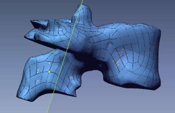
3rd International Conference on Advances in Mechanical Engineering (ICAME 2020)            IOP Publishing
IOP Conf. Series: Materials Science and Engineering 912 (2020) 022021 doi:10.1088/1757-899X/912/2/022021

A biomechanical study of the scoliotic thoracolumbar spine

                     D Raja1, Shraddha R Iyer2, Kunal Pandey2, Appaji Krishnan3 and Shantanu
                     Patil4
                     1
                       Department of Mechanical Engineering, SRM Institute of Science and Technology,
                     Kattankulathur, Chennai, India.
                     2
                       Student, Department of Mechanical Engineering, SRM Institute of Science and
                     Technology, Kattankulathur, Chennai, India.
                     3
                       Consultant Spine Surgeon, SIMS Hospital Vadapalani, Chennai
                     4
                       Department of Translational Medicine and Research, SRM Institute of Science and
                     Technology, Chennai, India.

                     E-mail: rameshshraddha14@gmail.com

                     Abstract. Scoliosis is a disease of the spine which leads to corkscrew curvature occurring due
                     to a combination of genetic and environmental factors. The abnormal curve is generally
                     observed during the growth spurt just before puberty. Scoliosis has been classified into three
                     different forms namely idiopathic, congenital and neuromuscular. When no specific cause for
                     spinal defect is identified, the deformity is called idiopathic scoliosis. The patient specific scan
                     model falls in the category congenital scoliosis. Mild cases of scoliosis can be treated by
                     physiological treatments. Severe cases of scoliosis may have vertebral twisting, vertebral
                     fusion and semi developed vertebral deformation. Severe cases of scoliosis could lead to
                     adjacent organ damage, especially the heart and lungs. Large number of patients experience
                     various back problems rendering day to day activities and normal physiological motion
                     difficult. In most of the cases, scoliosis needs multiple surgical corrections with various
                     implant rods and screws attached to the vertebrae. The purpose of this study is to investigate
                     the effect of upper body load on the scoliosis affected region located at the junction of thoracic
                     and lumbar region of the spine before surgery. The CT scan of the model is segmented and
                     meshed to conduct studies such as stress concentration analysis, strain analysis and deflection
                     in the segments.

1. Introduction
The human spine or the vertebral column is a component of the axial skeleton. It provides support and
strength and permits the human body to lean, bend stretch and rotate. The human spine is vulnerable to
many injuries and diseases like scoliosis, whiplash and low back pain [1]. The vertebral column is
divided into four curved regions namely cervical curve, thoracic curve, lumbar curve, and the sacral
curve. The cervical region comprises of seven vertebrae, the thoracic curve comprises of twelve
vertebrae and the Lumbar curve consists of five vertebrae. The sacral curve comprises of sacrum and
coccygeal vertebrae. The intervertebral discs separate two vertebrae. Spinal canal is housed by the
vertebral column. Spinal canal protects and encloses the spinal cord. Spine can be damaged and lead
too many problems, in that scoliosis was one of the most complex problem and it is treatable.
    Scoliosis is a deformity of the spine, complex in nature due to vertebral rotation and deviation of
the coronal plane from the median which forms the lateral curvature [2]. In addition to this kyphosis
and physiological lordosis on the sagittal plane may increase or decrease [3]. While scoliosis can be

              Content from this work may be used under the terms of the Creative Commons Attribution 3.0 licence. Any further distribution
              of this work must maintain attribution to the author(s) and the title of the work, journal citation and DOI.
Published under licence by IOP Publishing Ltd                          1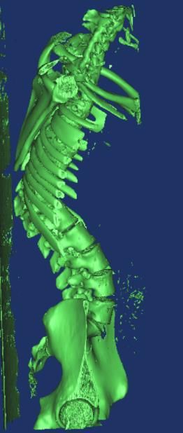
3rd International Conference on Advances in Mechanical Engineering (ICAME 2020) IOP Publishing
IOP Conf. Series: Materials Science and Engineering 912 (2020) 022021 doi:10.1088/1757-899X/912/2/022021
caused by conditions such as cerebral palsy and muscular dystrophy, the cause of most cases of
scoliosis is unknown [4, 5]. The patient affected by severe case of scoliosis is not only physiologically
affected but has far reaching effects such as the psychology of the patient and his family because of
negative emotions, depression and psychological trauma experienced [6]. Evolution of instrumentation
over the past 4 decades has made surgical management of the scoliotic spine possible. [7] Scoliosis
needs surgical correction with implant rods and screws attached to the vertebrae [8]. Severe cases of
scoliosis could lead to adjacent organ damage, especially the heart and lungs, apart from back
problems rendering day to day activities difficult. Surgical treatment is difficult and is prone to high
risks and postoperative complications. Therefore, the comprehensive analysis from the coronal, axial
and sagittal planes of intraoperative orthopaedics is a must [9].
In the proposed study the three-dimensional model of the Computed Tomography scan of the
patient with congenital scoliosis was segmented. The model was further used for analysis using Finite
Element Analysis. Finite elemental models help understand the underlying mechanisms of dysfunction
and injury which leads to better diagnosis, prevention and improved treatment of the clinical problems
of the spine. Finite Elemental models are categorised into two types namely static study models and
dynamic study models. The results of the patient specific research study provided for future studies
and research.
2. Materials and methods
The de-identified DICOM files from a CT scan of a 17-year-old female patient with severe congenital
scoliosis were obtained after IRB approval. As part of pre-surgical evaluation, Full length MRI of the
spine, CT scans along with the full-length lateral and positive radiographs of the spine of the patient
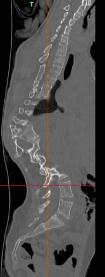
were obtained. Computed tomography scan was obtained from SIMS hospital Chennai. The thoracic
and lumbar region of the patient’s spine in the scan showed deviation in comparison to a healthy spine
scan, which indicated the affected region.
2.1. Three dimensional FEA model construction
The CT scan images were saved in standard digital imaging and communications in medicine
(DICOM) format. Computed Tomography images imported: The CT images and the data about the
vertebral column of the chosen patient was imported to MIMICS 14. The distance between each of the
361 slices (slice interval) of the CT scan images was 0.5 mm. In order to ensure no errors were in the
top, bottom, front and back positions of the images appropriate value of bone tissue grey was selected,
and a range of 0-3000 after submission of the images to the Mimics image processing software (figure
1, 2). Each vertebra was cropped and segmented.
Figure 1. Sagittal view of the scan. Figure 2. Anterior view of the scan.
The models of vertebrae from T1 to T12 in the thoracic region and L1 to L5 of the lumbar region
was generated and their STL files were generated to import it into Geomagics Design X64 software of
2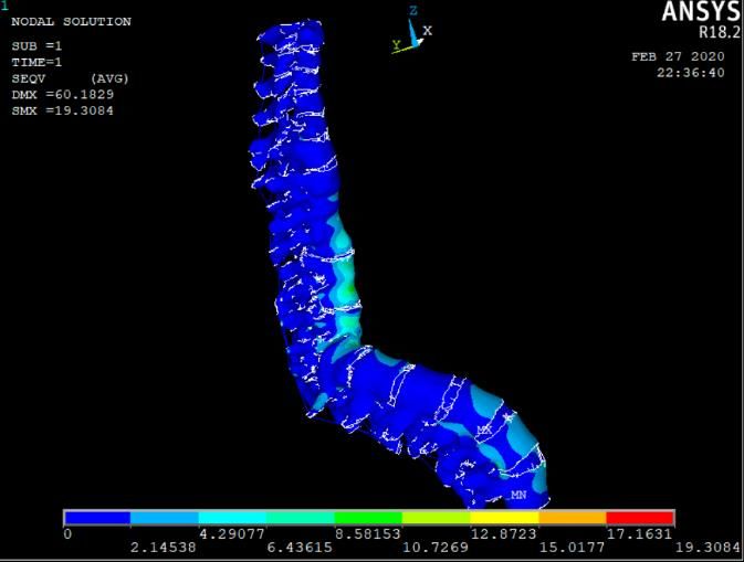
3rd International Conference on Advances in Mechanical Engineering (ICAME 2020)            IOP Publishing
IOP Conf. Series: Materials Science and Engineering 912 (2020) 022021 doi:10.1088/1757-899X/912/2/022021

3 D systems. The imported files were refined using the command subdivide and smooth so that the
rough edges and rough surfaces are smoothened to create surface model was shown in figure 3-5. 3D
model was developed and meshed in ANSYS 19.0. Finite element model of the ligamentous
thoracolumbar spine comprised of vertebrae, intervertebral discs and posterior elements along with the
following ligaments: supra-spinous (SSL), ligamentum flavum (LF), interspinous (ISL), transverse
(TL), anterior longitudinal (ALL), posterior longitudinal (PLL), and capsular (CL) was shown in
figure 6. It was assumed that the material properties were homogeneous and isotropic, and the data
was obtained from previously published literature [10-12] and shown in Table 1. In order to simulate
the ligaments, two-node link elements along with resistance tension only. The elements were then
arranged in anatomical direction. 20-node solid element used for the modelling of the cortical bone,
disc and cancellous bone. The disc nucleus comprises of gel like substance whereas the disc anulus
consists of fibres embedded in the ground substance. The region of facet joint was considered as a
problem of nonlinear three-dimensional contact by using surface to surface contact element. The
coefficient of friction was set to 0.1.

2.2. Boundary and Loading Condition
In the thoracolumbar spinal model all the inferior surfaces of the bottom most vertebra L5 were fixed
completely. The thoracolumbar spine was subjected to various loads at various levels. 50N weight as
the load of the head at T1, 350N weight of the upper extreme on T6 and 450N at T8 were applied on
the superior surfaces of the mentioned vertebrae (figure 7).

Table 1. Material properties of the FEM of thoracolumbar spine [10-12].
   Components        Young’s modulus       Poisson’s ratio     Cross-sectional             Element
                           (MPa)                                 area (mm2)
Cortical bone               1200                 0.26                 -
Cancellous bone             100                  0.2                  -
Posterior elements          3500                 0.25                 -
                                                                                          Solid 185
Disc
     Nucleus                  1                 0.499                 -
Ground substance             4.2                 0.45                 -
       Fiber                450                  0.3                0.76                   Link 10
Ligament
       ALL                   20                                     63.7
       PLL                   20                                      20
        TL                  58.7                                     3.6
         LF                 19.5                                     40                    Link 10
        ISL                 11.6                                     40
       SSL                   15                                      30
        CL                  32.9                                     60

                                                    3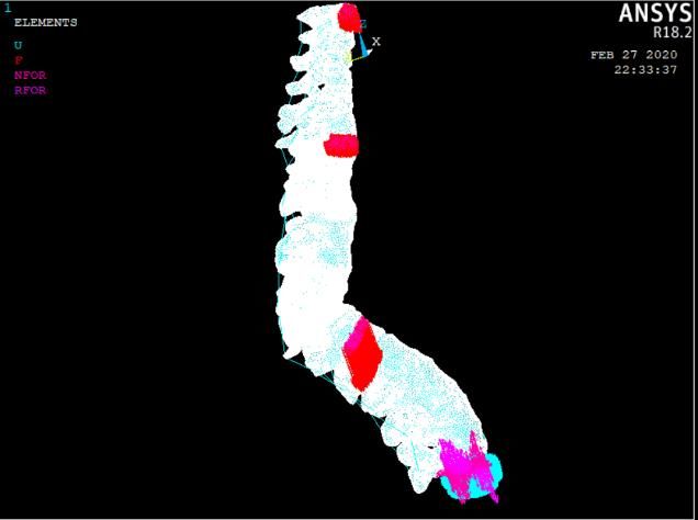
3rd International Conference on Advances in Mechanical Engineering (ICAME 2020)            IOP Publishing
IOP Conf. Series: Materials Science and Engineering 912 (2020) 022021 doi:10.1088/1757-899X/912/2/022021

                         Figure 3. Conversion from CT to Segmented model.

         Figure 4. Vertebra solid model.                Figure 5. Surface model.

                                                        Figure 7. Model with Loading and boundary
      Figure 6. Meshed model with discs.
                                                        conditions.

3. Results and discussion
The three-dimensional patient specific finite element model of scoliosis affected vertebrae was
developed from T1 to L5 and analysed for stress distribution, displacement in vertebrae and disc.
Special attention was given to the scoliotic region during analysis to understand the severity and the
root cause of the problem. In this study, the meshed model was subjected to only static load and the
deformation and stress concentration on the model was investigated. Compared to other vertebrae, T8
– T10 segments showed maximum stress concentration due to its shape deviation from the normal
spine axis. The von mises stress and deformation plot is shown in figure 8 and 9 after analysis.
                                                    4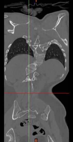
3rd International Conference on Advances in Mechanical Engineering (ICAME 2020)            IOP Publishing
 IOP Conf. Series: Materials Science and Engineering 912 (2020) 022021 doi:10.1088/1757-899X/912/2/022021

Figure 8. Lateral view of analysed model.                Figure 9. Anterior view of analysed model.

  After applying the uniformly distributed load on the vertebrae, results showed unpredictable variation
 in the displacement and stress pattern in the individual vertebrae and disc. Due to non-uniform
 deformity in the spine curvature the displacement was high in T7 and L4 vertebrae. While, comparing
 the disc displacement D5 and D8 shown more displacement compare to other discs. The T7 vertebrae
 and D8 disc displacement were high because that segment is the starting stage of the scoliotic region.
 Further, L4 vertebrae and D5 disc also showed higher displacement may be due to non-uniform
 deformity of spine curvature. The displacement plot for vertebrae and discs are shown in figure 10, 11.

  Figure 10. Displacement in vertebrae from T1            Figure 11. Displacement in discs from D1 –
  – L5.                                                   D13.

Figure 12. Stress distribution in vertebrae from         Figure 13. Stress distribution in discs from D1–
T1–L5.                                                   D13.
                                                     5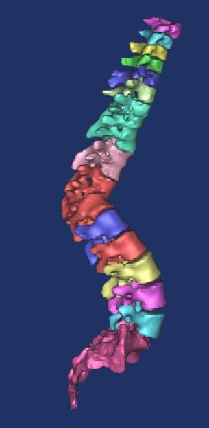
3rd International Conference on Advances in Mechanical Engineering (ICAME 2020)            IOP Publishing
IOP Conf. Series: Materials Science and Engineering 912 (2020) 022021 doi:10.1088/1757-899X/912/2/022021

   Similarly, the stress distribution was more in the scoliotic region and in the lower level lumbar
segment. Von mises stress values at T9 and T10 vertebrae along with the adjacent lower level
vertebrae showed high values. This proves that the scoliotic region experiences high stress values.
There is sudden raise in the value of von mises stress value for the disc lying in between D8 and D10.
Especially, since D9 showed very high stress value compared to other discs. However, the Disc D11
adjacent to scoliotic region disc does not experience the same level of stress value. But, last two disc
again showed the stress raiser. This proves that the stress concentration is maximum at the end of the
thoracic and lumber region in the scoliotic condition. The stress distribution for Vertebrae and discs
are shown in figure 12 & 13.

4. Conclusion
The spine with congenital scoliosis was modelled and analysed. FE analysis on the pre-surgery model
help understand the stress concentration and displacement patterns. The study revealed high stress
concentration on the vertebra and the disc comprised in the scoliotic region. The extracted model was
subject specific and the spine curvature was more complex, ergo difficult to segment the normal and
defected vertebrae from the CT scan. During the past two decades, many research works have
focussed on post-surgery condition. Author felt that, this study may pave pathways for future studies
in this field of pre-surgery condition.

Acknowledgements
This work was supported by Translational Medicine and Research (TMR) of SRM Medical College
Hospital & Research Centre and Biomechanics lab of SRM institute of science and technology.

5. References
[1] Tho K, Gibson I and Gao Z 2012 Development of a Detailed Human Spine Model with Haptic
         Interface Haptics Rendering and Applications: InTech. 9 165-94
[2] Illes T, Tunyogi Csapo M and Somoskeoy S 2011 Breakthrough in three-dimensional scoliosis
         diagnosis: significance of horizontal plane view and vertebra vectors European spine journal
         20 135-43
[3] Bruno A G, Anderson D E, D'Agostino J, Bouxsein M L 2012 The effect of thoracic kyphosis
         and sagittal plane alignment on vertebral compressive loading J. Bone Miner Res. 27 2144-
         51
[4] Taft E and Francis R 2003 Evaluation and management of scoliosis Journal of Pediatric Health
         Care 17 42-4
[5] Berven S and Bradford D S 2002 Neuromuscular Scoliosis: Causes of Deformity and Principles
         for Evaluation and Management Seminars in Neurology 22 167-78
[6] Chen X, Cai H, Zhang G, Zheng F, Wu C and Lin H 2020 The construction of the scoliosis 3D
         finite element model and the biomechanical analysis of PVCR orthopaedy Saudi J. Biol. Sci.
         27 695-700
[7] Kurtz S M and Devine J N 2007 PEEK biomaterials in trauma, orthopedic, and spinal implants
         Biomaterials 28 4845-69
[8] Salmingo R, Tadano S, Fujisaki K, Abe Y and Ito M 2012 Corrective force analysis for
         scoliosis from implant rod deformation Clinical Biomechanics 27 545-50
[9] Kirkham B. Wood W L, Darren S. Lebl and Avraam Ploumis 2014 Management of
         thoracolumbar spine fractures The Spine Journal 14 A18
[10] Pitzen T, Matthis D and Steudel W I 2002 The effect of posterior instrumentation following
         PLIF with BAK cages is most pronounced in weak bone Acta Neurochirurgica 144 121-8
[11] Polikeit A, Ferguson S J, Nolte L P and Orr T E 2003 Factors influencing stresses in the lumbar
         spine after the insertion of intervertebral cages: finite element analysis European spine
         journal 12 413-20
[12] Chiang M F, Zhong Z C, Chen C S, Cheng C K, Shih S L 2006 Biomechanical Comparison of
         Instrumented Posterior Lumbar Interbody Fusion With One or Two Cages by Finite Element
         Analysis Spine 31 682-9
                                                    6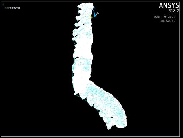

You can also read