Massive Gouty Tophi Presenting as Pseudotumor of the Elbow: A Rare Presentation - Cureus
←
→
Page content transcription
If your browser does not render page correctly, please read the page content below
Open Access Case
                                         Report                                                DOI: 10.7759/cureus.6769

                                         Massive Gouty Tophi Presenting as
                                         Pseudotumor of the Elbow: A Rare
                                         Presentation
                                         Divesh Jalan 1 , Deepak Kumar Maley 2 , Abhay Elhence 3 , Poonam Elhence 4 , Princi Jain 5

                                         1. Central Institute of Orthopaedics, Vardhman Mahavir Medical College and Safdarjung Hospital, New
                                         Delhi, IND 2. Orthopaedics, MediCiti Institute of Medical Sciences, Hyderabad, IND 3. Orthopaedics, All
                                         India Institute of Medical Sciences, Jodhpur, IND 4. Pathology, All India Institute of Medical Sciences,
                                         Jodhpur, IND 5. Internal Medicine, Atal Bihari Vajpayee Institute of Medical Sciences and Ram Manohar
                                         Lohia Hospital, New Delhi, IND

                                         Corresponding author: Divesh Jalan, dvsh_jalan@yahoo.com

                                         Abstract
                                         Gout is a systemic metabolic disorder characterized by hyperuricemia and deposition of
                                         monosodium urate crystals in joints and other extra-articular tissues. Poorly controlled cases
                                         progress to chronic gout with tophi, which can sometimes assume massive sizes. We report one
                                         such case of a 39-year-old male with poorly controlled polyarticular tophaceous gout
                                         presenting with a massive swelling of the left elbow simulating a soft tissue tumor. Subsequent
                                         investigations confirmed it to be a massive tophus which was then surgically excised, as the
                                         mass was not responding to the medical management. At the latest follow-up after two years,
                                         the patient has full function of the elbow and gout is well controlled with medications.

                                         Categories: Rheumatology, Orthopedics, Oncology
                                         Keywords: gout, tophi, pseudotumor, arthritis

                                         Introduction
                                         Gout is a systemic metabolic disorder with elevated serum urate levels and deposition of
                                         monosodium urate crystals in synovial and non-articular tissues resulting in repeated attacks
                                         of arthritis [1]. The cutaneous manifestations of gout are represented by intradermal lesions or
                                         subcutaneous nodules called tophi, commonly seen in avascular tissue over the ears, olecranon
                                         and prepatellar bursae or in acral sites, often associated with tendons. Although tophi are seen
                                         in 10% of the patients with chronic gout, literature is sporadic on massive gouty tophi [2,3]. We
                                         are presenting a case of massive elbow tophi simulating a tumor in a known patient of chronic
                                         gouty arthritis.
Received 12/16/2019
Review began 12/23/2019
Review ended 01/22/2020                  Case Presentation
Published 01/25/2020
                                         A 39-year-old male patient, a known case of chronic gouty arthritis, presented to the
© Copyright 2020                         orthopaedic clinic with massive swelling over the extensor aspect of left elbow, measuring
Jalan et al. This is an open access
                                         18x10 cm. which was progressively increasing over the past 10 years.The swelling was insidious
article distributed under the terms of
the Creative Commons Attribution
                                         in onset, slowly progressive with a waxing and waning course with occasional pain. There was
License CC-BY 3.0., which permits        no associated fever or any other constitutional symptoms. Past and family history were not
unrestricted use, distribution, and      significant.
reproduction in any medium, provided
the original author and source are
credited.                                On physical examination, the skin over the swelling was tense, shiny with venous prominence
                                         and superficial ulceration (Figure 1).

                                         How to cite this article
                                         Jalan D, Maley D, Elhence A, et al. (January 25, 2020) Massive Gouty Tophi Presenting as Pseudotumor
                                         of the Elbow: A Rare Presentation . Cureus 12(1): e6769. DOI 10.7759/cureus.6769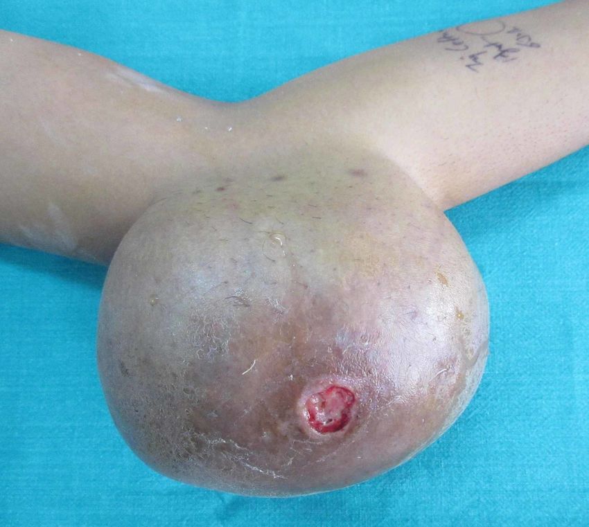
FIGURE 1: Clinical photograph of the left elbow showing
                                      massive swelling with central ulceration.

                                   There was chalky white discharge from the ulceration. The patient had full, pain free range of
                                   motion of the elbow joint without any neurovascular deficit. The patient had multiple small
                                   swellings over the right pinna, bilateral hands, ankle and both feet. The systemic examination
                                   was unremarkable.

                                   Hematological investigations revealed raised serum uric acid level (11 mg/dl), erythrocyte
                                   sedimentation rate of 38 mm/hour for the first hour and highly sensitive C-reactive protein
                                   level of 44.92 mg/l. Radiographs of the left elbow showed a huge soft tissue shadow with
                                   calcification (Figure 2).

2020 Jalan et al. Cureus 12(1): e6769. DOI 10.7759/cureus.6769                                                                2 of 10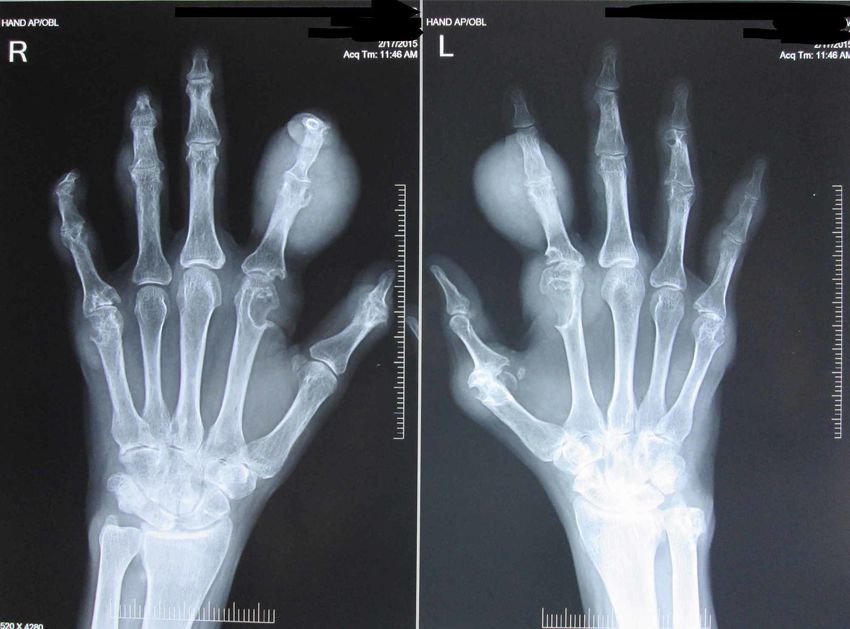
FIGURE 2: AP and lateral radiograph of the left elbow showing
                                      a huge soft tissue shadow with calcifications.

                                   Radiographs of bilateral hands, ankle and feet showed similar soft tissue shadows in phalanges
                                   and periarticular punched-out erosions (Figure 3).

2020 Jalan et al. Cureus 12(1): e6769. DOI 10.7759/cureus.6769                                                               3 of 10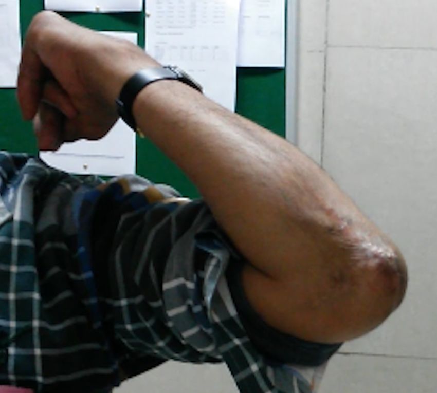
FIGURE 3: AP radiograph of bilateral hands showing soft
                                      tissue shadows in phalanges and periarticular punched-out
                                      erosions.

                                   Direct fine needle aspiration cytology was done from the left elbow swelling, which yielded
                                   blood mixed brownish material, and from the right index finger swelling, which yielded whitish
                                   chalky material. On microscopic examination, smears from both the sites showed similar
                                   cytological material comprising of numerous scattered and aggregates of non-branching
                                   needle-shaped urate crystals in a fluffy to amorphous dirty background. Few scattered
                                   histiocytes and occasional lymphocytes were also visualized along with red blood cells.

                                   The patient was initially put on dietary restrictions, plenty of fluids and drug therapy in the
                                   form of anti-inflammatory medications and oral allopurinol 100 mg three times daily for three
                                   months. At the end of three months, the serum uric acid levels reduced to 6.6 mg/dl, tophi over
                                   the pinna disappeared, the swellings over the hand and feet decreased in size, but the swelling
                                   and ulceration over the left elbow tophi continued to increase in size.

                                   After informed consent, the patient was planned for surgical excision of the massive left elbow
                                   tophi. The patient was positioned in a right lateral position under general anesthesia and en
                                   bloc excision of the swelling was performed through a standard posterior approach. The excised
                                   mass weighing around 1.5 kg was sent for histopathological examination and the wound was
                                   closed after excising the redundant skin margins (Figure 4).

2020 Jalan et al. Cureus 12(1): e6769. DOI 10.7759/cureus.6769                                                                4 of 10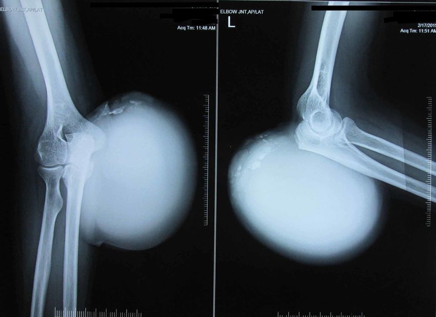
FIGURE 4: Excised mass weighing around 1,500 g.

                                   Imprint smear of the exudative material from the specimen confirmed gouty tophi with
                                   negatively birefringent needle-shaped sodium urate crystals (Figure 5).

2020 Jalan et al. Cureus 12(1): e6769. DOI 10.7759/cureus.6769                                                            5 of 10
FIGURE 5: Imprint smear of the exudative material showing
negatively birefringent needle-shaped sodium urate crystals.
Gross examination of soft tissue specimen revealed a skin-covered globular mass measuring
16x16x8 cm with an area of ulceration. On sectioning, a thick paste-like brownish material with
whitish chalky deposits was observed. Microscopic examination showed skin with
hyperkeratosis, parakeratosis and an ulcer covered by acute inflammatory exudate, crystalline
deposits and fibrin and dense perivascular lymphocytic infiltrate in the dermis. Extensive
crystal deposition (mainly needle-shaped crystals present in sheaves and bunches) and
associated calcification within the dermis and fibrocollagenous areas were associated with
multinucleated giant cell reaction and chronic inflammatory cells. These features were
consistent with urate arthropathy (Figure 6).
2020 Jalan et al. Cureus 12(1): e6769. DOI 10.7759/cureus.6769 6 of 10FIGURE 6: Histopathology image showing crystalline deposits
(marked *) associated with multinucleated giant cells (marked
arrow) and chronic inflammatory cells (H&E ×40).
Postoperatively, the wound healed uneventfully and at the last follow-up after two years, the
patient had full range of motion without any neurovascular deficit (Figure 7).
2020 Jalan et al. Cureus 12(1): e6769. DOI 10.7759/cureus.6769 7 of 10FIGURE 7: Clinical image showing healed scar and full
function at the elbow.
Discussion
Gout (also known as Podagra) is a systemic disorder of purine metabolism resulting in
increased uric acid levels with recurrent attacks of arthritis [4,5]. The male-to-female ratio is
3.6:1 [6]. Chronic gouty arthritis is often associated with tophi, which are deposits of
monosodium urate crystals in and around the joints and tendons. Distal extremities are most
commonly involved with predilection for extensor surfaces. These tophi are considered
pathognomonic for gout [7]. Conservative management with uricosuric drugs and xanthine
oxidase inhibitors is effective in stabilizing and reducing the size of the tophi. However, 5%-
10% of the patients do not respond completely to the conservative treatment [8].
The underlying pathology is invasion and destruction of skin, ligament, tendon, cartilage, and
bone by deposition of urates. The process is accompanied by an acute or chronic inflammatory
response at the site of involvement. The stages of gout include asymptomatic hyperuricemia,
acute gouty arthritis, intercritical gout and chronic tophaceous gout which develops after 10
years of the intercritical gout phase [9,10].Tophi vary from semiliquid to inspissated, chalk-like
deposits. These chalky materials reveal negatively birefringent needle-shaped crystals.
Early diagnosis and treatment prevent severe crippling from the disease. Although majority of
the cases respond to conservative management, relative surgical indications in chronic
tophaceous gout are unsightly painful tophi, infection, impairment of tendon function, nerve
2020 Jalan et al. Cureus 12(1): e6769. DOI 10.7759/cureus.6769 8 of 10compression due to tophi, impending skin necrosis, ulceration and discharging sinus, painful
destruction of joint, a decrease in uric acid level in the body by excision of massive tophi and
cosmesis [11-13]. Curettage and debridement can be done to remove tophi; however, it is
associated with high rates of delayed wound healing and skin necrosis [14]. Other surgical
methods include shaving, hydrotherapy and en bloc excision. We chose surgical treatment in
our case as the mass was progressively increasing despite treatment, has developed ulceration
and was cosmetically disfiguring.
Postoperative wound healing is generally poor after excision of large tophi because of
decreased circulation to the overlying skin which may require skin grafting [11]. In our case, the
wound healed uneventfully as entire avascular skin was excised along with the mass.
Conclusions
This case highlights one of the rare presentations of chronic tophaceous gout with a massive
elbow tophus weighing around 1.5 kg and locally mimicking a soft tissue tumor. A high index of
suspicion is required in these cases, and a simple test like aspiration of the material for crystal
analysis and cytology can help to distinguish this from soft tissue sarcoma.
Additional Information
Disclosures
Human subjects: Consent was obtained by all participants in this study. Conflicts of interest:
In compliance with the ICMJE uniform disclosure form, all authors declare the following:
Payment/services info: All authors have declared that no financial support was received from
any organization for the submitted work. Financial relationships: All authors have declared
that they have no financial relationships at present or within the previous three years with any
organizations that might have an interest in the submitted work. Other relationships: All
authors have declared that there are no other relationships or activities that could appear to
have influenced the submitted work.
References
1. Schumacher HR, Chen LX: Gout and other crystal-associated arthropathies. Harrison's
Principles of Internal Medicine 17th ed. Fauci AS, Braunwald E, Kasper DL (ed): McGraw-Hill,
New York; 2008. 2:2165-2169.
2. Nakayama DA, Barthelemy C, Carrera G, Lightfoot RW Jr, Wortmann RL: Tophaceous gout: a
clinical and radiographic assessment. Arthritis Rheum. 1984, 27:468-471.
10.1002/art.1780270417
3. Resnick D, Niwayama G: Gouty arthritis. Diagnosis of Bone and Joint Disorders. 4th ed.
Forrester DM (ed): WB Saunders, Philadelphia; 2002. 2:1519-1559.
https://doi.org/10.1002/art.1780250431
4. Eggebeen AT: Gout: an update . Am Fam Physician. 2007 , 76:801-808.
5. Richette P, Bardin T: Gout. Lancet . 2010, 375:318-328. 10.1016/s0140-6736(09)60883-7
6. Tang CY, Fung B: The last defence? Surgical aspects of gouty arthritis of hand and wrist . Hong
Kong Med J. 2011, 17:480-486.
7. Ragab G, Elshahaly M, Bardin T: Gout: an old disease in new perspective—a review . J Adv Res.
2017, 8:495-511. 10.1016/j.jare.2017.04.008
8. Burns CM, Wortmann RL: Latest evidence on gout management: what the clinician needs to
know. Ther Adv Chronic Dis. 2012, 3:271-286. 10.1177/2040622312462056
9. Schlesinger N: Management of acute and chronic gouty arthritis: present state-of-the-art .
Drugs. 2004, 64:2399-2416. 10.2165/00003495-200464210-00003
10. Yu KH, Ho HH, Chen JY, Luo SF: Gout complicated with necrotizing fasciitis--report of 15
cases. Rheumatology (Oxford). 2004, 43:518-521. 10.1093/rheumatology/keh097
11. Kumar S, Gow P: A survey of indications, results and complications of surgery for tophaceous
2020 Jalan et al. Cureus 12(1): e6769. DOI 10.7759/cureus.6769 9 of 10gout. N Z Med J. 2002, 115:U109.
12. Larmon WA, Kurtz JF: The surgical management of chronic tophaceous gout . J Bone Joint
Surg Am. 1958, 40:743-772. https://doi.org/10.2106/00004623-195840040-00001
13. Lee SS, Sun IF, Lu YM, Chang KP, Lai CS, Lin SD: Surgical treatment of the chronic
tophaceous deformity in upper extremities: the shaving technique. J Plast Reconstr Aesthet
Surg. 2009, 62:669-674. 10.1016/j.bjps.2007.12.021
14. Lee JH, Park JY, Seo JW, Oh DY, Ahn ST, Rhie JW: Surgical treatment of subcutaneous
tophaceous gout. J Plast Reconstr Aesthet Surg. 2010, 63:1933-1935.
10.1016/j.bjps.2010.03.019
2020 Jalan et al. Cureus 12(1): e6769. DOI 10.7759/cureus.6769 10 of 10You can also read