Synchronous Primary Heart Liposarcoma and Papillary Renal Carcinoma - a Case Report
←
→
Page content transcription
If your browser does not render page correctly, please read the page content below
Pol J Pathol 2003, 54, 2, 153-159                                                                            PL ISSN 1233-9687

Krystyna Gała˛zka1, Marcin Cie˛żarek 2, Jerzy Soja 2, Marek Krzanowski 2, Artur Szlubowski 2,
Katarzyna Sydor 3, Wojciech Adamczyk 3, Janusz Grodecki 3, Krzysztof Sładek 2

Synchronous Primary Heart Liposarcoma and Papillary Renal
Carcinoma - a Case Report
1
  Department of Clinical and Experimental Pathomorphology,
2
  Invasive Pulmonology Ward, Department of Internal Medicine, Collegium Medicum, Jagiellonian University,
3
  Cardiology Ward, G. Narutowicz’s Municipal Hospital, Kraków

     A case of synchronous primary cardiac dedifferen-            of circulatory disorder symptoms; for last 3 years he has been
tiated liposarcoma and papillary renal carcinoma is               treated with oral hypoglycemic drugs. At admission an
presented. The occurrence of typical areas of round cell          electrocardiogram revealed atrial fibrillation with ventricu-
liposarcoma made the pathological diagnosis of the sarcoma
                                                                  lar rate of 100, additionally tachypnoe 20 per minute and
relatively easy; however the neoplasm was not diagnosed
                                                                  normal blood pressure were observed. By auscultation bilat-
correctly before the autopsy. Cardiac liposarcoma is a very
rare primary malignant neoplasm and its diagnosis based           eral crepitations over lower lung areas were found. The liver
on image procedures may be extremely difficult especially         was slightly enlarged and bilateral lower leg edema was
at non-advanced stage of disease.                                 visible. Laboratory tests showed only mild elevation of
                                                                  glycaemia and hepatic enzymes - AspAT - 78U/l, AlAT -
                                                                  139U/l. X-ray examination of the chest revealed bilateral
Introduction                                                      blurring of the costophrenic angles due to the presence of
      The human heart is relatively rare location for malignant   pleural effusion and enlargement of the cardiac silhouette.
neoplasms. Among them the most frequent are metastases            Echocardiography showed normal atrial and ventricular vol-
of carcinomas and primary neoplasms of other chest organs         ume, normal contractility of cardiac muscle as well as a
infiltrating secondarily the myocardium. The development          pericardial effusion and dense, thicker pericardial echo. The
of mesenchymal malignant neoplasm originating from tissue         pericardial effusion increased, so two pericardial puncture
being a component of the heart is also possible. The fre-         were performed. During each of that procedures about
quency of primary heart mesenchymal tumors found during           1000ml of hemorrhagic fluid were obtained. Cytological
autopsies was estimated as high as 0.0017 - 0.28% [12] and        examination of pericardial effusion and transudate pleural
most of them (about 75%) are benign - myxomas, rhabdo-            effusion obtained following drainage punctures did not re-
myomas, lipomas, fibromas. Among malignant non-epithe-            veal atypical cells. The patient was firstly treated with sal-
lial neoplasms of the heart the most frequent are teratomas,      icylates, and then with corticosteroids as viral infection of
rhabdomyosarcomas and fibrosarcomas in children up to 16          the heart was suspected. However, during hospitalization
years old and angiosarcomas in adults. Primary heart lipo-        deterioration was observed - increasing symptoms of right
sarcoma is extremely rare - about 1% of primary heart             heart failure, recurrent pericardial effusion, and chest CT
sarcomas [5, 6, 12]. Till 1996 only 18 such neoplasms were        revealed tumorous thickening of interatrial septum. Ultra-
reported [8]. We present a case of primary heart liposarcoma      sound and CT examinations of abdominal cavity indicated a
correctly diagnosed during post-mortem procedure, syn-            1cm-tumor in the left kidney. Control laboratory tests
chronous with papillary renal carcinoma.                          showed gradual increase of hematocrit value (from 33.6%
                                                                  to 48.3%), hemoglobin concentration (from 11.4g/l to
                                                                  16.4g/l), erythrocyte and leukocyte count (from 3.96/mm3
A Case Description                                                to 5.67/mm3, from 8.7/mm3 to 13.9/mm3, respectively), and
     A 58-year-old man was admitted to Cardiology Ward            C-reactive protein (CRP), while blood platelets, serum levels
of Municipal Hospital in Kraków because of lower tolerance        of cholinesterase, total protein and albumins gradually de-
of exercise, subfebrile condition and non-characteristic chest    creased. As the condition of the patient deteriorated he was
pain lasting for few days. In the past he had never complained    transferred to the Invasive Pulmonology Unit, Department

                                                                                                                           153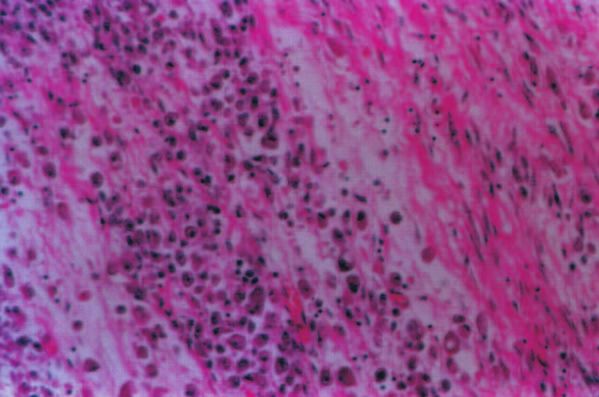
K. Gała˛zka et al

Fig. 1. Transthoracic echocardiography, apical 4-chamber view. Marked irregular thickening of interatrial septum and tissues behind the
heart (arrows). PK - right ventricle, LK - left ventricle, PP - right atrium, LP - left atrium.

Fig. 2. CT scan of the chest at the level of the heart. A tumor within the right atrium adhering to the interatrial septum. A pericardial effusion
is visible.

154
Primary heart liposarcoma
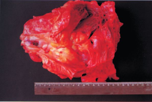
Fig. 3. Opened right heart ventricle and atrium. Yellow-whitish tumorous mass protruding into atrial lumen with distortion of the tricuspid
valve (on the left - opened superior vena cava).
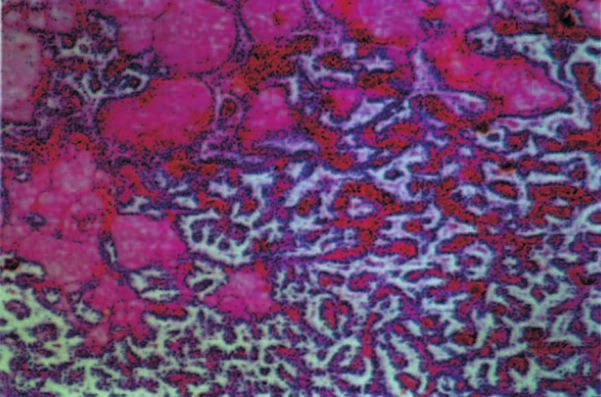
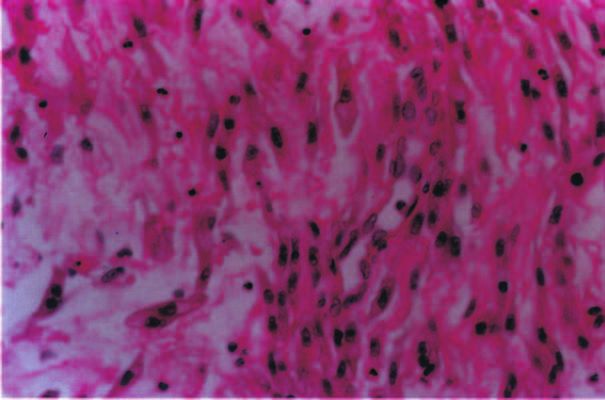
Fig. 4. The tumor composed of round cells. HE.
of Internal Medicine. At admission to the Ward the patient in the interatrial septum and mediastinum narrowing the left
was cachectic and confused. Transthoracic echocardio- inferior pulmonary vein (Figs. 1 and 2). Mild pericardial
graphy and CT showed a tumorous immobile mass located effusion and decreased ejection fraction were also found.
155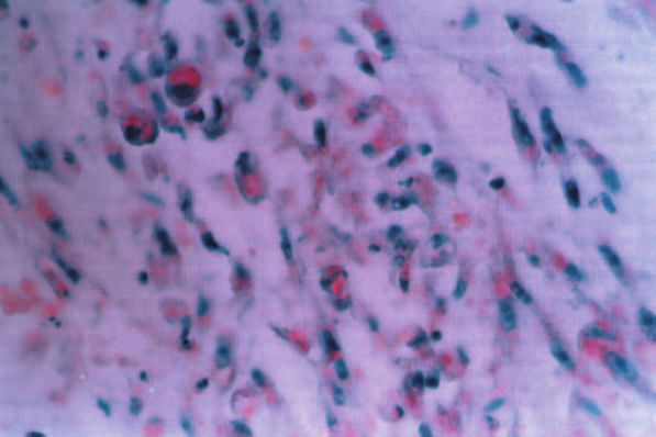
K. Gała˛zka et al

Fig. 5. Spindle cell fibrosarcoma-like pattern of the liposarcoma. HE.

Fig. 6. Empty vacuoles in the cytoplasm of elongated neoplastic cells. HE.

Three days after the admission the patient died and resusci-                 The autopsy was performed at the Department of Patho-
tation efforts were ineffective. The course of the disease was           morphology (No 122554). The pleural cavities contained
very rapid, ca 3 months from the first symptoms.                         abundant cloudy, yellowish fluid. Pleura of both lungs was

156
Primary heart liposarcoma

Fig. 7. Fat droplets in the cytoplasm of liposarcoma cells. Sudan.

Fig. 8. Classic picture of papillary renal carcinoma - the tumor of the left kidney. HE.

thickened up to 3 - 4mm, especially in lower parts of the                faces of lungs as well as the pleura with the pericardial sac.
lungs, grayish-yellowish in color and covered by fibrinous               Dispersed nodular infiltrates up to 6mm in thickness, yel-
exudate. Adhesions joined parietal and visceral pleura sur-              lowish on the cut section were present within the right

                                                                                                                                  157
K. Gała˛zka et al
parietal pleura, mainly in the lower part of the chest. The appeared shortly before the patient’s death. Certainly, the
pericardial sac was thickened up to 2cm in upper portion, course of the neoplasm was relatively rapid, but the first
nodular and mottled, focally yellowish, focally grayish and diagnostic procedures did not reveal any tumorous mass.
red with rough visceral and parietal surfaces, covered by Even so effective image procedure as CT may fail to
abundant fibrinous and slightly hemorrhagic exudate with reveal developing liposarcoma, as its density may be close
formation of weak adhesions. On the cut section of it nodular, to normal fatty tissue, and without evident distortion of
whitish foci were visible, focally infiltrating the subepicar- normal organ structure it is difficult to interpret properly
dial layer of cardiac muscle. After opening of the right heart the CT scans of mediastinum and the heart, usually rich
the irregular tumor up to 8cm in diameter was visible pro- in fatty tissue. A lipomatous hypertrophy of the interatrial
truding into the lumen of right atrium and upper part of the septum and arrhythmogenic right ventricular dysplasia
right ventricle, with distortion of the tricuspid valve (Fig. 3). also belong to fat-containing cardiac lesions and must be
The irregular infiltrate of the lower anterior mediastinum, considered in differential diagnosis of CT pictures [2].
pericardial sac and lower portions of both lung pleura layers Liposarcoma belongs to tumors characterized by weak
was in continuity with this tumor. The mediastinal lymph dispersion of cells, so the diagnosis based on cytological
nodes appeared to be free from the infiltrate. On the cut examination of neoplastic cells in body cavity effusions
section the infiltrate was solid, with slightly mottled appear- may be delayed. However, even early proper diagnosis of
ance - focally white-gray, in another areas rather yellowish, primary sarcoma of the heart does not allow usually
focally myxomatous. The heart was dilated, especially the effective treatment. Only single cases were reported with
right ventricle and atrium. 1 - 3-year survival after combined therapy - surgery/radio-
Moreover during the autopsy a solid yellow tumor 2cm /chemotherapy [5]. But even benign primary neoplasm of
in diameter was found in the cortex of the left kidney. From the heart frequently are life-threatening due to their loca-
the remaining findings congestion of internal organs as well tion and the malignant ones are additionally usually ag-
as of the lungs ("wet shock lung") was noticeable. gressive in their behavior, especially when they are
Histopathological examination of multiple specimens characterized by dismal histology as in our case. Most
obtained from the right heart tumor and infiltrate of the primary sarcomas and pericardial mesotheliomas are di-
mediastinum, pericardial sac and both pleuras revealed ma- agnosed just during the autopsy [13].
lignant neoplasm composed from round and spindle cells In our case the correct pathological diagnosis was rela-
with small areas resembling fibrosarcoma (Figs. 4 and 5). tively easy as multiple pieces of the tumor tissue were
The infiltrate was diffuse, without formation of any organoid available, encompassing besides the spindle cell areas also
structures. In the relatively abundant pink cytoplasm of the round cell neoplasm with intracytoplasmic vacuoles. The
round neoplastic cells small vacuoles were focally visible differential diagnosis must include malignant mesothelioma
(Fig. 6). Differential diagnosis included mainly two neo- because of the location, picture of diffuse infiltration and the
plasm - liposarcoma and malignant mesothelioma, due to biphasic histological pattern. Lack of cytokeratin and EMA
diffuse involvement of serosal surfaces. Immunohisto- expression in neoplastic cells excluded this cancer. Positive
chemically the cells were cytokeratin- and EMA-negative Sudan staining appeared to be the proof for the nature of the
and showed focal positivity for S-100 protein. Intracytoplas- neoplasm. Because of rarity of primary cardiac tumors the
mic vacuoles were mucicarmine-negative but orange stain- metastatic nature always must be excluded. Though meta-
ing of them was observed in Sudan method, what pointed to stases to the heart are more frequent from carcinomas, single
the presence of fat droplets (Fig. 7). Final diagnosis of the cases of metastatic liposarcoma were reported [7, 14]. In our
neoplasm was - liposarcoma, dedifferentiated because of the case detailed autopsy examination did not reveal any sarco-
presence of fibrosarcoma-like areas. ma in soft tissues.
The tumor of the left kidney presented microscopically A coincidence of primary liposarcoma and renal
characteristic pattern of papillary renal carcinoma (Fig. 8). papillary carcinoma seems to be interesting and an ac-
No metastases of it were found. cidental finding. We are unable to find any explanation
for the synchronous development of heart liposarcoma
and renal carcinoma. The genetic background may in-
Discussion
volve the same chromosome - trisomy 16 may occur in
The presented case in noteworthy as primary liposarco- sporadic papillary renal carcinoma and t(12;16) in myx-
ma of the heart is a very rare neoplasm, only single cases oid and round cell type of liposarcoma [10]. However
were reported [4, 8, 9, 11]. Clinical symptoms and radiologi- the detailed genetics of both tumors is not known and
cal signs of primary heart tumor are not characteristic [1, 3], there are no proofs for the involvement of the same
thus its diagnosis may be difficult in a patient at early stage region of chromosome 16. So far common risk factors
of the disease. In our case the suspicion of the heart tumor also are unknown.
158Primary heart liposarcoma
References 9. Pinelli G,. Trihn A, Carteaux JP, Mertes PM, Dopff C, Hubert T,
Villenot JP: Primary liposarcoma of the left ventricle. Apropos of
1. Black MD, Masters RG, Walley VM, Keon WJ: Hemoptysis: two a case and review of the literature. Arch Mal Coeur Vaiss 1996,
unusual causes. Can J Cardiol 1990, 6(1), 27-30. 89(2), 257-260.
2. Gaerte SC, Meyer CA, Winer-Muram HT, Tarver RD, Con- 10. Robbins Pathologic Basis of Disease. Contran RS, Kumar V, Collins
ces DJ Jr: Fat-containing lesions of the chest. Radiographics 2002, T, eds. 6th ed. WB Saunders Company 1999, pp. 992, 1261.
22, S61-S78. 11. Stamm C, Felderhoff T, Herse B, Dalichau H: Giant primary cardiac
3. Garrique S, Robert F, Rondaut R, Bonnet J: Assessment of non-in- liposarcoma vascularized via the circumflex coronary artery. Eur J
vasive new imaging techniques in the diagnosis of heart liposarco- Cardiothorac Surg 1999, 16(3), 367-370.
ma. Eur Heart J 1995,16(1), 139-141. 12. Vander Salm TJ: Unusual primary tumors of the heart. Semin
4. Macedo-Dias JA, Queiroz Machado F, Vouga L, Gonclaves V, Thorac Cardiovasc Surg 2000, 12(2), 89-100.
Gomes R: Liposarcoma of the heart. A case report. Am J Cardiovasc 13. Venditti FJ Jr, Pins MR: Weekly clinicopathological exercise: case
Pathol 1990, 3(3), 259-263. 19-1997: a 57-year-old man with a bloody pericardial effusion.
5. Molina JE, Edwards JE, Ward HB: Primary cardiac tumors: ex- N Engl J Med 1997, 336(25), 1812-1819.
perience at the University of Minnesota. Thorac Cardiovasc Surg 14. Wong SP, Ng CS, Wan S, Lee TW, Wan IY, Yim AP, Arifi AA: Giant
1990, 38(suppl 2),183-191. metastatic myxoid liposarcoma causing cardiac tamponade: a case
6. Nechaenko MA, Sheremeteva GF, Kniazeva GD, Rogov KA, Anto- report. Jpn J Clin Oncol 2002, 32(11), 480-482.
nov MV: Primary heart neoplasms. Khirurgiia (Mosk) 1994, 6,
8-13.
7. Ng C, Stebbing J, Judson I: Cardiac metastasis from a myxoid
liposarcoma. Clin Oncol (R Coll Radiol) 2001, 13(5), 384-385. Address for correspondence and reprint requests to:
8. Paraf F, Bruneval P, Balaton A, Deloche A, Mikol J, Maitre F, K. Gała˛zka M.D.
Scholl JM, De Saint-Maur PP, Camilleri JP: Primary liposarcoma Department of Pathomorphology CMUJ
of the heart. Am J Cardiovasc Pathol 1990, 3(2), 175-180. Grzegórzecka 16, 31-531 Kraków
159You can also read