Fetal Sinus Bradycardia Is Associated with Congenital Hypothyroidism: An Infant with Ectopic Thyroid Tissue - J-Stage
←
→
Page content transcription
If your browser does not render page correctly, please read the page content below
Tohoku J. Exp. Med., 2019, 248, 307-311
                                     Congenital Hypothyroidism with Antenatal Bradycardia                                         307

       Fetal Sinus Bradycardia Is Associated with Congenital
       Hypothyroidism: An Infant with Ectopic Thyroid Tissue
             Aya Nakanomori,1 Nobuhiko Nagano,1 Ayako Seimiya,1 Aya Okahashi1 and
             Ichiro Morioka1
              1
               Department of Pediatrics and Child Health, Nihon University School of Medicine, Tokyo, Japan

   Hypothyroidism is rarely included in the differential diagnosis for fetal sinus bradycardia. We report an
   infant with congenital hypothyroidism caused by ectopic thyroid tissue, who showed antenatal bradycardia.
   The baseline fetal heart rate was 100-110 bpm at 30 weeks of gestation, and fetal echocardiography
   revealed sinus bradycardia but no cardiac anomalies. Maternal thyroid function was normal (thyroid-
   stimulating hormone [TSH] 2.03 μ IU/ml, free T3 2.65 pg/ml, and free T4 0.99 ng/dl) when measured at 31
   weeks of gestation. Her serum anti SS-A and SS-B antibodies, anti-thyroglobulin, and microsomal
   antibodies were negative. A male infant without cardiac anomalies was delivered at 35 weeks and 4 days
   of gestation and admitted for prematurity and respiratory distress syndrome. The infant’s heart rate was
   70-110 bpm (normal: 120-160 bpm) on admission. On 8 days of age, thyroid function tests revealed that
   the infant had severe hypothyroidism (TSH 903.3 μ IU/ml, free T3 1.05 pg/ml, and free T4 0.26 ng/dl). The
   prolonged jaundice assumed to be due to hypothyroidism. Oral levothyroxine sodium hydrate (10 μ g/kg/
   day) was immediately started on day 8. After the treatment, the heart rate was gradually increased to
   130-140 bpm as the infant’s thyroid function was improved (TSH 79.8 μ IU/ml, free T3 2.95 pg/dl, and free
   T4 1.66 ng/dl on day 22). The infant was diagnosed ectopic thyroid tissue because of the high
   thyroglobulin level (85.9 μ g/l). In conclusion, congenital hypothyroidism should be included in the
   differential diagnosis in cases of fetal bradycardia without cardiac anomalies or maternal autoimmune
   diseases.

   Keywords: congenital hypothyroidism; ectopic thyroid tissufree; fetal bradycardia; levothyroxine sodium hydrate;
   maternal autoimmune disease
   Tohoku J. Exp. Med., 2019 August, 248 (4), 307-311. © 2019 Tohoku University Medical Press

                                                                        Jaeggi and Öhman 2016). A previous study has reported
                        Introduction                                    that congenital hypothyroidism was not predicted from a
      Fetal bradycardia is defined as a heart rate less than            decreased fetal heart rate (Miyai et al. 1979). On the other
110 bpm (Macones et al. 2008). The main mechanisms of                   hand, there are some cases that congenital hypothyroidism
perinatal bradycardia are sinus bradycardia, complete heart             presented with fetal bradycardia, such as autoimmune thy-
block, and functional atrioventricular block (Jaeggi and                roiditis with severe hypothyroidism and severe congenital
Öhman 2016). Fetal complete heart block is strongly asso-               hypothyroidism induced by maternal blocker antibodies
ciated with maternal connective tissue disease, such as                 (Kara et al. 2013; Marzuillo et al. 2016). In general, how-
Sjögren’s syndrome and systemic lupus erythematosus, and                ever, hypothyroidism is rarely included in the differential
the presence of maternal anti-SS-A/Ro or anti-SS-B/La                   diagnosis for fetal sinus bradycardia.
antibodies also increases the risk of complete heart block in                 Our patient had congenital hypothyroidism with fetal
the fetus (Izmirly et al. 2011). A pregnant woman with fetal            bradycardia, and the heart rate of the patient was increased
bradycardia should undergo testing for autoimmune dis-                  when treatment for hypothyroidism was started after deliv-
ease.                                                                   ery. We therefore propose that the fetal bradycardia is
      Here we report prenatal sinus bradycardia in an infant            attributed in this case to hypothyroidism. The institutional
whose mother did not have autoimmune disease. Fetal                     review board of Nihon University Itabashi Hospital
sinus bradycardia may be secondary to fetal distress, pla-              approved this case study with informed consent (approval
cental insufficiency, anatomic abnormality of the sinus                 number: RK-190709-5).
node, or long QT syndrome (Kleinman and Nehgme 2004;
       Received July 17, 2019; revised and accepted August 16, 2019. Published online August 28, 2019; doi: 10.1620/tjem.248.307.
       Correspondence: Nobuhiko Nagano, M.D., Ph.D., Department of Pediatrics and Child Health, Nihon University School of Medicine,
  30-1 Oyaguchi, Kami-Cho, Itabashi-ku, Tokyo 173-8610, Japan.
       e-mail: nagano.nobuhiko@nihon-u.ac.jp

                                                                  307
308 Aya Nakanomori et al.
Hospital course
Case Report Artificial surfactant was administered, and then venti-
A 31-year-old primigravida with an unremarkable past lation was started for respiratory distress syndrome.
medical history was admitted for threatened preterm labor Respiratory status was improved gradually, and the patient
at 30 weeks of gestation. The baseline fetal heart rate was was extubated on 1 day of age. Oxygen supplementation
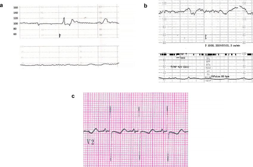
100-110 bpm (< 3% percentile; Fig. 1a). For comparison, was no longer needed by 7 days of age. The infant was
normal fetal heart rate is shown in Fig. 1b (140-160 bpm). noted to have hyperbilirubinemia, which persisted despite
Fetal echocardiography revealed sinus bradycardia but no phototherapy being started on 3 days of age. Routine neo-
cardiac anomalies. The mother has no signs of thyroid dis- natal screening was performed on 5 days of age and showed
ease and no history of hysterosalpingography or excess decreased thyroid function (TSH > 81.8 μIU/ml and free T4
iodine. Her thyroid function was normal (thyroid-stimulat- 0.28 ng/dl). On 8 days of age, thyroid function tests at our
ing hormone [TSH] 2.03 μIU/ml, free T3 2.65 pg/ml, and hospital revealed that the infant had severe hypothyroidism
free T4 0.99 ng/dl) at 31 weeks of gestation. Serum anti (TSH 903.3 μIU/ml, free T3 1.05 pg/ml, and free T4 0.26
SS-A and SS-B antibodies, anti-thyroglobulin, and micro- ng/dl). Radiographic examination of the lower limbs
somal antibodies were negative. A male infant was deliv- showed that both knee epiphyses were absent (Fig. 2a: this
ered by spontaneous vaginal delivery after premature rup- case and 2b: a normal newborn). Furthermore, ultrasonog-
ture of membranes at 35 weeks and 4 days of gestation and raphy revealed that the thyroid gland was not at the ortho-
admitted for prematurity and respiratory distress syndrome. topic site in the neck (Fig. 2c: this case and 2d: a normal
The Apgar scores were six and nine at one and five minutes infant). The thyroglobulin level in our case was 85.9 μg/l.
after birth. The heart rate was 70-110 bpm on admission. A previous study has reported the thyroglobulin level
The electrocardiogram showed sinus bradycardia but no ranged from < 1.0 to 18.7 μg/l in cases with athyreosis and
other abnormal findings, such as an irregular RR or pro- the level from 12.2 to 123 μg/l in cases with ectopic thyroid
longed QT interval (Fig. 1c). Echocardiography on admis- tissue (Beltrao et al. 2010). This case was, therefore, diag-
sion did not indicate a cardiac anomaly or any signs of heart nosed as ectopic thyroid tissue based on the thyroglobulin
failure. Laboratory findings did not reveal a cause of bra- level. The prolonged jaundice assumed to be a symptom of
dycardia, such as electrolyte imbalance (Table 1). hypothyroidism. There were no other symptoms of hypo-
thyroidism, including constipation, dry skin, poor general
Fig. 1. The cardiotocogram and electrocardiogram on admission.
a. The cardiotocogram at 30 weeks of gestation in our case. The fetal heart rate baseline is 100-110 bpm. b. The car-
diotocogram at 38 weeks of gestation in a normal fetus. The fetal heart rate baseline is 140-160 bpm. c. The electrocar-
diogram with sinus bradycardia on admission in our case (110 bpm).Congenital Hypothyroidism with Antenatal Bradycardia 309
Table 1. Laboratory data on admission.
Discussion
WBC 10,500 /µl
This case demonstrates that congenital hypothyroidism
RBC 370 ×104 /µl may cause fetal sinus bradycardia. We therefore suggest
Hemoglobin 14.1 g/dl that congenital hypothyroidism should be included in the
differential diagnosis for fetal sinus bradycardia.
4
Plt 31.3 ×10 /µl The fetal thyroid gland appears by 10 weeks of gesta-
T-bil 2.17 mg/dl tion, by which time it has already developed the ability to
concentrate radioiodine and synthesize iodothyronines
D-bil 0.55 mg/dl (Fisher and Klein 1981; Burrow et al. 1994). The fetal pitu-
AST 53 U/l itary gland also develops at about the same time, when TSH
and T4 are measurable in fetal serum. At gestational 18-22
ALT 6 U/l weeks, the pituitary and serum TSH concentrations start to
LDH 453 U/l increase. Peak serum TSH levels occur at gestational 20-24
weeks and gradually decrease until term. In contrast, fetal
ALP 627 U/l serum T4 and free T4 concentrations were increased from
CK 644 U/l mid-gestation, peaking between 20 and 30 weeks’ gestation.
In our case, the fetal bradycardia became noticeable from
BUN 9.4 mg/dl 30 weeks’ gestation. We suspect that congenital hypothy-
Cr 0.65 mg/dl roidism in a fetus manifests as bradycardia with a low T4
from that time onwards. However, little has been reported
Na 140 mmol/l on the relationship between fetal bradycardia and congeni-
K 3.8 mmol/l tal hypothyroidism. The human placenta acts as a barrier to
maternal-fetal transfer of thyroid hormones (Fisher 1997).
Cl 108 mmol/l In the past, the placenta was thought to be impermeable to
Ca 8.6 mg/dl thyroid hormones (Fisher and Klein 1981). However,
Vulsma et al. (1989) showed that the cord serum total T4
P 7.1 mg/dl level in the human fetus with thyroid agenesis is approxi-
Mg 2.2 mg/dl mately 30% of the concentration in the normal fetus. It is
presumed that there is limited but significant T4 transfer
TP 4.2 g/dl between the mother and fetus via the placenta. We specu-
Alb 2.9 g/dl late that the reason why there are so few reports of congeni-
tal hypothyroidism manifesting as fetal bradycardia is that
CRP < 0.1 mg/dl thyroid function in the fetus is supplemented by maternal
IgG 501 mg/dl thyroid hormones. Moreover, it was reported that infants
with TSH > 100 μIU/ml have significantly slower heart rate
IgA 3 mg/dl than those with lower TSH levels (Öner et al. 2015).
IgM 9 mg/dl Although there are limited relevant postnatal data, we con-
sider that neonates with markedly high TSH might have had
The laboratory data on admission
bradycardia at the fetal stage. It can be inferred from the
was normal and did not suggest a
cause of bradycardia.
present case that bradycardia as a symptom of low T4 at the
fetal stage occurs because placental transfer of thyroid hor-
mones is insufficient to fully supplement fetal thyroid func-
condition, cold extremities, and delayed closure of posterior tion. Congenital hypothyroidism should be considered
fontanelle. However, the infant’s weight had not return to when a fetus shows bradycardia in the absence of a cardiac
the birth weight by 8 days of age. Oral levothyroxine anomaly or maternal autoimmune disease.
sodium hydrate (10 μg/kg/day) was started from 8 days of Thyroid hormone is critical for normal growth and
age. After the treatment, the infant’s heart rate was gradu- brain development, and hypothyroidism in infancy is a
ally increased to 130-140 bpm as his thyroid function was leading cause of intellectual impairment. If the diagnosis of
improved (TSH 79.8 μIU/ml, free T3 2.95 pg/dl, and free hypothyroidism is made and treatment started within a few
T4 1.66 ng/dl on day 22). The hyperbilirubinemia was also weeks of birth, the neurodevelopmental outcome is gener-
improved gradually after treatment was initiated. The ally normal (LaFranchii and Austin 2007). Most newborns
infant showed sufficient weight gain and was discharged on with congenital hypothyroidism do not have a distinctive
27 days of age (Fig. 3). appearance and the symptoms of hypothyroidism are not
specific; namely, early diagnosis of hypothyroidism is chal-
lenging. Congenital hypothyroidism became easier to diag-310 Aya Nakanomori et al.
Fig. 2. Radiography of lower limbs and ultrasonography of neck.
a. Radiography of lower limbs of our case at 8 days of age: absence of both knee epiphyses (Arrows indicate abnormal
findings). b. Radiography of lower limbs of a normal newborn at 21 days of age: presence of both knee epiphyses. c.
Ultrasonography of neck of our case at 8 days of age: the thyroid gland is not at the orthotopic site. d. Ultrasonography
of neck of a normal infant at 1 month of age: the thyroid gland is at the orthotopic site.
OCA4=B=;!2=II!IPDCC434L !" #$!
67.!089:12;5
./ 012345
#)$
67.!Q!+#N+!89:12; !#-$$$
67.!,$&N&!89:12;
#($ !,$$
!+$$
#'$
!*$$
#&$
!)$$
#%$
!($$
##$
67.!&(,N*!89:12; !'$$
#$$
!&$$
,$ !%$$
67.!*,N++!89:12;
+$ !#$$
$
S !"
$ # % & ' ( ) * + , #$ ## #% #& #' #( #) #* #+ #, %$ %# %% %& %' %( %) %*
R6&!0EL12;5! !#N$(! !&N%)! !%N,(
R6'!04L1J;5!! !$N%+! !$N%)! !#N)*! !#N))
?@ABAB@CD=E>
FCGAB@>DAH34C!IAJ3K2!@>JD=BC!#$8L1ML1J=>
Fig. 3. Hospital course.
The heart rate was gradually increased as his thyroid function was improved.
FT3, free T3; FT4, free T4.
nose when neonatal screening was introduced. However, diagnosis.
the neonatal screening sample must be sent to a centralized In conclusion, we report a case of congenital hypothy-
laboratory for examination, and it takes time to the result. roidism caused by ectopic thyroid tissue that manifested as
We believe that consideration of congenital hypothyroidism fetal bradycardia, which was resolved after treatment with
when sinus bradycardia is detected in a fetus could aid early levothyroxine sodium hydrate. We suggest that congenitalCongenital Hypothyroidism with Antenatal Bradycardia 311
hypothyroidism should be included in the differential diag- disorders of thyroid function in the newborn. N. Engl. J.
nosis in cases of fetal bradycardia without cardiac anoma- Med., 304, 702-712.
Izmirly, P.M., Saxena, A., Kim, M.Y., Wang, D., Sahl, S.K.,
lies or maternal autoimmune diseases. Llanos, C., Friedman, D. & Buyon, J.P. (2011) Maternal and
fetal factors associated with mortality and morbidity in a
Acknowledgments multi-racial/ethnic registry of anti-SSA/Ro-associated cardiac
This work was supported by Grants-in-Aid for Young neonatal lupus. Circulation, 124, 1927-1935.
Jaeggi, E. & Öhman, A. (2016) Fetal and neonatal arrhythmias.
Scientists (grant number: 19K20194) of JSPS KAKENHI, the
Clin. Perinatol., 43, 99-112.
Practical Research Project for Intractable Diseases from Japan
Kara, S., Tayman, C., Tonbul, A., Andiran, N., Tatli, M. & Türkay,
Agency for Medical Research and Development, AMED
S. (2013) Congenital hypothyroidism presenting with post-
(19ek0109265h0003), and Nihon University Research Grant for
partum bradycardia. J. Coll. Physicians Surg. Pak., 23, 214-
Social Implementation for 2019. The authors received no other
215.
financial support for the research, authorship, and publication of
Kleinman, C.S. & Nehgme, R.A. (2004) Cardiac arrhythmias in
this article. the human fetus. Pediatr. Cardiol., 25, 234-251.
LaFranchi, S.H. & Austin, J. (2007) How should we be treating
Author Contributions children with congenital hypothyroidism? J. Pediatr. Endo-
A.N. and N.N. wrote the case report and discussion section crinol. Metab., 20, 559-578.
of the manuscript. R.A., M.H., K.F., A.S., K.K., R.K. and A.O. Macones, G.A., Hankins, G.D., Spong, C.Y., Hauth, J. & Moore, T.
contributed to the care provided during the hospital course. N.N. (2008) The 2008 National Institute of Child Health and
and I.M. interpreted the clinical data. I.M. critically reviewed Human Development workshop report on electronic fetal
the manuscript and assisted with editing. All authors contributed monitoring: update on definitions, interpretation, and research
to the intellectual content of this manuscript and approved the guidelines. Obstet. Gynecol., 112, 661-666.
final version of the manuscript for submission. Marzuillo, P., Grandone, A., Perrotta, S., Ruggiero, L., Capristo, C.,
Luongo, C., Miraglia Del Giudice, E. & Perrone, L. (2016)
Very early onset of autoimmune thyroiditis in a toddler with
Conflict of Interest severe hypothyroidism presentation: a case report. Ital. J.
The authors declare no conflict of interest. Pediatr., 42, 61.
Miyai, K., Mizuta, H., Amino, N., Tanizawa, O., Nose, O. & Oura,
References T. (1979) Fetal heart-rate in congenital hypothyroidism.
Lancet, 2, 693-694.
Beltrao, C.B., Juliano, A.G., Chammas, M.C., Watanabe, T., Öner, T., Ozdemir, R., Doksoz, O., Yozgat, Y., Karadeniz, C.,
Sapienza, M.T. & Marui, S. (2010) Etiology of congenital Demirpence, S., Yilmazer, M.M., Buyukinan, M., Mese, T. &
hypothyroidism using thyroglobulin and ultrasound combina- Tavli, V. (2015) Cardiac function in newborns with congenital
tion. Endocr. J., 57, 587-593. hypothyroidism: association with thyroid-stimulating hormone
Burrow, G.N., Fisher, D.A. & Larsen, P.R. (1994) Maternal and levels. J. Clin. Res. Pediatr. Endocrinol., 7, 307-311.
fetal thyroid function. N. Engl. J. Med., 331, 1072-1078. Vulsma, T., Gons, M.H. & de Vijlder, J.J. (1989) Maternal-fetal
Fisher, D.A. (1997) Fetal thyroid function: diagnosis and manage- transfer of thyroxine in congenital hypothyroidism due to a
ment of fetal thyroid disorders. Clin. Obstet. Gynecol., 40, total organification defect or thyroid agenesis. N. Engl. J.
16-31. Med., 321, 13-16.
Fisher, D.A. & Klein, A.H. (1981) Thyroid development andYou can also read