An Autopsy-Proven Case of Limbic-Predominant Age-Related TDP-43 Encephalopathy
←
→
Page content transcription
If your browser does not render page correctly, please read the page content below
Case Report
Yonsei Med J 2020 Aug;61(8):731-735
https://doi.org/10.3349/ymj.2020.61.8.731 pISSN: 0513-5796 · eISSN: 1976-2437
An Autopsy-Proven Case of Limbic-Predominant
Age-Related TDP-43 Encephalopathy
Soo Hyun Cho1, Seong-Min Choi1,2, Byeong C. Kim1,2, Won-Young Song2,
Hyung-Seok Kim2,3, and Kyung-Hwa Lee2,4
1
Department of Neurology, Chonnam National University Medical School and Hospital, Gwangju;
2
Chonnam National University Hospital Brain Bank, Gwangju;
3
Department of Forensic Medicine, Chonnam National University Medical School, Gwangju;
4
Department of Pathology, Chonnam National University Medical School and Hwasun Hospital, Hwasun, Korea.
Limbic-predominant age-related TDP-43 encephalopathy (LATE) is a recently established neurodegenerative disease entity.
LATE neuropathological change (LATE-NC) is characterized by a TDP-43 proteinopathy that mainly involves the amygdala and
medial temporal structures, with or without hippocampal sclerosis. LATE-NC is typically observed in individuals aged 80 years or
older and manifests clinically as amnestic memory decline. Herein, we report a case of LATE diagnosed by brain autopsy in an
82-year-old male who had an 11-year history of memory impairment. Pathological examination revealed high Alzheimer disease
neuropathological changes, as well as amygdala-predominant Lewy body pathology. In addition, immunohistochemistry for
TDP-43 revealed neuronal and glial cytoplasmic inclusions in the dentate gyrus of the hippocampus, amygdala, and inferior tem-
poral cortex. Increasing awareness of the newly defined entity LATE will enhance our understanding of the neurodegenerative
processes that occur in the oldest individuals.
Key Words: TDP-43 proteinopathy, limbic system, Alzheimer disease, Lewy body disease, autopsy
INTRODUCTION alopathy (LATE) has recently been introduced.5 LATE neuro-
pathological change (LATE-NC) is defined pathologically as
Transactive response DNA binding protein of 43 kDa (TDP-43) TDP-43 proteinopathy, in which phosphorylated TDP-43 accu-
is a ubiquitinated protein associated with diseases, such as amy- mulates in the cytoplasm or nuclei of neurons or in the cyto-
otrophic lateral sclerosis and certain variants of frontotemporal plasm of glial cells localized primarily to the limbic structures.
lobar degeneration.1,2 TDP-43 proteinopathy is frequently iden- The clinical characteristics of LATE include amnestic cogni-
tified in the limbic area in people aged 80 years and older, and tive decline progressing to advanced cognitive deficits. Indi-
is associated with Alzheimer disease neuropathologic changes viduals with both ADNC and LATE-NC manifest more rapid
(ADNC) and/or hippocampal sclerosis.3,4 and severe cognitive impairment than the persons with ‘pure’
A new term, limbic-predominant age-related TDP-43 enceph- LATE-NC.5 Herein, we present the brain autopsy findings of a
patient who presented with persistent cognitive decline and
Received: April 7, 2020 Revised: May 25, 2020 was found to have both high ADNC and LATE-NC.
Accepted: June 16, 2020
Corresponding author: Kyung-Hwa Lee, MD, PhD, Department of Pathology,
Chonnam National University Medical School and Hwasun Hospital, 264 Seoyang-
ro, Hwasun 58128, Korea.
Tel: 82-61-379-7075, Fax: 82-61-379-7099, E-mail: mdkaylee@jnu.ac.kr
CASE REPORT
•The authors have no potential conflicts of interest to disclose. An 82-year-old male with a known history of dementia was re-
© Copyright: Yonsei University College of Medicine 2020 ferred to Chonnam National University Hospital brain bank
This is an Open Access article distributed under the terms of the Creative Com- (CNUHBB) for brain autopsy. His memory began to decline at
mons Attribution Non-Commercial License (https://creativecommons.org/licenses/
by-nc/4.0) which permits unrestricted non-commercial use, distribution, and repro- the age of 71 years and further deteriorated thereafter. After the
duction in any medium, provided the original work is properly cited. clinical diagnosis was determined, the patient was prescribed
www.eymj.org 731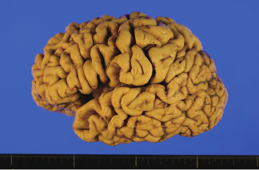
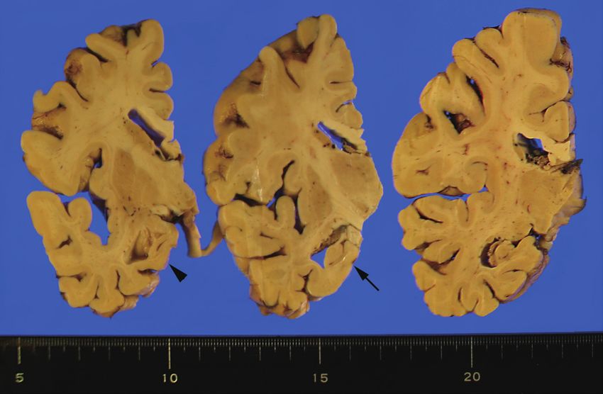
Autopsy Case of LATE

an acetylcholinesterase inhibitor. At the age of 73 years, he had              mography images revealed mild atrophy in the bilateral me-
trouble writing and finding his way home. He also had difficul-                dial temporal and frontal lobes, probably meeting the criteria
ty remembering words and names, and his orientation to time                    for Alzheimer disease (AD) (Fig. 1).6 His cognition continued to
worsened. He further demonstrated impairment in daily living                   deteriorate rapidly: MMSE scores of 17 at 74, 13 at 76, 5 at 77, 4
activities by failing to pay after eating at a restaurant and by ex-           at 80, and 0 at 82 years of age. The patient died of aggravated
periencing difficulties with hygiene management. At this time,                 pneumonia at the age of 82 years. Written informed consent
he recorded a Mini-Mental State Examination (MMSE) score                       was obtained from a legal surrogate of the patient.
of 20 (patient’s score/maximum score): temporal orientation                       The major pathological findings of his autopsy were sugges-
(4/5), spatial orientation (3/5), registration (3/3), attention and            tive AD. Grossly, atrophy was severe in the neocortex and hip-
calculation (2/5), remote memory (1/3), language (7/8), and                    pocampus (Fig. 2). Microscopically, neuronal loss and gliosis
copy the diagram (0/1). No family history of dementia or other                 were severe in the hippocampus and moderate in the neocor-
neurologic diseases was reported. Axial brain computed to-                     tex and amygdala (Fig. 3A). Superficial microvacuolation in

Fig. 1. Brain computed tomography (CT) scan acquired 10 years before death. Axial brain CT image demonstrating cortical and mild hippocampal atrophy.

 A                                                                           B
Fig. 2. Gross findings of a case with comorbid high Alzheimer disease neuropathologic changes, amygdala-predominant Lewy body disease, and limbic-
predominant age-related TDP-43 encephalopathy (LATE) neuropathological changes. (A) The lateral view shows diffuse neocortical atrophy with remark-
able involvement of the frontal cortex and temporal pole. (B) Coronal sections show thinning of the medial temporal lobe with dilatation of the temporal
horn of the lateral ventricle (arrowhead) and severe atrophy of the hippocampus (tailed arrow).

732                                                                                                               https://doi.org/10.3349/ymj.2020.61.8.731
Soo Hyun Cho, et al.

             A                                                                 B

             C                                                                 D

             E                                                                  F

             G                                           H                                             I
Fig. 3. Histopathological features of a case with LATE-NC. (A) The amygdala shows remarkable neuronal loss and gliosis. (B) Superficial microvacuola-
tion in cortical layer II (black arrows) is observed in the inferior temporal cortex (A and B, hematoxylin and eosin; original magnification, ×100). (C) The
hippocampus shows an abundance of neurofibrillary tangles and neuritic plaques, consistent with high Alzheimer disease neuropathologic changes
(Gallyas silver stain; original magnification, ×40). (D) Alpha-synuclein immunostaining highlights abundant Lewy bodies solely in the amygdala, consistent
with amygdala-predominant Lewy body disease. (E) Immunohistochemistry for TDP-43 indicates neuronal cytoplasmic inclusions (NCIs) in the dentate
gyrus of the hippocampus. (F-H) Abundant NCIs and a few dystrophic neurites are observed in the amygdala (F), entorhinal cortex (G), and hippocampus
(H). (I) NCIs are sparsely present in the insula (D-F, original magnification, ×200; G-I, original magnification, ×400).

https://doi.org/10.3349/ymj.2020.61.8.731                                                                                                              733
Autopsy Case of LATE
cortical layer II was present in the inferior temporal area (Fig. and show severe symptoms, such as behavioral problems or
3B). Neurofibrillary tangles were frequent in the entorhinal cor- language issues.12 LATE-NC typically involves the limbic re-
tex, hippocampus, and neocortex (Fig. 3C). Neuritic plaques gions, whereas FTLD-TDP affects the neocortices more widely.
were frequent in the neocortex. Amyloid deposition extended There were several limitations to the current case report. First,
up to CA4 of the hippocampus. Based on scores of A3 (Thal am- the interval between brain imaging and brain autopsy was long,
yloid phase 4), B3 (Braak neurofibrillary tangle stage VI), and and brain imaging was not performed close to the death of the
C3 [Consortium to Establish a Registry for Alzheimer’s Disease patient. Second, no other examinations, such as amyloid posi-
(CERAD) neuritic plaque score frequent], the pathological find- tron emission tomography or genetic analysis, were carried out
ings were compatible with high ADNC. α-Synuclein immunos- to elucidate the etiology of LATE. Nevertheless, this case is note-
taining revealed multiple Lewy bodies in the amygdala, but worthy, because it is the first autopsy-proven case of LATE to
none in the midbrain or cingulate (Fig. 3D), consistent with be documented in South Korea and will assist other neurolo-
amygdala-predominant Lewy body disease (LBD). Immuno- gists and pathologists in identifying LATE cases.
histochemistry for TDP-43 revealed neuronal cytoplasmic in- In conclusion, we suggest that LATE-NC can occur as a com-
clusions (NCIs) in the dentate gyrus of the hippocampus (Fig. bined pathology or a single entity. When brain autopsy is per-
3E). Abundant NCIs and some glial cytoplasmic inclusions were formed in cases with amnestic memory decline, routine TDP-43
observed in the amygdala, entorhinal cortex, hippocampus, su- immunohistochemistry should also be performed in critical
biculum, and inferior temporal cortex (Fig. 3F, G, and H). NCIs anatomic locations. Because there is no biofluid or neuroimag-
were sparsely present in the insula (Fig. 3I). The frontal cortex, ing biomarker for determining LATE status in vivo, acquisition
basal ganglia, and midbrain lacked TDP-43-positive inclusions. of postmortem data would facilitate the diagnosis and treatment
The TDP-43 pathology of the case indicated LATE-NC stage 2.5 of cognitive impairment among the oldest individuals, which
is important given the continual aging of society.
DISCUSSION ACKNOWLEDGEMENTS
We report the autopsy findings of a patient with severe amnes- This study was supported by the KBRI Basic Research Program
tic syndrome, who in fact had LATE-NC. The brain pathology through the Korea Brain Research Institute funded by the
exhibited high ADNC, amygdala-predominant LBD, and LATE- Ministry of Science and ICT (19-BR-03-04 for BCK), a National
NC. Previous studies have indicated that cases with coexisting Research Foundation of Korea grant funded by the Korean gov-
ADNC and LATE-NC are more likely to show hippocampal at- ernment (2019R1A2B5B01070598 for LKH), and the Chonnam
rophy than subjects with ADNC only.7 Indeed, structural alter- National University Hospital Biomedical Research Institute
ation of the amygdala reportedly indicates underlying LATE-NC (BCRI20012 for SHC). CNUHBB would like to acknowledge the
and may be linked to cognitive decline.8 The current case showed generosity shown by the donor and donor families in donating
severe hippocampal atrophy in association with both high ADNC brain tissue to the CNUHBB.
and LATE-NC. The amygdala showed notable neuronal loss
and gliosis in association with ADNC, LBD, and LATE-NC. AUTHOR CONTRIBUTIONS
Although TDP-43 proteinopathy largely confined to medial
temporal areas has been reported,4,9 a consensus definition of Conceptualization: Byeong C. Kim and Kyung-Hwa Lee. Funding ac-
LATE has been reached only recently. The consensus staging quisition: Soo Hyun Cho, Byeong C. Kim, and Kyung-Hwa Lee. Inves-
tigation: Soo Hyun Cho and Seong-Min Choi. Methodology: Hyung-
scheme for LATE-NC is a three-tier system encompassing hi-
Seok Kim. Project administration: Won-Young Song. Supervision:
erarchical spreading of TDP-43 proteinopathy from the amyg- Byeong C. Kim and Kyung-Hwa Lee. Validation: Seong-Min Choi and
dala (stage 1) to the hippocampus (stage 2) and middle frontal Hyung-Seok Kim. Visualization: Soo Hyun Cho and Kyung-Hwa Lee.
gyrus (stage 3),5 which represents a simpler system than the pre- Writing—original draft: Soo Hyun Cho. Writing—review & editing:
viously proposed six-stage scheme.9 To prevent under-recogni- Kyung-Hwa Lee. Approval of final manuscript: all authors.
tion of LATE-NC during brain autopsy, it is critical to perform
TDP-43 immunohistochemistry on three anatomical sections, ORCID iDs
as proposed previously.10 The prevalence of LATE-NC increases
gradually with age, while that of severe ADNC decreases.11 Con- Soo Hyun Cho https://orcid.org/0000-0002-4262-1468
Seong-Min Choi https://orcid.org/0000-0003-3138-1881
sequently, LATE plays a pivotal role in amnestic-type cognitive
Byeong C. Kim https://orcid.org/0000-0001-6827-6730
impairment among the rapidly growing oldest old population.5 Won-Young Song https://orcid.org/0000-0003-2331-4013
For confirmation of LATE-NC, it is important to differentiate Hyung-Seok Kim https://orcid.org/0000-0002-8297-9747
it from frontotemporal lobar degeneration with TDP-43 pro- Kyung-Hwa Lee https://orcid.org/0000-0002-3935-0361
teinopathy (FTLD-TDP). In comparison to those with LATE-NC,
most patients with FTLD-TDP are diagnosed at a younger age
734 https://doi.org/10.3349/ymj.2020.61.8.731Soo Hyun Cho, et al.
REFERENCES zheimer’s Association workgroups on diagnostic guidelines for Al-
zheimer’s disease. Alzheimers Dement 2011;7:263-9.
1. Neumann M, Sampathu DM, Kwong LK, Truax AC, Micsenyi MC, 7. Dawe RJ, Bennett DA, Schneider JA, Arfanakis K. Neuropathologic
Chou TT, et al. Ubiquitinated TDP-43 in frontotemporal lobar de- correlates of hippocampal atrophy in the elderly: a clinical, patho-
generation and amyotrophic lateral sclerosis. Science 2006;314: logic, postmortem MRI study. PLoS One 2011;6:e26286.
130-3. 8. Makkinejad N, Schneider JA, Yu J, Leurgans SE, Kotrotsou A, Evia
2. Bigio EH. TDP-43 variants of frontotemporal lobar degeneration. AM, et al. Associations of amygdala volume and shape with trans-
J Mol Neurosci 2011;45:390-401. active response DNA-binding protein 43 (TDP-43) pathology in a
3. Uchino A, Takao M, Hatsuta H, Sumikura H, Nakano Y, Nogami A, community cohort of older adults. Neurobiol Aging 2019;77:104-11.
et al. Incidence and extent of TDP-43 accumulation in aging hu- 9. Josephs KA, Murray ME, Whitwell JL, Tosakulwong N, Weigand SD,
man brain. Acta Neuropathol Commun 2015;3:35. Petrucelli L, et al. Updated TDP-43 in Alzheimer’s disease staging
4. Amador-Ortiz C, Lin WL, Ahmed Z, Personett D, Davies P, Duara scheme. Acta Neuropathol 2016;131:571-85.
R, et al. TDP-43 immunoreactivity in hippocampal sclerosis and 10. Lee KH, Seo SW, Lim TS, Kim EJ, Kim BC, Kim Y, et al. Proposal
Alzheimer’s disease. Ann Neurol 2007;61:435-45. guidelines for standardized operating procedures of brain autop-
5. Nelson PT, Dickson DW, Trojanowski JQ, Jack CR, Boyle PA, Arfa- sy: brain bank in South Korea. Yonsei Med J 2017;58:1055-60.
nakis K, et al. Limbic-predominant age-related TDP-43 encepha- 11. Nag S, Yu L, Boyle PA, Leurgans SE, Bennett DA, Schneider JA.
lopathy (LATE): consensus working group report. Brain 2019;142: TDP-43 pathology in anterior temporal pole cortex in aging and
1503-27. Alzheimer’s disease. Acta Neuropathol Commun 2018;6:33.
6. McKhann GM, Knopman DS, Chertkow H, Hyman BT, Jack CR Jr, 12. Schneider JA, Nelson PT. Reply: Limbic-predominant age-related
Kawas CH, et al. The diagnosis of dementia due to Alzheimer’s dis- TDP-43 encephalopathy (LATE). Brain 2019;142:e43.
ease: recommendations from the National Institute on Aging-Al-
https://doi.org/10.3349/ymj.2020.61.8.731 735You can also read