Light chain proteinuria revealing mu-heavy chain disease: an atypical presentation of Waldenstrom macroglobulinemia in two cases
←
→
Page content transcription
If your browser does not render page correctly, please read the page content below
Light chain proteinuria revealing mu-heavy chain disease:
an atypical presentation of Waldenstrom
macroglobulinemia in two cases

by Hélène Vergneault, Djaouida Bengoufa, Aline Frazier-Mironer, Isabelle Brocheriou,
Samuel Bitoun, Camille Villesuzanne, Alexis Talbot, Stéphanie Harel, Bertrand Arnulf
and Bruno Royer

Haematologica 2021 [Epub ahead of print]

Citation: Hélène Vergneault, Djaouida Bengoufa, Aline Frazier-Mironer, Isabelle Brocheriou,
Samuel Bitoun, Camille Villesuzanne, Alexis Talbot, Stéphanie Harel, Bertrand Arnulf,
and Bruno Royer. Light chain proteinuria revealing mu-heavy chain disease: an atypical presentation
of Waldenstrom macroglobulinemia in two cases.
Haematologica. 2021; 106:xxx
doi:10.3324/haematol.2020.277137

Publisher's Disclaimer.
E-publishing ahead of print is increasingly important for the rapid dissemination of science.
Haematologica is, therefore, E-publishing PDF files of an early version of manuscripts that
have completed a regular peer review and have been accepted for publication. E-publishing
of this PDF file has been approved by the authors. After having E-published Ahead of Print,
manuscripts will then undergo technical and English editing, typesetting, proof correction and
be presented for the authors' final approval; the final version of the manuscript will then
appear in print on a regular issue of the journal. All legal disclaimers that apply to the
journal also pertain to this production process.
Light chain proteinuria revealing mu-heavy chain disease: an atypical presentation of
Waldenström macroglobulinemia in two cases

Hélène Vergneault1, Djaouida Bengoufa2, Aline Frazier-Mironer3, Isabelle Brocheriou4,
Samuel Bitoun1, Camille Villesuzanne1, Alexis Talbot1,5, Stéphanie Harel1, Bertrand Arnulf1,
Bruno Royer1

   1.   Immuno-hematology Department, Saint-Louis Hospital, APHP, Paris, France
   2.   Immunology Laboratory, Saint-Louis Hospital, APHP, Paris, France
   3.   Rheumatology Department, Lariboisière Hospital, APHP, Paris, France
   4.   Pathology Laboratory, La Pitié Salpêtrière Hospital, APHP, Paris, France
   5.   INSERM U976 Équipe 5, Institut de Recherche Saint Louis, Université de Paris, Paris,
        France

Running head : Mu-HCD, an atypical presentation of WM
Corresponding author:

Bruno Royer, MD, PhD

Immuno-hematology Department

Assistance Publique - Hôpitaux de Paris, Saint-Louis Hospital

1 avenue Claude Vellefaux, 75010 Paris

Email : bruno.royer@aphp.fr

Manuscript word count: 1496

Number of figure: 1

Financial support: None

Disclosures: No author has a potential conflict of interest, financial or otherwise, to disclose

regarding this study

Authors contributions : HV, BA and BR initiated the study, analyzed the data and wrote the

manuscript; DB performed immunological analyzes; IB interpreted renal biopsy; HV, AT,

SB, CV, BA, AFM, SH and BR took care of patients; and all authors analyzed the data,

reviewed and approved the final manuscript .
Heavy chain diseases (HCDs) are rare mature B-cells proliferative disorders first
described in 19641 and characterized by the production of a paraprotein consisting of
truncated heavy chains devoid of bound light chains. A normal immunoglobulin is composed
of two heavy chains and two light chains joined by disulfide bonds at the heavy chain
constant domain 1 (CH1). In the absence of light chain, the heat shock protein BiP binds to
CH1 and retains the heavy chain in the endoplasmic reticulum2. In HCD various mutations are
responsible of splicing error leading to complete or partial deletion of CH1 and preventing
therefore the binding of heavy chains to light chains as well as BiP 3,4. Three HCD involving
the main immunoglobulin classes have been described: α-HCD, -HCD and µ-HCD which is
the least common. A single case of δ-HCD has been reported. Mu-HCD is often associated
with a B cell lymphoid disorder such as chronic lymphocytic leukemia with
hepatosplenomegaly. It has also been described in association with myelodysplasia, cirrhosis
and auto-immune disease 5.
Waldenström macroglobulinemia (WM) is a lymphoplasmacytic lymphoma secreting
monoclonal IgM gammopathy mainly κ and strongly associated with MYD88 L265P somatic
mutation6. Patients may be asymptomatic or may present symptoms related either to bone
marrow infiltration and/or to the IgM gammopathy physico-chemical properties
(hyperviscosity, auto-immune hemolytic anemia, cryoglobulinemia, anti-MAG neuropathy).
Serum-free light chains (sFLC) rarely reach high levels in WM and complications related to
7
light chains like cast nephropathy or amyloidosis are uncommon compared to multiple
myeloma.
We report here two cases with IgM kappa monoclonal gammopathy composed of a
small peak on serum protein electrophoresis (SPEP) contrasting with a high level of sFLC
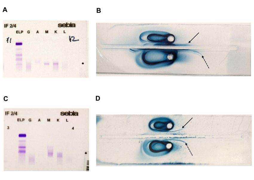
revealing a µ-heavy chain disease associated with WM.Case 1. In December 2018, a 79 year-old man with chronic renal failure from unknown origin presented with acute renal failure (creatinine 1160 µmol/L) associated with nephrotic syndrome (proteinuria 2,9g/24h and albumin 27 g/L) leading to end-stage renal failure requiring hemodialysis and normocytic non-regenerative anemia. The blood count was as following : hemoglobin 6g/dL, platelets 208000/mm3, neutrophils 3100/mm3, lymphocytes 900/mm3. Kidney biopsy showed interstitial fibrosis and tubular atrophy associated with linear Congo red-negative deposits of kappa-light chains along the basement membrane suggestive of Randall-type monoclonal immunoglobulin deposition disease (MIDD). sFLC- kappa were elevated at 2585 mg/L with a kappa-to-lambda ratio (κ/λ ratio) at 57,36. Gammaglobulins were at 6,5 g/L and no peak was detected on SPEP but immunofixation was positive for monoclonal IgM kappa (Figure 1A). Serum immuno-selection confirmed the presence of µ-heavy chain (Figure 1B). Bone marrow aspiration and biopsy showed lympho- plasmocytic infiltration with 19% of lymphoid and plasma cells on aspiration and 25% of CD19+ CD20+ cells with monotypic expression of kappa light chain on flow cytometry. Immuno-histochemistry on biopsy identified kappa monotypic population consisting of 20% of mature plasma cells CD138+ and lymphoid cells CD20+ CD79a+ CD5+ CD10- CD23-. The screening for L265P mutation of MYD88 was positive. No adenomegaly nor splenomegalia was found on whole body-CT scan. Cardiac markers were increased (Troponin 199 ng/l and NT-pro BNP 13664 ng/L) and echocardiography suggested infiltrative cardiomyopathy confirmed by cardiac MRI. Although imaging features could not distinguish between amyloid and Randall type deposits, endomyocardial biopsy was not performed because of histological evidence of MIDD in kidney. This infiltrative cardiomyopathy was attributed to probable Randall type light chain deposition. A treatment combining rituximab 375mg/m² + cyclophosphamide 750 mg/m² + dexamethasone 20 mg was initiated allowing
partial hematological response after 7 cycles. Weekly subcutaneous injections of bortezomib

1,3mg/m² were then added, leading to kappa-to-lambda ratio normalization after 1 cycle of

bortezomib, so that cyclophosphamide was discontinued. At date of follow-up in October

2020, the patient had received 13 cycles of rituximab and 8 courses cycles of bortezomib and

had achieved a sustained complete hematological response.

Case 2: In March 2019, a 74 year-old woman presented to Rheumatologic clinic for diffuse

pains associated with joints swelling leading to the diagnosis of articular chondrocalcinosis. In

the   context    of    osteo-articular   pains,    a    SPEP     was     performed     revealing

hypogammaglobulinemia at 2,7 g/L. sFLC-kappa were elevated at 3260 mg/L with a κ/λ ratio

of 1461. The blood count was as following: hemoglobin 12 g/dL, platelets 374 000/mm3,

neutrophils 7660/mm3, lymphocytes 2790/mm3. There was no hypercalcemia. Urine protein-

to-creatinine ratio was at 29 mg/mmol (corresponding to 0,29 g/24h of proteinuria) and renal

function was normal. Urine protein immuno-electrophoresis revealed Bence Jones proteinuria.

Beta-2-microglobuline was 2,6 mg/L. Free light chain multiple myeloma was suspected and

bone marrow aspiration was performed which revealed lymphocytic infiltration consisting of

54% of mature lymphocytes associated with 3% plasma cells frequently containing vacuoles.

This cytologic aspect of low-grade lymphoma was suggestive of Waldenström disease. Flow

cytometry on bone marrow confirmed this diagnosis with large monoclonal kappa-B cells

population CD19+, CD20+, CD22+, FMC7+, CD200+, CD5-, CD23-, CD10-, CD43-, CD38-

. L265P mutation of MYD88 was detected.

                                18
Whole-body CT-scan and           FDG-PET scanner showed no lytic bone lesion or

hepatosplenomegaly or lymphadenopathy. Monoclonal IgM kappa was detected on serum

immunofixation (Figure 1C) and immuno-selection confirmed the presence of µ-heavy chains

(Figure 1D).
In the absence of clinical impact, no specific treatment was introduced other than sodium
bicarbonate to prevent cast nephropathy.
Few cases of γ-HCD at diagnosis or during the evolution of WM have been described8
and Wahner-Roedler et al reported in 1992 three cases of WM among the 27 first cases of µ-
HCD11. Unlike γ-HCD and α-HCD, µ-HCD is characterized by secretion of sFLC most often
kappa in the urine in one-half to two-third of patients with a risk of cast nephropathy or
amyloidosis4,5. Besides, the abnormal immunoglobulin is not detected by SPEP in two-third
of µ-HCD5 .
These two new cases illustrated uncommon presentation of WM like light-chain
multiple myeloma with hypogammaglobulinemia, elevated sFLC and proteinuria revealing
finally µ-HCD. The dissociation between sFLC level and the absence of peak on SPEP was
unusual. Moreover, the presence of vacuolated plasma cells in bone marrow was highly
suggestive of µ-HCD. To detect heavy chains devoid of light chains on immuno-
electrophoresis, immuno-selection techniques and the use of specific anti-light chains anti-
serum are required. The serum samples were electrophoresed in agar containing anti-kappa
and anti-lambda antibodies trapping free lights chains and complete immunoglobulins. The
throughs contained anti-µ antiserum revealing mobile free µ-heavy chain through precipitin
line (Figure 1B and 1D). Bone marrow aspiration, immune phenotyping of B-cells and the
presence of monoclonal IgM on immunofixation easily clarified the diagnosis of WM, just as
MYD88 mutation. Of note this is to our knowledge the first report of such mutation in patients
with µ-HCD. The association of these two conditions raises the question of the underlying
pathophysiology and may suggest a continuum between WM and µ-HCD: the secretion of
truncated monoclonal IgM would be secondary to alteration of immunoglobulin gene within
lympho-plasmocytic cells. Unfortunately we did not have sufficient biological sample to
perform DNA sequencing.Serum-free light chains are part of the monitoring of multiple myeloma especially
oligo-secretory myeloma and light-chain myeloma as well as AL amyloidosis. It has been
recently suggested that sFLC could be a reliable marker in WM for prognosis and therapeutic
response10,11 but currently, their routine use is not recommended in WM. Although µ-HCD is
a rare condition and renal complications are even more infrequent, it could be cost-effective to
screen for proteinuria or even to measure sFLC and light chain proteinuria at diagnosis of
lymphoplasmatic lymphoma, especially if the paraprotein is not detected on electrophoresis,
because of the possible harmful renal and systemic consequences of sFLC increase. Indeed,
cases of cast nephropathy12 and systemic amyloidosis13 associated with µ-HCD have been
reported and here we described the first case of MIDD.
Patient 1 presented Randall-type light chains deposition disease (LCDD) with no
heavy chains deposition disease (HCDD). Even though both conditions, HCDD and HCD are
due to CH1 deletion, µ-HC protein never cause kidney or another organ damage. This
difference could be explained by the fact that in µ-HCD, CH1 deletion is associated with
deletions of variable region which seems to be involved in tissue precipitation. Indeed, it has
been reported that sequence analysis of HCDD proteins revealed amino acid substitution in
the variable region responsible of charge and hydrophobicity modifications14.
Because of the rarety of this condition, there is no prospective studies and therefore
no guidelines for the management of µ-HCD which is based on case reports. In asymptomatic
patients such as patient 2, simple monitoring seems reasonable. For symptomatic patients, the
chemotherapy targets the underlying clone as proposed in the MGCS fields 15. In this report
the use of rituximab associated with bortezomib and cyclophosphamide + dexamethasone
allowed complete response in patient 1.In summary, low levels of IgM protein with the presence of light chain proteinuria and high level of sFLC in WM patients are highly suggestive of µ-HCD, even more if bone marrow examination reveals vacuolated plasma cells, and should alert physician to the possibility of kidney damage. Finally, our report suggests that µ-HCD associated with lymphoplasmatic proliferation and MYDD mutation can be regarded as particular subgroup of WM.
REFERENCES

1. Franklin EC, Lowenstein J, Bigelow B, Meltzer M. Heavy chain disease—A new disorder
   of serum γ-globulins: Report of the first case. Am J Med. 1964;37(3):332-350.

2. Hendershot L, Bole D, Köhler G, Kearney JF. Assembly and secretion of heavy chains
   that do not associate posttranslationally with immunoglobulin heavy chain-binding
   protein. J Cell Biol. 1987;104(3):761-767.

3. Bakhshi A, Guglielmi P, Siebenlist U, Ravetch JV, Jensen JP, Korsmeyer SJ. A DNA
   insertion/deletion necessitates an aberrant RNA splice accounting for a mu heavy chain
   disease protein. Proc Natl Acad Sci U S A. 1986;83(8):2689-2693.

4. Seligmann M, Mihaesco E, Preud’homme J-L, Danon F, Brouet J-C. Heavy Chain
   Diseases: Current Findings and Concepts. Immunol Rev. 1979;48(1):145-167.

5. Fermand J-P, Brouet J-C. HEAVY-CHAIN DISEASES. Hematol Oncol Clin North Am.
   1999;13(6):1281-1294.

6. Treon SP, Xu L, Yang G, et al. MYD88 L265P somatic mutation in Waldenström’s
   macroglobulinemia. N Engl J Med. 2012;367(9):826-833.

7. Uppal NN, Monga D, Vernace MA, et al. Kidney diseases associated with Waldenström
   macroglobulinemia. Nephrol Dial Transplant. 2019;34(10):1644-1652.

8. Presti BC, Sciotto CG, Marsh SG. Lymphocytic Lymphoma with Associated Gamma
   Heavy Chain and IgM-Lambda Paraproteins: An Unusual Biclonal Gammopathy. Am J
   Clin Pathol.1990;93(1):137-141.

9. Wahner-Roedler DL, Kyle RA. μ-heavy chain disease: Presentation as a benign
   monoclonal gammopathy. Am J Hematol. 1992;40(1):56-60.

10. Leleu X, Xie W, Bagshaw M, et al. The Role of Serum Immunoglobulin Free Light Chain
    in Response and Progression in Waldenstrom Macroglobulinemia. Clin Cancer Res.
    2011;17(9):3013-3018.

11. Itzykson R, Le Garff-Tavernier M, Katsahian S, Diemert M-C, Musset L, Leblond V.
    Serum-free light chain elevation is associated with a shorter time to treatment in
    Waldenstrom’s macroglobulinemia. Haematologica. 2008;93(5):793-794.

12. Preud’homme JL, Bauwens M, Dumont G, Goujon JM, Dreyfus B, Touchard G. Cast
    nephropathy in mu heavy chain disease. Clin Nephrol. 1997;48(2):118-121.

13. Kinoshita K, Yamagata T, Nozaki Y, et al. Mu-heavy chain disease associated with
    systemic amyloidosis. Hematol Amst Neth. 2004;9(2):135-137.

14. Khamlichi AA, Aucouturier P, Preud’homme JL, Cogné M. Structure of abnormal heavy
    chains in human heavy-chain-deposition disease. Eur J Biochem. 1995;229(1):54-60.

15. Witzig TE, Wahner-Roedler DL. Heavy chain disease. Curr Treat Options Oncol.
   2002;3(3):247-254.
FIGURE LEGEND Figure 1. Immunological tests. On the left side, serum protein electrophoresis and immuno- fixation for patient 1 (A) and patient 2 (C) who had both monoclonal IgM kappa. On the right side, immuno-electrophoresis with immuno-selection for patient 1 (B) and patient 2 (D). Black arrow indicate precipitine line consisting of mu-heavy chain (continuus arrow for patients, dotted arrow for positive control)
You can also read