GASTRIC IGG4-RELATED DISEASE PRESENTING AS A MASS LESION AND MASQUERADING AS A GASTROINTESTINAL STROMAL TUMOR
←
→
Page content transcription
If your browser does not render page correctly, please read the page content below
Journal of Pathology and Translational Medicine 2020; 54: 258-262
https://doi.org/10.4132/jptm.2020.02.10 CASE STUDY
Gastric IgG4-related disease presenting as a mass lesion and
masquerading as a gastrointestinal stromal tumor
Banumathi Ramakrishna1, Rohan Yewale2, Kavita Vijayakumar1, Patta Radhakrishna3, Balakrishnan Siddartha Ramakrishna2
Departments of 1Pathology, 2Medical Gastroenterology, and 3Surgical Gastroenterology, SRM Institutes for Medical Science, Vadapalani, India
IgG4-related disease of the stomach is a rare disorder, and only a few cases have been reported. We present two cases that were iden-
tified over a 2-month period in our center. Two male patients aged 52 and 48 years presented with mass lesion in the stomach, which
were clinically thought to be gastrointestinal stromal tumor, and they underwent excision of the lesion. Microscopic examination re-
vealed marked fibrosis, which was storiform in one case, associated with diffuse lymphoplasmacytic infiltration and an increase in IgG4-
positive plasma cells on immunohistochemistry. Serum IgG4 level was markedly elevated. Although rare, IgG4-related disease should
be considered in the differential diagnosis of gastric submucosal mass lesions.
Key Words: IgG4-related disease; Stomach; Autoimmune diseases; Gastrointestinal stromal tumors
Received: December 4, 2019 Revised: February 5, 2020 Accepted: February 10, 2020
Corresponding Author: Banumathi Ramakrishna, MBBS, MD, Department of Pathology, SRM Institutes for Medical Science, 1 Jawaharlal Nehru Road, Vadapalani, Chennai
600026, India
Tel: +91-44-20002001, Fax: +91-44-49211455, E-mail: banu_ramakrishna@hotmail.com
IgG4-related disease (IgG4-RD) is a newly-recognized, im- (CT) of the kidney, ureter, and bladder revealed an incidental
mune-mediated fibroinflammatory disorder that most com- mass lesion in the posterior wall of the stomach. Upper gastro-
monly affects the pancreas [1]. It can also involve other organs intestinal endoscopy revealed submucosal lesions in the esopha-
such as the bile duct, liver, gallbladder, salivary glands, lacrimal gus and stomach that were clinically suspected to be GIST. The
glands, retroperitoneum, and lymph nodes, although involve- patient underwent endoscopic ultrasound-guided fine-needle
ment of the gastrointestinal tract is very rare [2,3]. The disorder aspiration biopsy of the esophageal lesion and endoscopic mucosal
can present as diffuse wall thickening or as polyp or mass-like biopsy of the stomach, both of which were inconclusive. The
lesion [2]. Histologically, this disease is characterized by stori- patient also underwent wedge resection of the gastric lesion. His-
form fibrosis, diffuse lymphoplasmacytic infiltrates, obliterative topathological examination revealed a wedge of the gastric wall
or non-obliterative phlebitis, increase in IgG4-positive plasma with a globular submucosal lesion measuring 6.5 × 6.0 × 4.0 cm.
cells on immunostaining, and often with elevated serum IgG4 The cut surface had a greyish white appearance with foci of cal-
level [1,3,4]. We report two cases of IgG4-RD of the stomach cification. Microscopically, there was marked fibrous expansion
that presented as a mass lesion, were identified over a two-month of the submucosa with collagenization extending through the
period, and were clinically suspected to be gastrointestinal stromal muscularis propria to the subserosa, diffuse infiltrates of predomi-
tumor (GIST) and surgically excised. nantly plasma cells arranged in cords and clusters admixed with
lymphocytes and eosinophils, and several lymphoid follicles with
CASE REPORT reactive germinal centers (Fig. 1A, B). Small foci of calcification
were also present. The muscle coat was disorganized and had mus-
Case 1 cular hypertrophy in foci. There was no storiform fibrosis or evi-
A 42-year-old male patient was admitted to our hospital with dence of obliterative or non-obliterative phlebitis. Based on these
a diagnosis of acute pyelonephritis. Computed tomography characteristics, a possible diagnosis of IgG4-RD was suggested.
© 2020 The Korean Society of Pathologists/The Korean Society for Cytopathology pISSN 2383-7837
This is an Open Access article distributed under the terms of the Creative Commons Attribution Non-Commercial License (https://creativecommons.org/licenses/
258 by-nc/4.0) which permits unrestricted non-commercial use, distribution, and reproduction in any medium, provided the original work is properly cited. eISSN 2383-7845Gastric IgG4-related disease mimicking GIST • 259
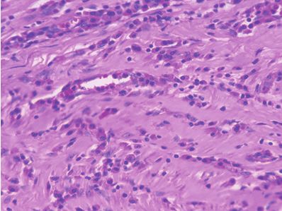
A B
C D
Fig. 1. (A) Gastric wall with marked submucosal fibrosis and prominent lymphoid follicles. (B) Diffuse plasma cell rich inflammation. (C) Stori-
form fibrosis with diffuse inflammation and a lymphoid follicle. (D) Many immunoreactive IgG4-positive plasma cells.
IgG4 immunohistochemistry (IHC) and serum IgG4 level hancing soft tissue lesion in the distal body of the stomach
were assessed. The IgG4 IHC showed 35–40 immunoreactive along the lesser curvature, which was suspected to be GIST or
plasma cells per high power field (hpf), and serum IgG4 level leiomyoma. He was advised to undergo follow-up and elective
was elevated (4.36 g/L). These tests confirmed the diagnosis of surgery. His gastric symptoms worsened over the next 10 months,
IgG4-RD. Subsequent serum levels after 3 and 4 months were and he underwent excision of the gastric submucosal lesion in
2.71g/L and 2.53 g/L, respectively. The patient was treated with October 2018. Histopathological examination revealed a well-
steroids and azathioprine. He experienced postsurgery complica- circumscribed globular mass measuring 4 × 3.3 × 3.5 cm, and
tions that required revision of the gastric anastomosis. During whorling was seen on the cut surface. Microscopically, the le-
follow-up, his prognosis while receiving medical treatment has sion was composed of extensively fibrotic and sclerotic stroma
been good. with a storiform pattern of fibrosis in foci (Fig. 1C). Discrete,
cords and clusters of plasma cells admixed with lymphocytes, a
Case 2 few eosinophils and a few scattered lymphoid aggregates and
The second patient was a 58-year-old male found to have erosive follicles were present. Perivascular aggregates of plasma cells
gastritis and a submucosal swelling in the body of the stomach were also present. There was no evidence of obliterative or non-
in December 2017, while undergoing upper gastrointestinal obliterative phlebitis. Bundles of smooth muscle were identified
endoscopy for investigation of dyspepsia. CT examination at the periphery on one aspect. The possibility of IgG4-RD of
showed a well-defined, 3 × 2.9 cm, round, homogeneous, en- the stomach was suggested, and IgG4 IHC and serum estima-
https://doi.org/10.4132/jptm.2020.02.10 http://jpatholtm.org/260 • Ramakrishna B et al.
tion were recommended. The IgG4 IHC revealed 20–30 im- diffuse or partial enlargement of the organ and histologically as
muno-reactive plasma cells/hpf (Fig. 1D). Serum IgG4 was ele- dense lymphoplasmacytic infiltration with an increase in IgG4
vated at 3.11g/L, well above the 1.35 g/L cut-off for diagnosis of plasma cells on immunostaining, a storiform pattern of fibrosis
IgG4-RD. The patient did not return for follow-up. and obliterative or non-obliterative phlebitis, and an increase in
serum IgG4 level.
Ethics statement IgG4-RD of the gastrointestinal tract is very rare and can
The authors certify that they obtained patient consent for present as diffuse wall thickening or as polyp or mass-like lesion
publication, and the study was approved by the Institutional [2,3]. Even though obliterative phlebitis was not present in
Review Board of Sri Ramaswamy Memorial Institutes for Med- these two cases, the presence of dense fibrosis, which was storiform
ical Science, Chennai, India (IEC NO: SIMS IEC/other/18/2019). in one case, and dense lymphoplasmacytic infiltration with lym-
phoid aggregates and follicles, presenting as a submucosal mass-
DISCUSSION like lesion, suggested the possibility of IgG4-RD, which was
confirmed by IHC and elevated serum IgG4 level. The presence
IgG4-RD is an immune-mediated fibroinflammatory lesion, of at least two histological features is required for confident diag-
first described in patients with sclerosing cholangitis associated nosis of IgG4-RD; in most cases, dense lymphoplasmacytic infil-
with autoimmune pancreatitis type 1 [5]. Later, it was identified trate and diffuse/storiform fibrosis are seen. Additional clinical,
in other organs including the liver, bile ducts, salivary glands, serological (serum level > 135 mg/dL or 1.35 g/L), or radiological
retroperitoneum, lymph nodes, and lungs. It is characterized by evidence is required to confirm IgG4-RD [1].
Table 1. Clinicopathological features of IgG4-related disease manifesting as gastric lesions
Age Endoscopic finding/ Size Serum
Case No. Sex Location Histopathology/IHC Treatment Study
(yr) Clinical diagnosis (mm) IgG4 levels
1 48 F Mass/GIST/NET Mid body 36 × 22
SF, LP, OP, IgG4 + 210/hpf, NA WR Woo et al. [3]
IgG4/IgG ratio about 85%
2 62 F Mass/gastic cancer Antrum 80 × 30 SF, LP, OP, IgG4 + ve Elevated DG Bulanov et al. [6]
lymphoplasmacytes > 50/hpf
3 59 F Mass/GIST NA 33 × 14 Abundant LP, SF, lymphoid Normal WR Kim et al. [7]
follicles, IgG4 > 50/hpf
4 56 F Mass/GIST NA 21 × 15 Abundant LP, SF, calcification, Normal WR Kim et al. [7]
IgG4 > 50/hpf
5 60 F Nodule/NA Fundus 10 × 15 Fibrosis, dense LP, Normal WR Chetty et al. [8]
IgG4 > 80/hpf
6 45 M Multiple nodules/NA Antrum Up to 22 LP, many eosinophils, NA DG Chetty et al. [8]
IgG4/IgG ratio 0.84
7 56 M Nodule/NA Body 8 SF, LP, IgG4-40-102/hpf NA ESR Na et al. [9]
IgG4/IgG ratio 80%–90%
8 58 M Nodule/AIP Fundus and body 14 Dense LP, extensive IgG and Normal Steroid Baez et al. [10]
IgG4 + staining
9 55 F Nodule/GIST Body 20 Dense hyalinization, LP, Normal ESR Zhang et al. [11]
IgG4/IgG ratio 41%
10 75 F Polyp/GIST Body 56 × 50 Fibrosis, LP, many eosinophils, Normal WR Rollins et al. [12]
IgG4–39/hpf
11 44 M Mass/GIST Body 20 × 18 Fibrosis, LP, IgG4 + ve Normal ESR Otsuka et al. [13]
lymphoplasmacytes
12 27 F Mass/GIST/NET Fundus 40 Dense fibrosis, LP, IgG4/IgG Normal WR Cheong et al. [14]
ratio 25.3%
13 29 F Mass/GIST Body 20 × 15 Fibrosis, LP, IgG4 + ve plasma NA WR Skorus et al. [15]
cells 150/hpf
14 43 M Mass/GIST Antrum 70 × 50 Dense LP, IgG4 + plasma cells Elevated WR + Present case 1
35–40/hpf Steroids
15 58 M Mass/GIST Distal body 45 × 40 LP, SP, IgG4 + ve plasma cells Elevated WR Present case 2
20–30/hpf
IHC, immunohistochemistry; GIST, gastrointestinal stromal tumor; NET, neuroendocrine tumor; SF, storiform fibrosis; LP, lymphoplasmacytic infiltrate; OP, ob-
literative phlebitis; hpf, high power field; NA, not available; WR, wedge resection; DG, distal gastrectomy; ESR, endoscopic submucosal resection; AIP, auto-
immune pancreatitis.
http://jpatholtm.org/ https://doi.org/10.4132/jptm.2020.02.10Gastric IgG4-related disease mimicking GIST • 261
The cut-off point for the presence of IgG4 plasma cells in tis- Balakrishnan Siddartha Ramakrishna:
sues varies and can range from > 30 plasma cells/hpf to > 50/hpf, https://orcid.org/0000-0001-5090-9501
which is highly specific [1,5]. In biopsy specimens, more than 10
IgG4 plasma cells/hpf were reported in one study [16]. However, Author Contributions
the cut-off points vary depending on organ system. Some stud- Conceptualization: BR.
ies have suggested that IgG4+/IgG+ plasma cell ratio > 0.4 is a Data curation: BR, RY.
marker of IgG4-RD in the presence of classic histopathological Investigation: BR, KV, PR, RY, BSR.
features and with a compatible clinical features [1,17]. Visualization: BR.
IgG4-RD can involve multiple organs or any sites in the body Writing—original draft: BR.
synchronously or metachronously [18]. Patients can present with Writing—review and editing: BR, RY, BSR.
non-specific symptoms and swelling or mass-like lesion. Patients
with biliary or pancreatic lesion can present with jaundice, weight Conflicts of Interest
loss, and vague abdominal pain. The disease can be an incidental The authors declare that they have no potential conflicts of
finding during radiological examination and can be mistaken for interest.
malignancy, as there are no specific radiological features charac-
teristic of this disease [18,19]. Most cases of gastric IgG4-RD Funding
have been reported in middle-aged patients, and both men and No funding to declare.
women are affected [3,6]. Both patients in this report were mid-
dle-aged men. REFERENCES
IgG4-RD of the stomach was first described in 2004 by Shinji
1. Deshpande V, Zen Y, Chan JK, et al. Consensus statement on the
et al. [20], presenting as a gastric ulcer. Because it is difficult to
pathology of IgG4-related disease. Mod Pathol 2012; 25: 1181-92.
diagnose clinically, especially in isolated cases, most of the reported
2. Koizumi S, Kamisawa T, Kuruma S, et al. Immunoglobulin G4-re-
patients have undergone surgery. Because this disease involves a
lated gastrointestinal diseases, are they immunoglobulin G4-related
submucosal lesion in the stomach, these cases are often misdi-
diseases? World J Gastroenterol 2013; 19: 5769-74.
agnosed as GIST [3,7,18,19] and are difficult to diagnose on en-
3. Woo CG, Yook JH, Kim AY, Kim J. IgG4-related disease presented
doscopic forceps biopsy, similar to our cases. Gastric lesions that
as a mural mass in the stomach. J Pathol Transl Med 2016; 50: 67-70.
have been mistaken for GIST or gastric cancer have been reported
4. Stone JH, Zen Y, Deshpande V. IgG4-related disease. N Engl J Med
in the literature (Table 1) [3,6-15]. IgG4-RD of the stomach that
2012; 366: 539-51.
involved the regional lymph nodes has also been reported [6].
5. Kamisawa T, Funata N, Hayashi Y, et al. A new clinicopathological
Although steroids are the first therapeutic option for treating
entity of IgG4-related autoimmune disease. J Gastroenterol 2003;
IgG4-RD, it is difficult to diagnose gastric IgG4-RD without
38: 982-4.
histopathological examination. Almost all cases have been reported
6. Bulanov D, Arabadzhieva E, Bonev S, et al. A rare case of IgG4-related
after surgical resection. Therefore, this disease should be considered
disease: a gastric mass, associated with regional lymphadenopathy.
in the differential diagnosis of gastric submucosal mass lesion.
BMC Surg 2016; 16: 37.
To conclude, we present two cases of IgG4-RD of the stom-
7. Kim DH, Kim J, Park DH, et al. Immunoglobulin G4-related inflam-
ach that presented as a mass lesion and were clinically suspected
matory pseudotumor of the stomach. Gastrointest Endosc 2012; 76:
to be GIST. The diagnosis was made only after histopathological
451-2.
examination of resection specimens. This highlights the impor-
8. Chetty R, Serra S, Gauchotte G, Markl B, Agaimy A. Sclerosing
tance of considering this disease in differential diagnosis to avoid
nodular lesions of the gastrointestinal tract containing large num-
surgical resection.
bers of IgG4 plasma cells. Pathology 2011; 43: 31-5.
9. Na KY, Sung JY, Jang JY, et al. Gastric nodular lesion caused by
IgG4-related disease. Pathol Int 2012; 62: 716-8.
ORCID
Banumathi Ramakrishna: 10. Baez JC, Hamilton MJ, Bellizzi A, Mortelé KJ. Gastric involvement
https://orcid.org/0000-0001-5635-1633 in autoimmune pancreatitis: MDCT and histopathologic features.
JOP 2010; 11: 610-3.
https://doi.org/10.4132/jptm.2020.02.10 http://jpatholtm.org/262 • Ramakrishna B et al.
11. Zhang H, Jin Z, Ding S. Gastric calcifying fibrous tumor: a case of troenterol 2007; 42 Suppl 18: 39-41.
suspected immunoglobulin G4-related gastric disease. Saudi J Gas- 17. Miyabe K, Zen Y, Cornell LD, et al. Gastrointestinal and extra-intes-
troenterol 2015; 21: 423-6. tinal manifestations of IgG4-related disease. Gastroenterology 2018;
12. Rollins KE, Mehta SP, O'Donovan M, Safranek PM. Gastric IgG4- 155: 990-1003.
related autoimmune fibrosclerosing pseudotumour: a novel location. 18. Seo HS, Jung YJ, Park CH, Song KY, Jung ES. IgG4-related disease
ISRN Gastroenterol 2011; 2011: 873087. in the stomach which was confused with gastrointestinal stromal
13. Otsuka R, Kano M, Hayashi H, et al. Probable IgG4-related scleros- tumor (GIST): two case reports and review of the literature. J Gastric
ing disease presenting as a gastric submucosal tumor with an intense Cancer 2018; 18: 99-107.
tracer uptake on PET/CT: a case report. Surg Case Rep 2016; 2: 33. 19. Inoue D, Yoneda N, Yoshida K, et al. Imaging and pathological fea-
14. Cheong HR, Lee BE, Song GA, Kim GH, An SG, Lim W. Immuno- tures of gastric lesion of immunoglobulin G4-related disease: A
globulin G4-related inflammatory pseudotumor presenting as a case report and review of the recent literature. Mod Rheumatol
solitary mass in the stomach. Clin Endosc 2016; 49: 197-201. 2019; 29: 377-82.
15. Skorus U, Kenig J, Mastalerz K. IgG4-related disease manifesting 20. Shinji A, Sano K, Hamano H, et al. Autoimmune pancreatitis is
as an isolated gastric lesion- a literature review. Pol Przegl Chir closely associated with gastric ulcer presenting with abundant
2018; 90: 41-5. IgG4-bearing plasma cell infiltration. Gastrointest Endosc 2004; 59:
16. Chari ST. Diagnosis of autoimmune pancreatitis using its five cardi- 506-11.
nal features: introducing the Mayo Clinic’s HISORt criteria. J Gas-
http://jpatholtm.org/ https://doi.org/10.4132/jptm.2020.02.10You can also read