Cholesterol crystal embolism to the gastrointestinal tract: a catastrophic case
←
→
Page content transcription
If your browser does not render page correctly, please read the page content below
Article / Clinical Case Report
Cholesterol crystal embolism to the gastrointestinal tract:
a catastrophic case
Miao Tiana , Karen E Matsukumaa
How to cite: Tian M, Matsukuma KE. Cholesterol crystal embolism to the gastrointestinal tract: a catastrophic case.
Autops Case Rep [Internet]. 2019;9(2):e2018082. https://doi.org/10.4322/acr.2018.082
ABSTRACT
Cholesterol crystal embolism is a rare and easily overlooked cause of colonic ischemia. The gastrointestinal tract is the
third most common organ system affected by cholesterol emboli, second only to kidney and skin. Here we present a
catastrophic case of gastrointestinal cholesterol crystal embolism leading to extensive post-operative bowel infarction and
ultimately death. For a practicing pathologist, careful attention to the vessels of any ischemic bowel and recognition of
the subtle but distinct angular imprint of cholesterol crystals facilitates prompt identification of the atheroemboli. In some
cases, early identification may help mitigate further tissue damage. In more acute and severe cases, identification of the
cholesterol crystal emboli may be important primarily for documentation of procedural complications.
Keywords
Cholesterol; Embolism; Gastrointestinal Tract; Ischemia.
CASE REPORT
A 62-year-old man underwent bilateral common was identified, and these segments were resected.
femoral endarterectomy and aortobifemoral bypass Intraoperatively, the patient became acidotic and
for severe claudication secondary to extensive hyperkalemic, and upon transport to the intensive care
atherosclerosis. On postoperative day 1, he developed unit, the patient expired.
melena and hematochezia. Emergency colonoscopy
demonstrated severe ischemia of the proximal
colon and severe circumferential ischemic proctitis. PATHOLOGIC RESULTS
Due to severe hypoxia, hypotension, and lactic
Gross examination of the small bowel and colon
acidosis, exploratory laparotomy was performed
resections revealed dusky serosa and dark red to grey
on postoperative day 2. Necrosis of the distal ileum
mucosa with attenuated folds (Figure 1).
and cecum was identified, and these segments were
subsequently resected. Dusky areas in the remainder On the histologic sections, extensive mucosal and
of the small bowel and colon were left intact, pending focal submucosal necrosis was present throughout
future operative evaluation. On the following day, the bowel (Figure 2). In the submucosa, numerous
the patient developed multi-organ failure, and a thin, needle-shaped clefts were noted in congested
second-look exploratory laparotomy was performed. arterioles. Fibrin thrombi were also seen in some of
Diffuse small bowel and ascending colon necrosis the arterioles (Figure 2A). Additionally, focal areas of
a
University of California, Davis Medical Center, Department of Pathology and Laboratory Medicine. Sacramento, CA, USA.
Autopsy and Case Reports. ISSN 2236-1960. Copyright © 2019. This is an Open Access article distributed
under the terms of the Creative Commons Attribution Non-Commercial License, which permits unrestricted
non-commercial use, distribution, and reproduction in any medium provided the article is properly cited.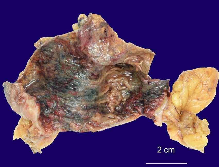
Cholesterol crystal embolism to the gastrointestinal tract: a catastrophic case

viable mucosa were noted on histologic sections. These                       DISCUSSION
areas were notable for absence of the aforementioned
vascular changes.                                                                 Needle-shaped clefts are the characteristic imprint
                                                                             of cholesterol crystals. The cholesterol itself is no longer
                                                                             present, as it dissolves during tissue processing (due
                                                                             to serial incubations in ethanol and xylene). When the
                                                                             needle-shaped clefts are present in small blood
                                                                             vessels in association with histologic features of tissue
                                                                             ischemia, the findings are diagnostic of cholesterol
                                                                             crystal embolism (CCE). 1-3 Fibrin thrombi develop
                                                                             secondarily as a result of obstructed blood flow.1,3
                                                                                  C C E a ls o re fe rre d to a s a th e ro e m b o l i s m
                                                                             or cholesterol embolization syndrome is a rare
                                                                             manifestation of the atherosclerotic disease.4 It occurs
                                                                             when an atherosclerotic plaque in the aorta or
                                                                             other major artery ruptures and releases cholesterol
                                                                             crystals and atheroma debris into the bloodstream.
Figure 1. Ascending colon showing dusky mucosa                               The crystals embolize to small and medium-sized
with necrosis, hemorrhage, and attenuated folding                            arteries and arterioles, resulting in end-organ damage.1
(formalin‑fixed specimen).                                                   The location of the disrupted atherosclerotic plaque

Figure 2. Photomicrograph of the ascending colon. A – Cholesterol crystal clefts (arrow) in an arteriole located
in the submucosa of the cecum. Note the fibrin thrombus associated with the cholesterol crystal cleft. Mucosal
necrosis is present; B – Cholesterol crystal clefts (arrows) in arterioles of varying sizes within the superficial and mid
layers of the submucosa. (All images digitally scanned at 40X).

2-5                                                                                           Autops Case Rep (São Paulo). 2019;9(2):e2018082
Tian M, Matsukuma KE
determines the pattern of end-organ damage and improve prognosis. 13 Nonetheless, overall mortality
thus its clinical manifestations. CCE can occur upon rates are still as high as 80%.14
spontaneous rupture of an atherosclerotic plaque, In the present case, the patient had multiple
after iatrogenic mechanical trauma (e.g., vascular atherosclerotic plaques (identified by imaging) along
surgery, angiography, or angioplasty), or as a side the entire abdominal aorta, placing him at risk for
effect of medications targeting the coagulation system multi-organ CCE. Indeed, besides the extensive
(e.g., anticoagulants, or thrombolytics).5 gastrointestinal tract involvement, acute renal and liver
The incidence of CCE is estimated to be less failure emerged after aortobifemoral bypass. Although
than 0.5% based on studies of unselected autopsy an autopsy was not performed, the widespread aortic
populations. 6 However, it has been reported to be atherosclerosis, diffuse bowel infarction, and acute
1.4% after coronary catheterization.7 The risk factors renal and liver dysfunction point to multi-organ CCE
as the ultimate cause of his demise. Nonetheless,
for cholesterol crystal formation are primarily those of
since no post-surgical angiography studies were
atherosclerosis (e.g., smoking, hypercholesterolemia,
performed, we cannot entirely exclude the possibility
hypertension, and obesity), while the risk factors for
that vasoconstriction of large arteries contributed
cholesterol crystal embolization include atherosclerosis,
to the vascular compromise, as has been described
vascular manipulation, anticoagulation, and
previously.15 It is also worth noting that the patient’s
thrombolytic therapy.5,8
abdominal aorta clamp time (35 minutes) and blood
The gastrointestinal tract is the third most loss (1.5 L) would be considered moderate16 and would
frequent organ system affected (13.4%), following not be expected to be the primary cause of the patient’s
kidney (31.5%) and skin (15.5%). 6 Within the massive vascular compromise.
alimentary tract, the colon is the most common From a histologic standpoint, the differential
site of involvement (42.3%), followed by small diagnosis of colonic ischemia includes infection
intestine (33%), stomach (12.3%), rectum (9.2%), (e.g., cytomegalovirus, E. coli O157:H7), vasculitis,
and esophagus (1.5%).9 CCE may also involve the mesenteric ischemia due to prolonged aortic
pancreas, liver, and gallbladder. 10,11 CCE involving clamping, mesenteric thrombosis, radiation-induced
the digestive tract often presents as abdominal pain, vasculopathy, mesenteric myointimal hyperplasia,17
diarrhea, and gastrointestinal bleeding. 5,8,12 Because and enterocolic lymphocytic phlebitis, 18 none of
the clinical presentation is not specific, the disease can which were observed in this case. Additionally, a
masquerade as other conditions (e.g., infection, tumor, thrombus overlying an atheromatous plaque can
inflammatory bowel disease). CCE can present as either become loose and occlude downstream large caliber
a chronic indolent disease that resolves over time or arteries (thromboembolism).3,4,19 Thromboembolism
an acute catastrophic multi-organ disorder with poor is distinguished from CCE in that it usually involves a
prognosis.5 Although histologic evaluation is the gold single target organ and the emboli are predominantly
standard for diagnosis, the co-occurrence of 3 clinical composed of fibrin, whereas CCE is characterized by
findings: (1) history of known risk factors for CCE (e.g., multiple cholesterol crystal emboli in small arterioles.
vascular surgery, anticoagulation), (2) acute onset Of note, some studies report the presence of CCE in
renal failure with creatinine elevation of greater than small vessels mostly less than 200 µm in diameter,2,20
50% of baseline, and (3) signs of cutaneous vascular whereas in the current case the size of the affected
compromise (e.g., livedo reticularis, purples toes) or arterioles ranged from 50 to 950 µm in diameter,
similar to the findings of Flory.1
funduscopic evidence of retinal atheroemboli, has
proved to be relatively specific for disseminated CCE.13
Thus, non-invasive procedures such as funduscopic CONCLUSION
examination can be useful. No specific treatment for
CCE exists; however, early and aggressive supportive CCE is a type of vasculopathy that occurs when an
therapy (e.g., use of blood pressure lowering agents - in atherosclerotic plaque ruptures and releases cholesterol
the context of cardiac failure, hemodialysis, nutritional crystals and atheroma debris into downstream arterioles.
support, discontinuation of inciting medications) can Although the digestive tract is a commonly affected
Autops Case Rep (São Paulo). 2019;9(2):e2018082 3-5Cholesterol crystal embolism to the gastrointestinal tract: a catastrophic case
site, CCE is a relatively uncommon cause of colonic 9. Moolenaar W, Lamers CB. Cholesterol crystal embolisation
ischemia overall and as such must be kept in mind to the alimentary tract. Gut. 1996;38(2):196-200. http://
dx.doi.org/10.1136/gut.38.2.196. PMid:8801196.
in the differential diagnosis of ischemia, particularly
in the setting of a recent vascular procedure and/or 10. Konstantinidis IT, Warshaw AL, Deshpande V, et al.
Cholesterol crystal embolization presenting as either solid
certain patient-specific risk factors (atherosclerosis, or cystic pancreatic lesion. J Surg Oncol. 2010;102(6):706-
smoking, hypercholesterolemia, hypertension, obesity). 8. http://dx.doi.org/10.1002/jso.21521. PMid:20976733.
Unfortunately, due to the relatively non-specific 11. Moolenaar W, Lamers CB. Cholesterol crystal
symptoms, a high degree of clinical suspicion is embolization to liver, gallbladder, and pancreas. Dig
necessary for establishing a timely diagnosis. For the Dis Sci. 1996;41(9):1819-22. http://dx.doi.org/10.1007/
BF02088752. PMid:8794801.
pathologist, attention to the character and content of
the vessels is critical for identification. If not viewed 12. Moolenaar W, Lamers CB. Cholesterol crystal embolization
and the digestive system. Scand J Gastroenterol
carefully, cholesterol clefts may be overlooked as
Suppl. 1991;188(Suppl 188):69-72. http://dx.doi.
artefactual fractioning of vessel lumina rather than org/10.3109/00365529109111232. PMid:1775943.
an indication of a potentially widespread and morbid
13. Belenfant X, Meyrier A, Jacquot C. Supportive treatment
pathologic process. improves survival in multivisceral cholesterol crystal
embolism. Am J Kidney Dis. 1999;33(5):840-50.
http://dx.doi.org/10.1016/S0272-6386(99)70415-4.
REFERENCES PMid:10213638.
14. Ghanem F, Vodnala D, K Kalavakunta J, et al. Cholesterol
1. Flory CM. Arterial occlusions produced by emboli from crystal embolization following plaque rupture: a
eroded aortic atheromatous plaques. Am J Pathol. systemic disease with unusual features. J Biomed Res.
1945;21(3):549-65. PMid:19970827. 2017;31(2):82-94. PMid:28808190.
2. Kassirer JP. Atheroembolic renal disease. N Engl J 15. Imanaka K, Kyo S, Ban S. Possible close relationship
Med. 1969;280(15):812-8. http://dx.doi.org/10.1056/ between non-occlusive mesenteric ischemia and
NEJM196904102801506. PMid:4887250. cholesterol crystal embolism after cardiovascular
surgery. Eur J Cardiothorac Surg. 2002;22(6):1032-4.
3. Eliot RS, Kanjuh VI, Edwards JE. Atheromatous
http://dx.doi.org/10.1016/S1010-7940(02)00590-0.
embolism. Circulation. 1964;30(4):611-8. http://dx.doi.
PMid:12467838.
org/10.1161/01.CIR.30.4.611. PMid:14211824.
16. Bruls S, Quaniers J, Tromme P, Lavigne JP, Van Damme
4. Saric M, Kronzon I. Aortic atherosclerosis and embolic H, Defraigne JO. Comparison of laparoscopic and open
events. Curr Cardiol Rep. 2012;14(3):342-9. http://dx.doi. aortobifemoral bypass in the treatment of aortoiliac
org/10.1007/s11886-012-0261-2. PMid:22437371. disease: results of a contemporary series (2003-2009).
5. Ben-Horin S, Bardan E, Barshack I, Zaks N, Livneh Acta Chir Belg. 2012;112(1):51-8. http://dx.doi.org/10.
A. Cholesterol crystal embolization to the digestive 1080/00015458.2012.11680795. PMid:22442910.
system: characterization of a common, yet overlooked 17. Yantiss RK, Cui I, Panarelli NC, Jessurun J. Idiopathic
presentation of atheroembolism. Am J Gastroenterol. myointimal hyperplasia of mesenteric veins: an uncommon
2003;98(7):1471-9. http://dx.doi.org/10.1111/j.1572- cause of ischemic colitis with distinct mucosal features.
0241.2003.07532.x. PMid:12873565. Am J Surg Pathol. 2017;41(12):1657-65. http://dx.doi.
org/10.1097/PAS.0000000000000905. PMid:28817406.
6. Moolenaar W, Lamers CB. Cholesterol crystal
embolization in the Netherlands. Arch Intern Med. 18. Louie CY, DiMaio MA, Charville GW, Berry GJ, Longacre
1996;156(6):653-7. http://dx.doi.org/10.1001/ TA. Gastrointestinal Tract Vasculopathy: Clinicopathology
archinte.1996.00440060081009. PMid:8629877. and Description of a Possible “New Entity” With
Protean Features. Am J Surg Pathol. 2018;42(7):866-
7. Fukumoto Y, Tsutsui H, Tsuchihashi M, Masumoto A, 76. http://dx.doi.org/10.1097/PAS.0000000000001060.
Takeshita A. The incidence and risk factors of cholesterol PMid:29624512.
embolization syndrome, a complication of cardiac
catheterization: a prospective study. J Am Coll Cardiol. 19. Tunick PA, Kronzon I. Atheromas of the thoracic aorta:
2003;42(2):211-6. http://dx.doi.org/10.1016/S0735- clinical and therapeutic update. J Am Coll Cardiol.
1097(03)00579-5. PMid:12875753. 2000;35(3):545-54. http://dx.doi.org/10.1016/S0735-
1097(99)00604-X. PMid:10716454.
8. Fries C, Roos M, Gaspert A, et al. Atheroembolic disease:
a frequently missed diagnosis: results of a 12-year 20. Quinones A, Saric M. The cholesterol emboli syndrome
matched-pair autopsy study. Medicine. 2010;89(2):126- in atherosclerosis. Curr Atheroscler Rep. 2013;15(4):315.
32. http://dx.doi.org/10.1097/MD.0b013e3181d5eb39. http://dx.doi.org/10.1007/s11883-013-0315-y.
PMid:20517183. PMid:23423524.
4-5 Autops Case Rep (São Paulo). 2019;9(2):e2018082Tian M, Matsukuma KE Author contributions: Tian M made the figures. Matsukuma KE and Tian M wrote the manuscript and developed the discussion. All authors collectively proofread the manuscript and approved it for publication. The manuscript is in accordance with our institutional ethical research committee. Conflict of interest: None Financial support: None Submitted on: February 1st, 2019 Accepted on: March 11th, 2019 Correspondence Karen E Matsukuma Department of Pathology and Laboratory Medicine - Davis Medical Center- University of California 4400 V Street – Sacramento/CA – USA PO Box: 95817 Phone: +1 (91) 6734-2529 kmatsukuma@ucdavis.edu Autops Case Rep (São Paulo). 2019;9(2):e2018082 5-5
You can also read