Evaluation of Chest Pain in 2019 - Mohit Bhasin MD, FACC Norfolk, VA 35 min +10 min questions - American College of Physicians
←
→
Page content transcription
If your browser does not render page correctly, please read the page content below
Evaluation of Chest
    Pain in 2019

Mohit Bhasin MD, FACC

      Norfolk, VA

35 min +10 min questions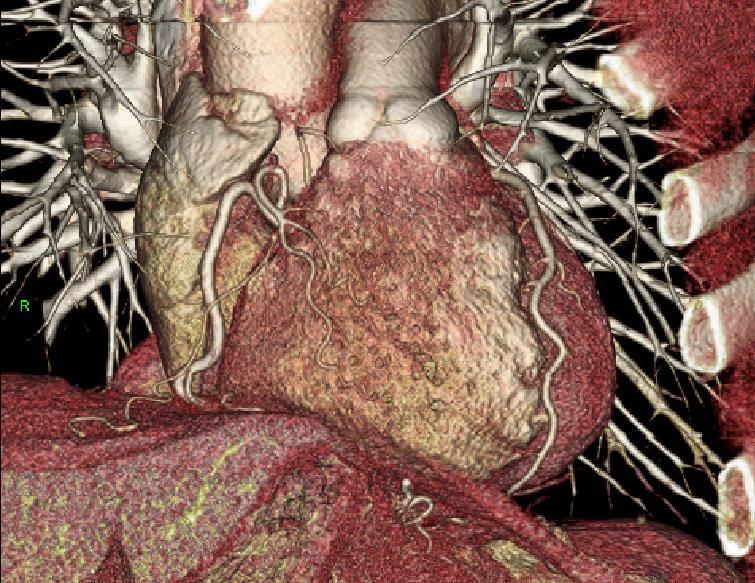
No Financial
Disclosures
Chest Pain is one of the most difficult yet
common symptoms evaluated by PCPs,
Emergency Physicians, Cardiologists, and
searches on the internet by patients.
Institute of Medicine

7 million Chest Pain patients present to U.S.
Emergency Departments yearly (National Hospital Medical Care
Survey), with benign to life-threatening causes, costing

$6 billion.
CMS Physician Compare
               Public-reported Data
      ?new iteration of incentives and punishments
• CMS physician spend per beneficiary to be publically reported, as well hospitals.
• “2017 MIPS scores will be public and posted on 'Physician Compare' on the CMS
  website as a result of MACRA. Performance will be ranked against peers.
• Non participation will give you the lowest possible score.
Chest Pain is a high risk Chief Complaint
  Current consensus is to exclude life-
 threatening causes of chest pain to less
 than 1% uncertainty (99% NPV), do it
     cost-effectively, efficiently and
      noninvasively when possible.
Many clinicians do not want to
    evaluate chest pain.
Epidemiology of chest pain
reflects selection bias
Diagnosis Primary Care: Primary Care: Emergency
USA Europe Department
Musculoskeletal conditions 36% 29% 7%
Gastrointestinal disease 19% 10% 3%
Serious Cardiovascular disease (Infarction, 16% 13% 54%
Unstable Angina, PE, HF)
Stable CAD 10% 8% 13%
Unstable CAD 1.5% 13%
Psychosocial or Psychiatric disease 8% 17% 9%
Pulmonary disease 5% 20% 12%
(Pneumonia, Pneumothorax, Lung Cancer)
Non-specific chest pain 16% 11% 15%
➢J Fam Pract. 1994 Apr;38(4):345-52.
➢Fam Pract 2001;18: 586-589.Acute Cardiopulmonary symptoms
(chest pain, dysnea,)
ACS AAS VTE Tamponade
Esophageal
STEMI Acute Venothrombo- Rupture
NSTEMI Aortic embolism
Unstable Angina Syndromes Spontaneous
tension PTX
16%
IMH PAU
Aortic Dissection
Penetrating Symptomatic
60% Intramural
Atherosclerotic Aneurysm
Hematoma
Ulcer (impending rupture)
(Type A 60%, B 40%) 5-25% 7-12%2018 FDA Recommendation Healthcare professionals should: • Avoid prescribing fluoroquinolone antibiotics to patients who have an aortic aneurysm or are at risk for an aortic aneurysm, such as patients with peripheral atherosclerotic vascular diseases, hypertension, certain genetic conditions such as Marfan syndrome and Ehlers-Danlos syndrome, and elderly patients. • Prescribe fluoroquinolones to these patients only when no other treatment options are available. • Stop fluoroquinolone treatment immediately if a patient reports side effects suggestive of aortic aneurysm or dissection.
• For Life-threatening causes,
  – Consider the entire thorax and abdomen.
• Know characteristics of noninvasive tools in
  your region
• Exclude life-threatening causes to at least
Outpatient Chest Pain
Up to 16% of outpatient chest pain
evaluations may never reach a definitive
diagnosis.

Before ordering a test elicit patient’s fears
(false negative stress testing, invasive
procedures), expectations (resume exercise
with confidence), and define a successful
chest pain workup together:
For a successful chest pain workup:
• Plan what happens if we don’t find heart or lung
  disease– it’s not intellectually satisfying but the
  outcome is better
• Exclude life-threatening causes including CAD
  sufficiently that ‘if you re-start exercising and
  feel chest discomfort, that you will have the
  confidence to push through’
  – This recommendation requires 100% NPV
• Plan to follow patients no matter what the tests
  show, to ask more questions and look for patterns
  that might suggest other diagnoses
If persistent chest discomfort, or evolution,
then start evaluating for rare causes-
 avoid ‘zebra retreat’
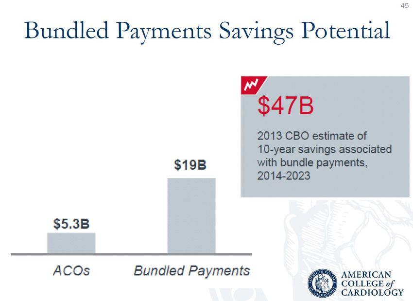
Chest Pain evaluation is in rapid evolution due to
• Recent multi-center trial data evaluating event-free
  survival for years after randomizing patients to
  testing alternatives
• cost pressure, including diagnostic bundle
  payments
• background medico-legal pressure – high rate of
  claims from missed MI, Acute aortic syndromes,
  and PE – pushes for tests with high sensitivity and
  high NPV.
Only “typical” angina helps
High likelihood of ACS:
• Chest pain with radiation to the arms or shoulders
• Chest pain associated with exertion, diaphoresis,
  nausea or vomiting.
• Chest pain described as pressure or similar to a
  previous MI.”

Low likelihood of ACS:
• Pleuritic or positional.

➢ JAMA. 2005 Nov
23;294(20):2623-9.
Important findings with chest pain
  New diastolic murmur
  Late-peaking systolic murmur
   BP differential
    between the arms
   Diminished pulses
   JVD and HJR
   Chest-wall tenderness on
     palpation (markedly reduces
     ACS probability in low
     pretest probability settings)
Tests become critically important
to diagnosis and subsequent
outcomesOrganize tools based on pre-test probability:
after History, Risk Factors, Exam, EKG, CXR, and a cardiac
enzyme, weigh the testing strategy sensitivity/NPV, renal
and radiation risks if young, and ability to improve patient
outcomesNTG Response
GI Cocktail Response
Stress Echo,
Plain ECG Treadmill
Empiric PPI Response
Chest Wall Palpation
Telemetry Observation Stress MRI
NUCLEAR SPECT
Stress MRI
Coronary CT CATH / IVUS / FFR
CAD
Post-test Probability of life-threatening disease
0 10% 50% 80% 100%
Pre-/Post test probability of CAD
(Calcium scoring is absent - only for asymptomatic persons for risk stratification)Exercise Treadmill Test (ETT)
• Non-imaging stress test
• Bruce protocol
• Duke Treadmill Score (DTS)
– DTS = exercise time – (5 x max ST deviation in mm) – (4 x
treadmill angina index)
– Risk stratification
• < -10: High risk
• -10 to +4: Intermediate risk
• ≥ +5: Low risk
• Limitations1
– False-positive and false-negative results
1. Gibbons RJ, et al. ACC/AHA 2002 guideline update for exercise testing. 2002.
Available at http://www.acc.org/clinical/guidelines/exercise/exercise_clean.pdf.ST Segment Depression
Diffuse Anterolateral T Wave Abnormalities
Exercise Treadmill Testing
• Abnormal (Positive) Test
  – 39% incidence of cardiac events over 6.3 years
  – 89% of these events were occurrence of angina
• Normal (Negative) Test
  – 5.3% incidence of cardiac event over 6.3 years
  – 73% of these events were myocardial infarction
    or sudden death
Echocardiography (stress echo) • No ionizing radiation exposure • Stress echo commonly performed with exercise or dobutamine
Correlation of Coronary Arteries
  and Regional Wall Motion
      LAD
                               LAD   Diag

                                     Cx
 Cx
                            RCA/Cx
            RCA/Cx

                     LAD
                                     LAD
                            RCA/Cx

 RCA/Cx
                       Cx
ACCURACY TO DETECT ischemia • Plain non-imaging exercise treadmill(ETT) – 60% • ECHO – Stress - 75-80% – Specific but not sensitive
Nuclear stress
What’s missed with stress testing?
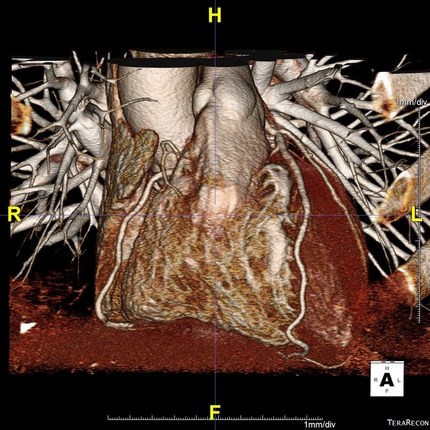
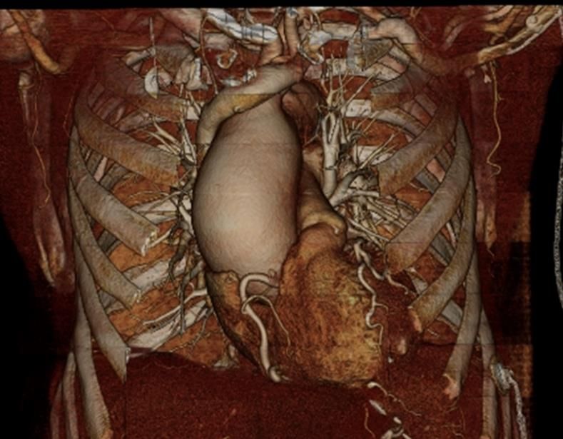
CT Heart and great vessels with contrast
Normal Cardiac CT has 100 % NPV
Coronary CT has been well-studied in the ED
setting, and recently in the outpatient setting,
with over a long-term follow-up, in 14 high-
quality randomized controlled trials and meta-
analyses.
CATCH Study

38
CATCH Study
                                                                          Coronary CT

                                                                           Stress test
                                 Coronary CT

                                 Stress test

Composite endpoint: cardiac death, MI, hospitalization for unstable angina, late
symptom-driven revascularization, & readmission for chest pain
Value of Cardiac CT in ED
NEJM Aug 2018
Death or MI after Coronary CT vs Stress Testing

                                       NEJM
                                       Aug 2018
Danish Registry
86,705 patients underwent either stress testing or coronary CTA followed
for 3.6 years.
Coronary CT lower risk of MI (hazard ratio: 0.71; 95% confidence
interval: 0.61 to 0.82).
Mads E. Jørgensen et al. JACC 2017;69:1761-1770NIH
PROMISE trial Economic Substudy:
Estimation of Initial Chest Pain Testing Costs
Dx Test Mean Cost* MD Fees** Total
Coronary CTA $285 $119 $404
Echo w/ exercise stress $428 $86 $ 514
Echo w/ pharmacologic stress $415 $86 $ 501
ECG-only nonimaging stress $137 $37 $174
Nuclear w/ exercise stress $829 $117 $ 946
Nuclear w/ pharmacologic stress $1015 $117 $ 1132
*based on costs in Premier database
**based on Medicare Fee ScheduleU.K. National Institute for Health and Care Excellence (NICE) clinical guideline on ‘Chest pain of recent onset’ • Coronary CT is the first line test before any consideration of invasive cath or stress testing. “Coronary CT has almost 100% accuracy in excluding significant coronary artery disease and when compared with stress tests or invasive angiography and is the lowest cost, and can be delivered at low radiation dose.”
U.K. National Institute for Health and Care Excellence
  (NICE) clinical guideline on ‘Chest pain of recent onset’
“The evidence has brought
new recommendations that
propose Cardiac CT as the
most clinically cost-effective
diagnostic first line test for all
patients presenting with chest
pain. ”
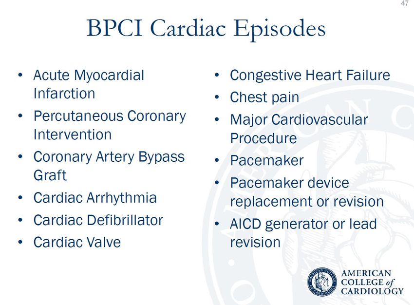
System Solution
      ACC Rapid Chest Pain Pathway

The American College of Cardiology endorsed Rapid Chest Pain
Assessment by CT heart to time to diagnosis, unnecessary admissions,
reduce total costs, and repeat evaluations for recurrent chest pain,
with near zero undetected cases of ACS, with a 5 year ‘warranty’ for
major adverse cardiac events after negative scans.
Green Pathway for Chest Discomfort with or without moderate suspicion of PE or Aortic Syndrome
How’s it work?

Patients need only one troponin before the scan; do not have
to be NPO

   – Operates continuously through the night: sensitive,
     safe cardiac diagnosis

   – Unlike stress testing, borderline troponin is not a
     contraindication given its safety
Normal aorta, pulmonary arteries, and LVEF. Proximal LAD and
Color20-30%
RCA 3D images,
mixed report,
density and cardiologist recommendation
plaques.
handed
Cardiologist to the patient ASA
Recommendation: for their
81mgPCP dailyat discharge
and atorvastatin 20mg
or rosuvastatin 10mg, with no further testing.
Intermediate 50-60% stenosis of the dominant RCA.
Cardiologist Recommendation: ASA 325mg and atorvastatin 80mg or
rosuvastatin 40mg, with consideration of antianginals if anginal quality
pain is noted and ischemic testing for these territories would be
reasonable. If symptoms are atypical for angina, could treat medically.
Normal Coronaries, LVEF, aorta and pulmonary arteries. Normal lungs.
Cardiologist Recommendation: No further testing. Return to full
exercise, discontinue aspirin. Primary prevention.Speeding test efficiency to bring down DRG spend • Syncope workups taking days (current strategy of echo, stress, cardiology consultation) – both LVEF/RWMA and stress information can be combined in one cardiac CT presenting 0700-2300 • Troponinemia (type II nstemi) slows discharges (current strategy of echo, stress, cardiology consultation) for dysnea,syncope, anemia, asthma, dizziness, weakness • Sob/Dysnea (CT PE then repeat CT coronary or stress and echo) – this current strategy is replaced by CT Triple rule out immediately • Because coronary CT is a much more sensitive test than stress echo, increase caths seen and increased PCI by 15-60%, net neutral on Nuclear stress, improved patient survival b/c no false negative stress, adherence to meds, other diagnoses.
Patient Survival with Coronary CT
How to Provide tests with few local
            imagers?
      Unlock efficiencies by
         telecardiology
Interstate Telecardiology Cardiologists and cardiac radiologists, across time zones, collaborating for accurate, cost-effective, and rapid diagnostics
National Physician Portal
 Connected by Cloud-Based 3D Software and a smartphone application

    Inter-state practice of medicine by physician colleagues
  Providing EKG quality assurance then coronary CT with as
needed clinical decision support to hospitals, free-standing Eds,
                       and imaging centers
Mon-Fri 0700-2300
Weekends 0700-1700 plus Mon-Fri 0700-2300
Interstate Medical Licensure Compact-
                 2017
              www.imlcc.org
Microhospitals and free-standing EDs, required to
stay open 24/7NTG Response
GI Cocktail Response
Stress Echo,
Plain ECG Treadmill
Empiric PPI Response
Chest Wall Palpation
Telemetry Observation Stress MRI
NUCLEAR SPECT
Stress MRI
Coronary CT CATH / IVUS / FFR
CAD
Post-test Probability of life-threatening disease
0 10% 50% 80% 100%
Pre-/Post test probability of CAD
(Calcium scoring absent—best for asymptomatic persons for risk stratification)You can also read